. 2024 Aug 21;16(8):1336.
doi: 10.3390/v16081336.

Bunyamwera Virus Infection of Wolbachia-Carrying Aedes aegypti Mosquitoes Reduces Wolbachia Density

Affiliations

Bunyamwera Virus Infection of Wolbachia-Carrying Aedes aegypti Mosquitoes Reduces Wolbachia Density

Daniella A Lefteri et al. Viruses. .

Abstract

Wolbachia symbionts introduced into Aedes mosquitoes provide a highly effective dengue virus transmission control strategy, increasingly utilised in many countries in an attempt to reduce disease burden. Whilst highly effective against dengue and other positive-sense RNA viruses, it remains unclear how effective Wolbachia is against negative-sense RNA viruses. Therefore, the effect of Wolbachia on Bunyamwera virus (BUNV) infection in Aedes aegypti was investigated using wMel and wAlbB, two strains currently used in Wolbachia releases for dengue control, as well as wAu, a strain that typically persists at a high density and is an extremely efficient blocker of positive-sense viruses. Wolbachia was found to reduce BUNV infection in vitro but not in vivo. Instead, BUNV caused significant impacts on density of all three Wolbachia strains following infection of Ae. aegypti mosquitoes. The ability of Wolbachia to successfully persist within the mosquito and block virus transmission is partially dependent on its intracellular density. However, reduction in Wolbachia density was not observed in offspring of infected mothers. This could be due in part to a lack of transovarial transmission of BUNV observed. The results highlight the importance of understanding the complex interactions between multiple arboviruses, mosquitoes and Wolbachia in natural environments, the impact this can have on maintaining protection against diseases, and the necessity for monitoring Wolbachia prevalence at release sites.

Keywords: Aedes; Bunyamwera; Wolbachia; arboviruses; mosquitoes; viruses.


Conflict of interest statement

The authors declare no conflicts of interest.

Figures

Figure 1
Figure 1
Wolbachia density is reduced by BUNV in Aa23 cells. Aa23 cells were infected with an MOI of 0.1. Samples were collected 24 h and 72 h p.i. N = 6 was used for each group. A line representing the sample mean has been included for each group. (A) A significant reduction in viral RNA in Wolbachia carrying lines can be observed at 72 h p.i. Cells were lysed in Trizol and analysed for viral RNA via qPCR. (B) Supernatant was collected and viral titres analysed via plaque assays. A significant reduction was observed in Wolbachia-carrying lines at 72 h p.i. (C) Wolbachia density of the cells was measured via qPCR. A significant reduction in density was observed in all three cell lines 72 h p.i. Bars represent the mean whilst error bars represent the standard deviation. * p < 0.05, ** p < 0.01, *** p < 0.001, **** p < 0.0001, ns = not significant.
Figure 2
Figure 2
2HPCD rescues BUNV infection in Aa23 cells. Aa23 cells were pre-treated with PBS, 0.1 mM or 1 mM of 2HPCD for 48 h prior to infection with an MOI of 0.1 of BUNV. N = 6 was used for each group. Samples were collected 24 h p.i and analysed via qPCR. A line representing the sample mean has been included for each group. (A) BUNV infection in cells is rescued when 1 mM 2HPCD was added. (B) Wolbachia density in cells was not rescued by the addition of 2HPCD.
Figure 3
Figure 3
Wolbachia does not block BUNV infection in Ae. aegypti mosquitoes. Ae. aegypti mosquitoes were infected via infectious blood meal containing 1 × 10⁷ PFU/mL of BUNV. Salivary glands and remaining carcass were collected at 9 and 12 days p.i. Each group has N = 20 with the exception for wAlbB at 9 days which has N = 18. A line representing the sample mean has been included for each group. (A) Salivary glands were placed in 250 uL serum free media and viral titres measured via plaque assays. (B) Carcass stored in 500 uL Trizol was analysed via qPCR. (C) Wolbachia density was measured by quantifying 16S via qPCR on mosquito carcass. Bars represent the mean whilst error bars represent the standard deviation. ** p < 0.01, *** p < 0.001, **** p < 0.0001, ns = not significant.
Figure 4
Figure 4
Ae. aegypti mosquitoes were infected with BUNV following feeding on an infectious blood meal containing 1 × 10⁷ PFU/mL of BUNV. Bloodfed mosquitoes were individualised and allowed to lay eggs. Eggs were collected and, following drying, they were hatched. Adult mosquitoes were collected 4–6 days post emergence. Both mothers and offspring were collected and placed into Trizol and analysed by qPCR. A line representing the sample mean has been included for each group. (A) Wolbachia density did not change between the groups. (B) BUNV was detected in the mothers but not the offspring.
Figure 5
Figure 5
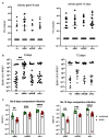
Ae. aegypti mosquitoes were infected with BUNV and/or SFV following feeding on an infectious blood meal containing 1 × 10⁷ PFU/mL of both viruses. Bloodfed mosquitoes were collected 7 d.p.i. Heads and thorax were separated from body and these samples were processed separately via qPCR. A line representing the sample mean has been included for each group. (A) SFV infection in the co-infected mosquitoes was quantified in the head and thorax and the bodies via qPCR. (B) BUNV infection in the co-infected mosquitoes was quantified sin the head and thorax and the bodies via qPCR. (C) wMel and wAlbB density of SFV only, BUNV only, or co-infected mosquitoes was quantified via qPCR in head and thorax and bodies. * p < 0.05.
Figure 6
Figure 6
Ae. aegypti mosquitoes were infected with BUNV and/or SFV following feeding on an infectious blood meal containing 1 × 10⁷ PFU/mL of both viruses. Bloodfed mosquitoes were collected 12 d.p.i. Heads and thorax were separated from body and these samples were processed separately via qPCR. (A) SFV infection in the head and thorax and the bodies of co-infected mosquitoes (B) BUNV infection in the head and thorax and the bodies of co-infected mosquitoes (C) SFV infection in non-Wolbachia-carrying mosquitoes infected with SFV only or alongside BUNV (D) BUNV infection in non-Wolbachia-carrying mosquitoes infected with BUNV only or alongside SFV (E) Wolbachia density in the head and thorax and bodies. * p < 0.05, ** p < 0.01, *** p < 0.001, **** p < 0.0001, ns = not significant.

Similar articles

References

    1. Ross P.A., Callahan A.G., Yang Q., Jasper M., Arif M.A.K., Afizah A.N., Nazni W.A., Hoffmann A.A. An elusive endosymbiont: Does Wolbachia occur naturally in Aedes aegypti? Ecol. Evol. 2020;10:1581–1591. doi: 10.1002/ece3.6012. - DOI - PMC - PubMed
    1. Walker T., Johnson P.H., Moreira L.A., Iturbe-Ormaetxe I., Frentiu F.D., McMeniman C.J., Leong Y.S., Dong Y., Axford J., Kriesner P., et al. The wMel Wolbachia strain blocks dengue and invades caged Aedes aegypti populations. Nature. 2011;476:450–453. doi: 10.1038/nature10355. - DOI - PubMed
    1. Moreira L.A., Iturbe-Ormaetxe I., Jeffery J.A., Lu G., Pyke A.T., Hedges L.M., Rocha B.C., Hall-Mendelin S., Day A., Riegler M., et al. A Wolbachia symbiont in Aedes aegypti limits infection with dengue, Chikungunya, and Plasmodium. Cell. 2009;139:1268–1278. doi: 10.1016/j.cell.2009.11.042. - DOI - PubMed
    1. Bian G., Xu Y., Lu P., Xie Y., Xi Z. The endosymbiotic bacterium Wolbachia induces resistance to dengue virus in Aedes aegypti. PLoS Pathog. 2010;6:e1000833. doi: 10.1371/journal.ppat.1000833. - DOI - PMC - PubMed
    1. Nazni W.A., Hoffmann A.A., NoorAfizah A., Cheong Y.L., Mancini M.V., Golding N., Kamarul G.M.R., Arif M.A.K., Thohir H., NurSyamimi H., et al. Establishment of Wolbachia Strain wAlbB in Malaysian Populations of Aedes aegypti for Dengue Control. Curr. Biol. 2019;29:4241–4248. doi: 10.1016/j.cub.2019.11.007. - DOI - PMC - PubMed

Publication types

LinkOut - more resources