. 2024 Aug 24:19:8695-8707.
doi: 10.2147/IJN.S470901. eCollection 2024.

Bone Regeneration Revolution: Pulsed Electromagnetic Field Modulates Macrophage-Derived Exosomes to Attenuate Osteoclastogenesis

Affiliations

Bone Regeneration Revolution: Pulsed Electromagnetic Field Modulates Macrophage-Derived Exosomes to Attenuate Osteoclastogenesis

Martina Trentini et al. Int J Nanomedicine. .

Erratum in

Abstract

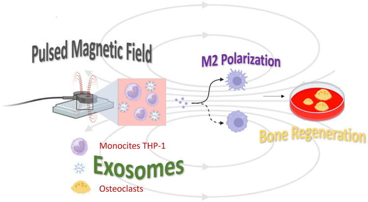
Introduction: In the process of bone regeneration, a prominent role is played by macrophages involved in both the initial inflammation and the regeneration/vascularization phases, due to their M2 anti-inflammatory phenotype. Together with osteoclasts, they participate in the degradation of the bone matrix if the inflammatory process does not end. In this complex scenario, recently, much attention has been paid to extracellular communication mediated by nanometer-sized vesicles, with high information content, called exosomes (EVs). Considering these considerations, the purpose of the present work is to demonstrate how the presence of a pulsed electromagnetic field (PEMF) can positively affect communication through EVs.

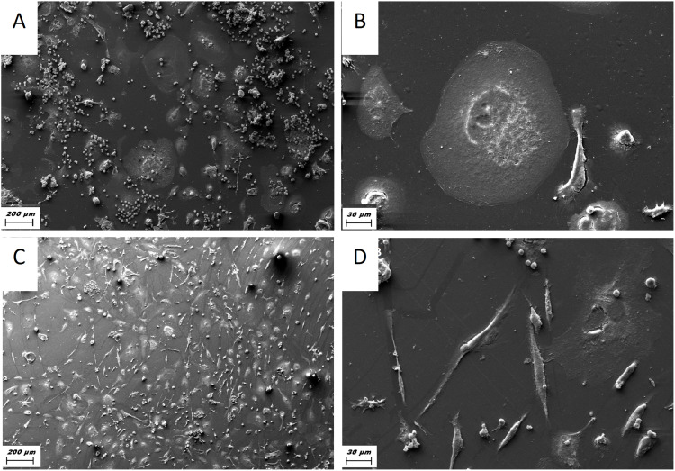
Methods: To this aim, macrophages and osteoclasts were treated in vitro with PEMF and analyzed through molecular biology analysis and by electron microscopy. Moreover, EVs produced by macrophages were characterized and used to verify their activity onto osteoclasts.

Results: The results confirmed that PEMF not only reduces the inflammatory activity of macrophages and the degradative activity of osteoclasts but that the EVS produced by macrophages, obtained from PEMF treatment, positively affect osteoclasts by reducing their activity.

Discussion: The co-treatment of PEMF with M2 macrophage-derived EVs (M2-EVs) decreased osteoclastogenesis to a greater degree than separate treatments.

Keywords: PEMF; bone; exosomes; macrophages; osteoclast.


Conflict of interest statement

Professor Shlomo Barak is the chairman of the board of Magdent Ltd. Dr Oleg Dolkart is a consultant for Magdent Ltd. The authors report no other conflicts of interest in this work.

Figures

None
Graphical abstract
Figure 1
Figure 1
Gene expression related to osteoclastogenesis activity of RAW264.7: CTSK, NFATC1, TRAP; miR-7, miR-1897. Orange bars are related to RANK-L treatment. Blue bars are related to the control (no RANK-L treatment). Grey bars are related to the presence of PEMF and RANK-L, yellow bars are related to PEMF. Statistical analysis of variance of the means between each group and the respective control was assessed by ANOVA and post hoc Bonferroni testing [#p < 0.0001].
Figure 2
Figure 2
SEM analyses of THP-1: (A and B) in normal conditions, cells acquire a round morphology typical of M1 phenotype; meanwhile, (C and D) in presence of PEMF, they acquire a fusiform morphology typical of M2 phenotype. (A and C) Scale Bar: 200 μm, (B and D) Scale Bar: 30 μm.
Figure 3
Figure 3
Heat map and histogram related to gene expression of principal genes expressed by THP-1 cells in presence of PEMF.
Figure 4
Figure 4
IPA canonical pathways of (A) down-regulated genes, and (B) up-regulated genes.
Figure 5
Figure 5
Analyses of EVs: (A) Particle size distribution and concentration of EVs analyzed by tunable resistive pulse sensing: mean diameter of 100 ± 20.9 nm, mode of 65 nm. (B) Representative image of EVs at TEM. EVs appear with the typical bilayer cup-shaped membrane structure. (C) Elisa for EVs showing positivity to surface markers: CD63 CD81 and CD90. (D) Representative image of the uptake of PKH26-labeled red fluorescent EVs from macrophage after 24 h of incubation. Nuclei stained with Hoechst 33342 (blue).
Figure 6
Figure 6
Gene expression related to osteoclastogenesis activity of RAW264.7: CTSK, NFATC1, TRAP; miR-7, miR-1897. Orange bars are related to RANK-L treatment. Blue bars are related to the control (no RANK-L treatment). Grey bars are related to the presence of PEMF and RANK-L, green bars are related to RANK-L plus EVs, red bars are related to RANK-L plus PEMF plus EVs, yellow bars are related to PEMF alone. Statistical analysis of variance of the means between each group and the respective control was assessed by ANOVA and post hoc Bonferroni testing [*p < 0.05; #p < 0.0001].

References

    1. Michalski MN, McCauley LK. Macrophages and skeletal health. Pharmacol Ther. 2017;174:43–54. doi:10.1016/j.pharmthera.2017.02.017 - DOI - PMC - PubMed
    1. Alvarez MM, Liu JC, Trujillo-de santiago G, et al. Delivery strategies to control inflammatory response: modulating M1-M2 polarization in tissue engineering applications. J Control Release. 2016;240:349–363. doi:10.1016/j.jconrel.2016.01.026 - DOI - PMC - PubMed
    1. Bai L, Du Z, Du J, et al. A multifaceted coating on titanium dictates osteoimmunomodulation and osteo/angio-genesis towards ameliorative osseointegration. Biomaterials. 2018;162:154–169. doi:10.1016/j.biomaterials.2018.02.010 - DOI - PubMed
    1. Zhao S, Mi Y, Guan B, et al. Tumor-derived exosomal miR-934 induces macrophage M2 polarization to promote liver metastasis of colorectal cancer. J Hematol Oncol. 2020;13(1):156. doi:10.1186/s13045-020-00991-2 - DOI - PMC - PubMed
    1. Guihard P, Danger Y, Brounais B, et al. Induction of osteogenesis in mesenchymal stem cells by activated monocytes/macrophages depends on oncostatin m signaling. Stem Cells. 2012;30(4):762–772. doi:10.1002/stem.1040 - DOI - PubMed