Review
. 2024 Aug 3:28:101167.
doi: 10.1016/j.mtbio.2024.101167. eCollection 2024 Oct.

Quantum dots for bone tissue engineering

Affiliations
Review

Quantum dots for bone tissue engineering

Ning Ding et al. Mater Today Bio. .

Abstract

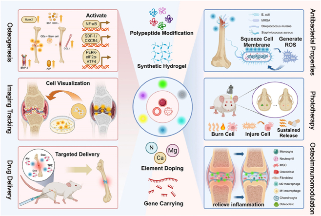
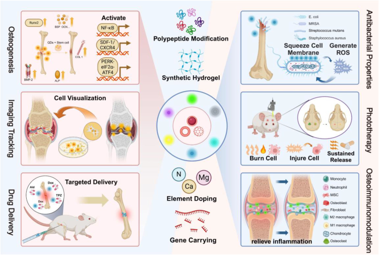
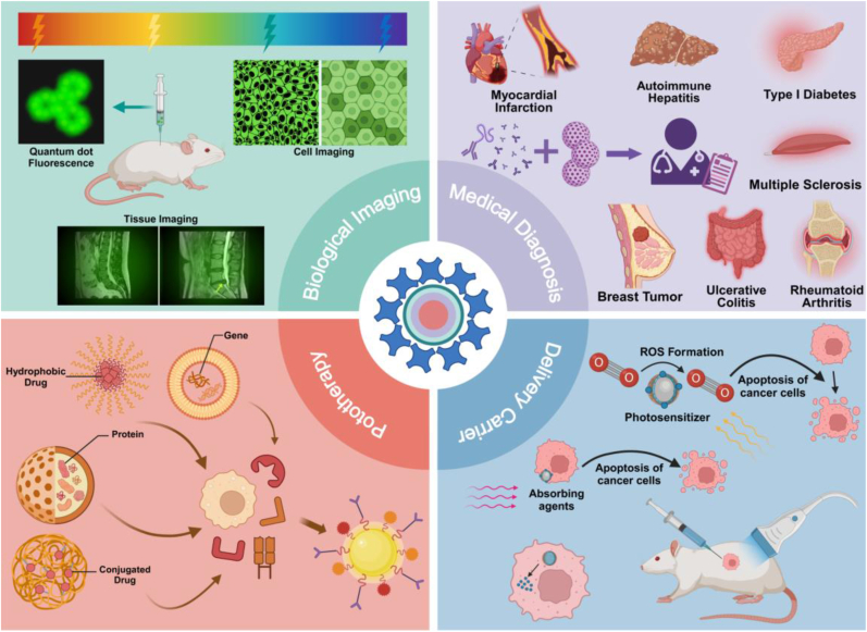
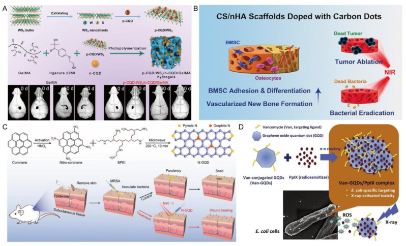
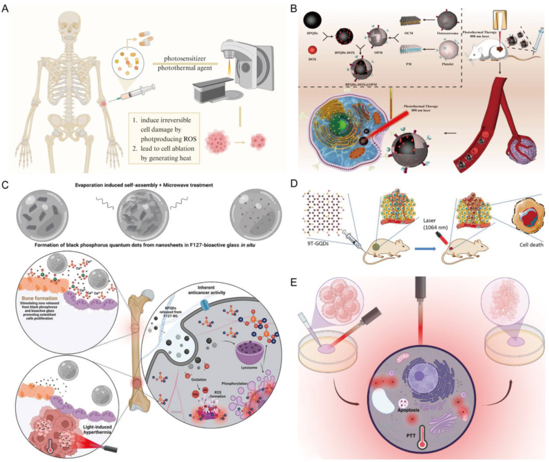
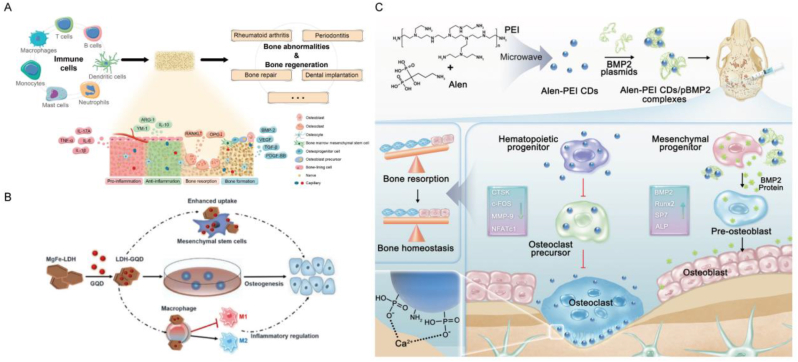
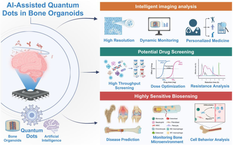
In confronting the global prevalence of bone-related disorders, bone tissue engineering (BTE) has developed into a critical discipline, seeking innovative materials to revolutionize treatment paradigms. Quantum dots (QDs), nanoscale semiconductor particles with tunable optical properties, are at the cutting edge of improving bone regeneration. This comprehensive review delves into the multifaceted roles that QDs play within the realm of BTE, emphasizing their potential to not only revolutionize imaging but also to osteogenesis, drug delivery, antimicrobial strategies and phototherapy. The customizable nature of QDs, attributed to their size-dependent optical and electronic properties, has been leveraged to develop precise imaging modalities, enabling the visualization of bone growth and scaffold integration at an unprecedented resolution. Their nanoscopic scale facilitates targeted drug delivery systems, ensuring the localized release of therapeutics. QDs also possess the potential to combat infections at bone defect sites, preventing and improving bacterial infections. Additionally, they can be used in phototherapy to stimulate important bone repair processes and work well with the immune system to improve the overall healing environment. In combination with current trendy artificial intelligence (AI) technology, the development of bone organoids can also be combined with QDs. While QDs demonstrate considerable promise in BTE, the transition from laboratory research to clinical application is fraught with challenges. Concerns regarding the biocompatibility, long-term stability of QDs within the biological environment, and the cost-effectiveness of their production pose significant hurdles to their clinical adoption. This review summarizes the potential of QDs in BTE and highlights the challenges that lie ahead. By overcoming these obstacles, more effective, efficient, and personalized bone regeneration strategies will emerge, offering new hope for patients suffering from debilitating bone diseases.

Keywords: Artificial intelligence; Bioimaging; Bone organoids; Bone tissue engineering; Drug delivery; Quantum dots.


Conflict of interest statement

We declare that we have no financial and personal relationships with other people or organizations that can inappropriately influence our work, there is no professional or other personal interest of any nature or kind in any product, service, and/or company that could be construed as influencing the position presented in, or the review of, the manuscript entitled, “Quantum Dots for Bone Tissue Engineering”.

Figures

Image 1
Graphical abstract
Fig. 1
Fig. 1
The application of QDs to bone repair in biomedicine. Multiple modifications of QDs and their application principles in osteogenesis, imaging tracking, drug delivery, antimicrobial properties, phototherapy, and osteoimmunomodulation.
Fig. 2
Fig. 2
Brief development history of QDs. Major events related to QDs since their discovery.
Fig. 3
Fig. 3
Structure and modification of QDs. A) Different types of QDs and their corresponding radiation. Reproduced and adapted with permission [29]. Copyright 2014, Tabriz University of Medical Sciences. B) CdSe QDs, CdSe/ZnS QDs and selectively integrated QDs models. Reproduced and adapted with permission [30]. Copyright 2020, Elsevier. C) Model diagram of CQDs and GQDs. Reproduced and adapted with permission [31]. Copyright 2023, Elsevier. D) The most common core-shell QDs model. Reproduced and adapted with permission [33]. Copyright 2016, Baqiyatallah University of Medical Sciences. E) Common interfacial chemical modifications and biological coupling of QDs. Reproduced and adapted with permission [26]. Copyright 2013, SAGE Publications Inc.
Fig. 4
Fig. 4
QDs in Biomedicine: Imaging, Diagnosis, and Therapy. Due to its unique optical and chemical properties, QDs is widely used in biological imaging, medical diagnosis, chemical phototherapy and as a drug carrier, and has diversified applications in the biomedical field.
Fig. 5
Fig. 5
QDs for osteogenesis in BTE. A) A rough sketch of QDs combining with MSCs to promote bone formation. B) The synthesis of metformin-coated CQDs and their mechanism in promoting periodontal bone regeneration. Reproduced and adapted with permission [113]. Copyright 2021, John Wiley and Sons Ltd. C) The preparation process of sulfonated glycosaminoglycan bioinspired CQDs for effective cell labeling and promotion of mesenchymal stem cell differentiation. Reproduced and adapted with permission [111]. Copyright 2020, Royal Society of Chemistry. D) Effect of GQDs on proliferation of PDLSCs in simulated pro-inflammatory environment medium and standard medium. Reproduced and adapted with permission [114]. Copyright 2023, BioMed Central. E) Construct composite scaffolds by binding of N-GQDs to Mg for bone defect repair. Reproduced and adapted with permission [115]. Copyright 2023, American Chemical Society.
Fig. 6
Fig. 6
QDs for imaging and fluorescence tracing in BTE. A) Overview of QDs for in vivo fluorescence imaging and diagnosis of stem cells. Reproduced and adapted with permission [121]. Copyright 2017, American Chemical Society. B) Synthesis of Alen-CDs and Alen-EDA-CDs and schematic diagram of their bone affinity. Reproduced and adapted with permission [125]. Copyright 2019, Royal Society of Chemistry. C) Nanohybrid system constructed based on QDs with fluorescent core for labeling and imaging of bone marrow stromal cells. Reproduced and adapted with permission [124]. Copyright 2014, Royal Society of Chemistry. D) QD-βCD-His was demonstrated in 2D culture and 3D hydrogel scaffolds to label and track MSCs and their differentiation into bone tissue. Reproduced and adapted with permission [126]. Copyright 2020, Future Medicine Ltd. E) Synthesis of PbS QDs and its imaging under SWIR fluorescence after intravenous injection. Reproduced and adapted with permission [92]. Copyright 2020, Royal Society of Chemistry. F) Monodentate ligands bind to the CD surface to chelate Ca2+ exposed at the site of microcracks, and are used for the detection of bone microcracks by CD fluorescence. Reproduced and adapted with permission [127]. Copyright 2018, American Chemical Society.
Fig. 7
Fig. 7
QDs for drug loading and delivery in BTE. A) CdSe MSQDs exhibit a mitigating effect on ROS production in neutrophils and macrophages in response to opsonized zymosan stimulation. Reproduced and adapted with permission [140]. Copyright 2022, Future Medicine Ltd. B) Nanoparticles prepared based on QDs can store TPZ and are modified with PEGyl-folate to target activated macrophages in RA. Reproduced and adapted with permission [146]. Copyright 2021, Elsevier. C) Construct bioconjugated carbon points for the delivery of siTnfα, which can enhance chondrogenesis of MSCs by inhibiting inflammation. Reproduced and adapted with permission [147]. Copyright 2019, John Wiley and Sons Ltd. D) HCPC NPs based on CD synthesis enhances the treatment of RA through passive targeting, M1 macrophage targeting, and reactive drug release. Reproduced and adapted with permission [148]. Copyright 2023, Elsevier Ltd.
Fig. 8
Fig. 8
QDs for antibacterial agent in BTE. A) The hydrogel scaffolds synthesized based on CQDs and their antibacterial properties were used for the regeneration of bone defects infected by multi-drug resistant bacteria. Reproduced and adapted with permission [165]. Copyright 2021, Elsevier Ltd. B) The composite scaffold based on CD synthesis promotes bone formation and anti-tumor, and also has obvious antibacterial properties against Staphylococcus aureus and Escherichia coli collected in clinic. Reproduced and adapted with permission [166]. Copyright 2018, American Chemical Society. C) The preparation process of N-GQDs and its excellent antibacterial and antibiofilm activity against MDR bacteria present under laser irradiation. Reproduced and adapted with permission [167]. Copyright 2022, Royal Society of Chemistry. D) GQDs coupled with vancomycin were assembled with protoporphyrin IX to construct a complex that can play a bactericidal role against E. coli. Reproduced and adapted with permission [168]. Copyright 2017, Elsevier BV.
Fig. 9
Fig. 9
QDs for phototherapy in BTE. A) Schematic diagram of the application of QDs to BTE under phototherapy. B) A schematic illustration of the preparation of BPQDs-DOX@OPM system and its combination with PTT enhanced drug therapy for OS. Reproduced and adapted with permission [177]. Copyright 2023, BioMed Central. C) Combined with NIR hyperthermia, a nanocomposite material for the treatment of bone cancer and bone tissue regeneration was constructed based on BPQDs. Reproduced and adapted with permission [179]. Copyright 2024, KeAi Communications Co. D) Imaging-guided PTT of 9T-GQDs irradiated under NIR-II laser. Reproduced and adapted with permission [180]. Copyright 2020, Elsevier BV. E) Schematic diagram of anti-cancer potential induced by F127-BG-BPQDs hyperthermia in vitro. Reproduced and adapted with permission [179]. Copyright 2024, KeAi Communications Co.
Fig. 10
Fig. 10
QDs for osteoimmunomodulation in BTE. A) Reproduced and adapted with permission [186]. Copyright 2023, MDPI (Basel, Switzerland). B) Schematic diagram of LDH-GQD promoting osteogenic differentiation ofrBMSCs by enhanced cellular uptake and inflammatory regulation. Reproduced and adapted with permission [189]. Copyright 2022, IOP Publishing Ltd. C) Multifunctional fluorescent Alen-PEI CDs can inhibit osteoclasts and significantly reverse the imbalance of bone homeostasis through the bidirectional mechanism of bone immunity. Reproduced and adapted with permission [190]. Copyright 2022, Wiley-Blackwell.
Fig. 11
Fig. 11
Further application of AI-assisted QDs in the study of bone organoids. AI-assisted quantum dots in bone organoid research can promote in-depth understanding of bone tissue development and disease mechanisms through precise imaging, drug analysis and screening, and high-sensitivity sensing, providing a powerful tool for personalized medicine and the development of new therapies.

Similar articles

Cited by

References

    1. Shrivats A.R., McDermott M.C., Hollinger J.O. Bone tissue engineering: state of the union. Drug Discov. Today. 2014;19:781–786. doi: 10.1016/j.drudis.2014.04.010. - DOI - PubMed
    1. Li Z., Liu J., Li C., Wu M., Li Y., Cui Y., Xiong W., Yang F., Liu B. Advances in the application of bone transport techniques in the treatment of bone nonunion and bone defects. Orthop. Surg. 2023;15:3046–3054. doi: 10.1111/os.13936. - DOI - PMC - PubMed
    1. Alonzo M., Primo F.A., Kumar S.A., Mudloff J.A., Dominguez E., Fregoso G., Ortiz N., Weiss W.M., Joddar B. Bone tissue engineering techniques, advances and scaffolds for treatment of bone defects. Curr. Opin. Biomed. Eng. 2021;17 doi: 10.1016/j.cobme.2020.100248. - DOI - PMC - PubMed
    1. Haugen H.J., Lyngstadaas S.P., Rossi F., Perale G. Bone grafts: which is the ideal biomaterial? J. Clin. Periodontol. 2019;46:92–102. doi: 10.1111/jcpe.13058. - DOI - PubMed
    1. Pérez-González F., Molinero-Mourelle P., Sánchez-Labrador L., Sáez-Alcaide L.M., Limones A., Cortés-Bretón Brinkmann J., López-Quiles J. Assessment of clinical outcomes and histomorphometric findings in alveolar ridge augmentation procedures with allogeneic bone block grafts: a systematic review and meta-analysis. Med. Oral. Patol. Oral. 2020;25:e291–e298. doi: 10.4317/medoral.23353. - DOI - PMC - PubMed

LinkOut - more resources