. 2024 Aug 27;13(1):2392897.
doi: 10.1080/2162402X.2024.2392897. eCollection 2024.

CD4+ tumor-infiltrating lymphocytes secreting T cell-engagers induce regression of autologous patient-derived non-small cell lung cancer xenografts

Affiliations

CD4+ tumor-infiltrating lymphocytes secreting T cell-engagers induce regression of autologous patient-derived non-small cell lung cancer xenografts

Anaïs Jiménez-Reinoso et al. Oncoimmunology. .

Abstract

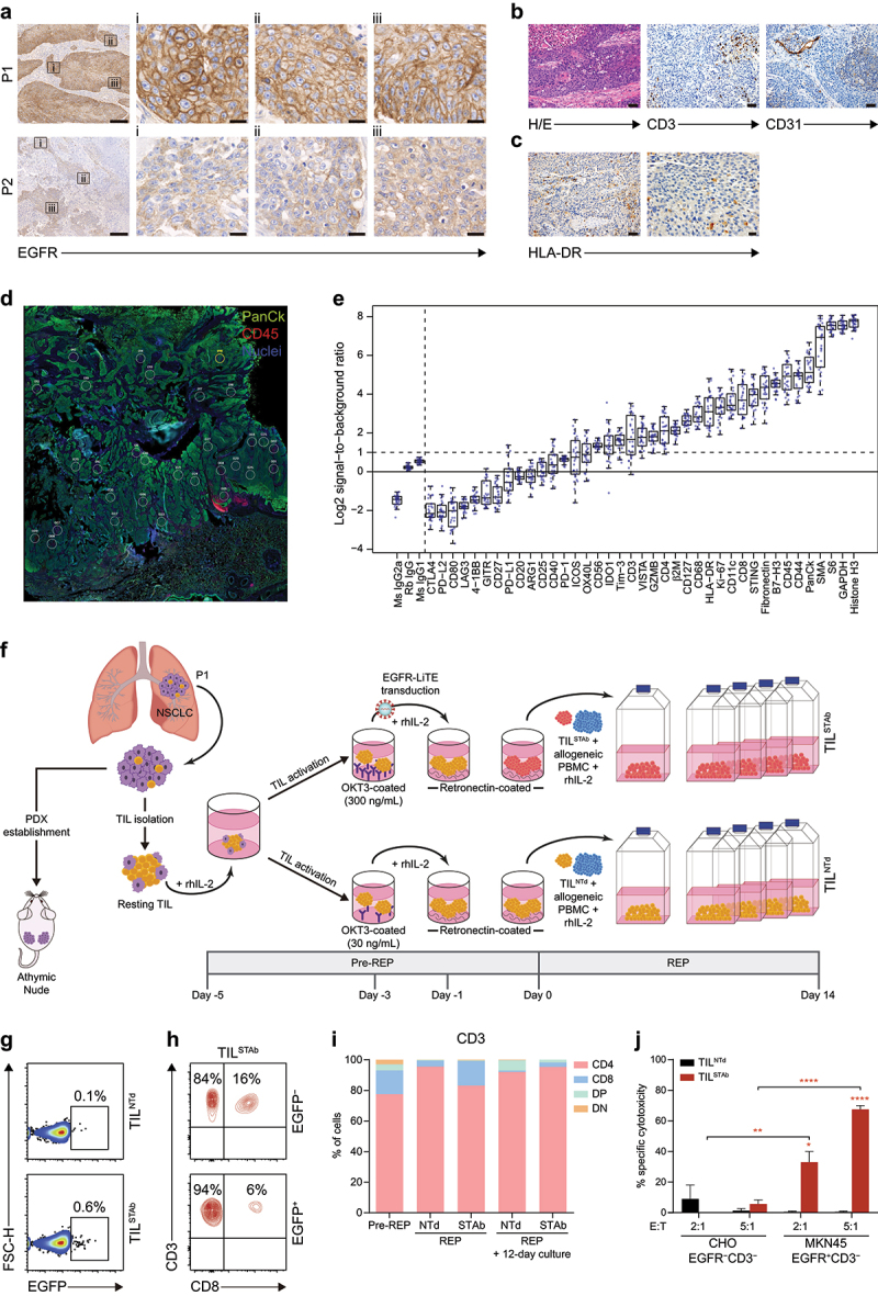
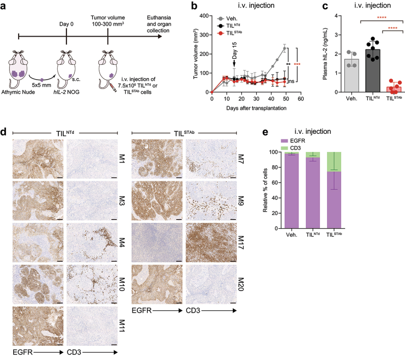
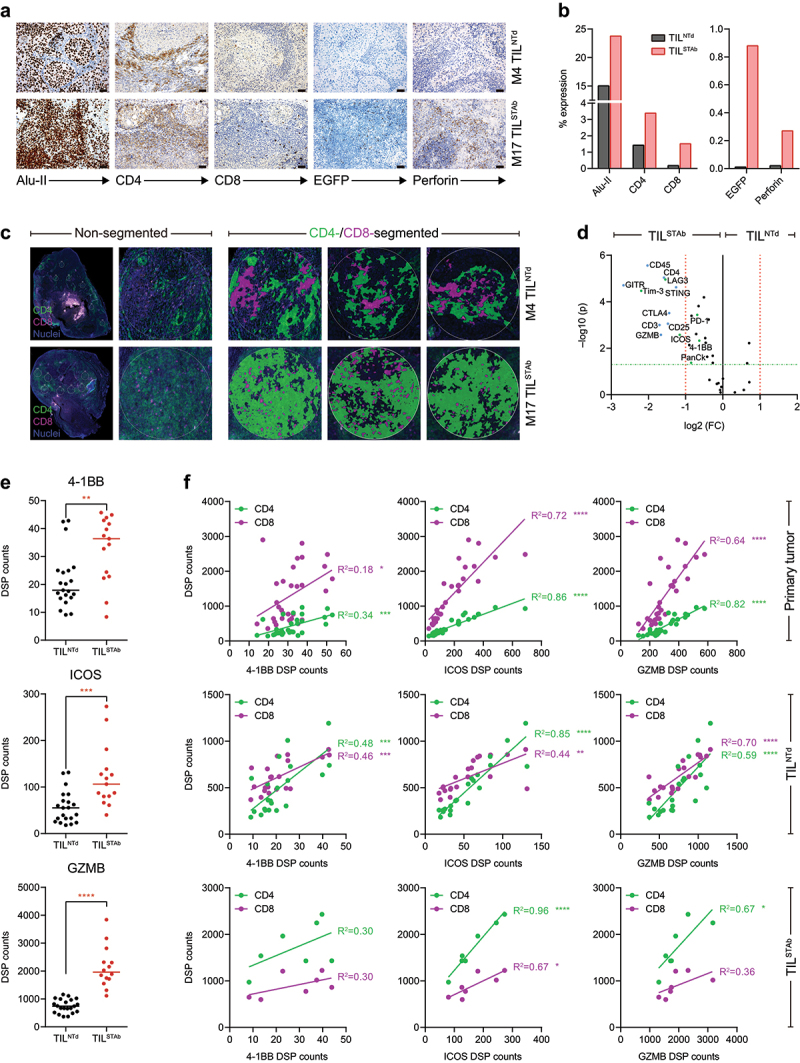
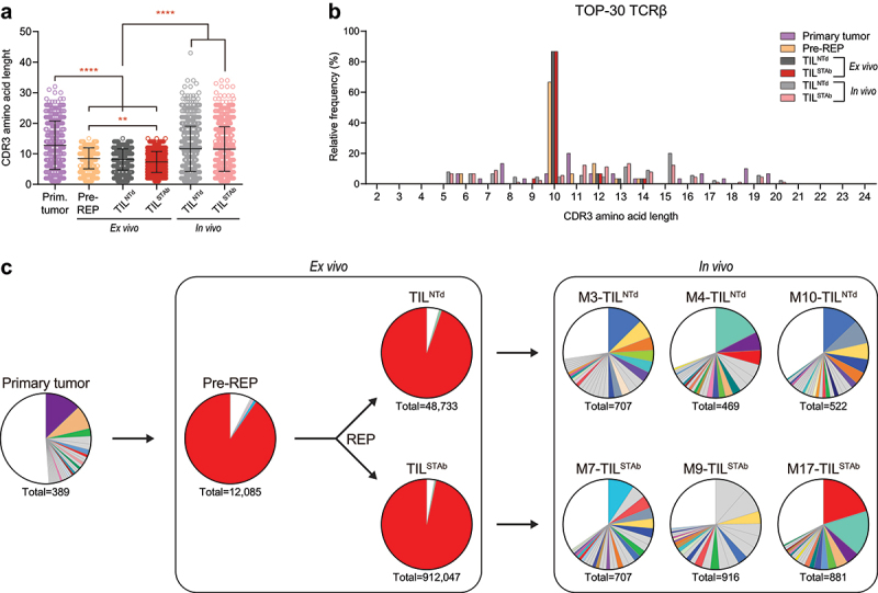
Adoptive transfer of tumor-infiltrating lymphocytes (TIL) has shown remarkable results in melanoma, but only modest clinical benefits in other cancers, even after TIL have been genetically modified to improve their tumor homing, cytotoxic potential or overcome cell exhaustion. The required ex vivo TIL expansion process may induce changes in the T cell clonal composition, which could likely compromise the tumor reactivity of TIL preparations and ultimately the success of TIL therapy. A promising approach based on the production of bispecific T cell-engagers (TCE) by engineered T cells (STAb-T therapy) improves the efficacy of current T cell redirection strategies against tumor-associated antigens in hematological tumors. We studied the TCRβ repertoire in non-small cell lung cancer (NSCLC) tumors and in ex vivo expanded TIL from two unrelated patients. We generated TIL secreting anti-epidermal growth factor receptor (EGFR) × anti-CD3 TCE (TILSTAb) and tested their antitumor efficacy in vitro and in vivo using a NSCLC patient-derived xenograft (PDX) model in which tumor fragments and TIL from the same patient were transplanted into hIL-2 NOG mice. We confirmed that the standard TIL expansion protocol promotes the loss of tumor-dominant T cell clones and the overgrowth of virus-reactive TCR clonotypes that were marginally detectable in primary tumors. We demonstrated the antitumor activity of TILSTAb both in vitro and in vivo when administered intratumorally and systemically in an autologous immune-humanized PDX EGFR+ NSCLC mouse model, where tumor regression was mediated by TCE-redirected CD4+ TIL bearing non-tumor dominant clonotypes.

Keywords: Adoptive cell therapy; bispecific T cell-engagers; cytotoxic CD4+ TIL; solid tumors; tumor-infiltrating lymphocytes.


Conflict of interest statement

B.B., L.P-A and L.A-V are co-founders and shareholders of STAb Therapeutics, a spin-off company from the imas12. B.B. and L.A-V are inventors on the patent EP21708942 pending. L.D-A. and L.A-V are inventors on the patent EP23383410.0 pending. L.A-V is cofounder and shareholder of Leadartis, a company focused on unrelated interest. L.A-V reports speaker honoraria from MSD, Merck KGaA, BMS, Janssen, GSK, and Miltenyi, and receives grant support from Merck KGaA, all outside the submitted work. J.Z. has served as a consultant for Sanofi, Pfizer, Astra Zeneca, BMS, Novartis, NanoString, and Guardant Health; reports speaker honoraria from Pfizer, BMS, Roche, Astra Zeneca, NanoString and Guardant Health; and receives grant support from Astra Zeneca, Roche, and BMS, all outside the submitted work. L.P-A. is shareholder of Altum Squencing and STAb Therapeutics, has served as a consultant for Lilly, MSD, Roche, Pharmamar, Merck KGaA, Astra Zeneca, Novartis, Amgen, Pfizer, Sanofi, Bayer, BMS, Mirati, GSK, Janssen, Takeda, Regeneron, and Sanofi. L.P-A. received grant support from MSD, Astra Zeneca, BMS, Pfizer and Pharmamar, all outside the submitted work. R.T. and E.P. are consultants for the company NISOLAB.

Figures

Figure 1.
Figure 1.
Phenotype of primary tumors and generation of patient-derived engineered EGFR-TCE-secreting TIL. (a) EGFR expression by IHC in P1 and P2 primary tumors. Magnification of three ROIs (i, ii, iii) from left images are shown. Scale bars: 200 µm and 20 µm in magnified images. (b) Histological detail of the P1 primary tumor (H/E), T cell infiltration (CD3+) and vascularization (CD31+) by IHC. The P1 tumor was a pleomorphic squamous cell carcinoma with atypical dyskeratotic cells and large necrotic areas. Scale bars: 50 µm. (c) HLA-dr expression by IHC in P1 primary tumor. Scale bars: 50 µm (left) and 20 µm (right). (d) DSP image showing the multiple circular ROI of a maximum of 500 µm from PanCk+CD45+ intertumoral areas in P1 primary tumor. (e) Signal-to-noise plot showing the log2-transformed signal (counts of each protein) relative to background levels (averaged counts of the three negative controls) of all markers included in the DSP immune-related protein panel in P1 primary tumor sample. (f) Engineered EGFR-STAb TIL (TILSTAb) small-scale REP generation and PDX establishment from P1 NSCLC primary tumor. (g) Percentage of reporter EGFP expression in non-transduced TIL (TILNTd) and TILSTAb after REP. (h) Percentages of CD4+ and CD8+ T cells within EGFP and EGFP+ TILSTAb cells. (i) Percentages of CD4+, CD8+, double positive (DP) and double negative (DN) T cells among pre-rep, REP and 12 days after REP cultured TILNTd and TILSTAb cells. (j) Specific cytotoxicity 12 days after REP of TILNTd or TILSTAb cells against egfr-negative (CHO) or egfr-positive (MKN45) cells at the indicated E:T ratios after 72 hours. The percentage of specific cytotoxicity was calculated by adding D-luciferin to detect bioluminescence. Data represent mean + SEM and statistical significance was calculated by two-way ANOVA test corrected by Tukey’s multiple comparisons test; *p < 0.05; **p < 0.01, ****p < 0.0001. TCE, bispecific T cell-engagers; IHC, Immunohistochemistry; P1, patient 1; P2, patient 2; ROI, region of interest; H/E: hematoxylin and eosin; REP, rapid expansion protocol; DSP, digital spatial profiling; SEM, standard error of mean.
Figure 2.
Figure 2.
Treatment of EGFR+ NSCLC PDX mouse model with autologous engineered TCE-secreting TIL. (a) Experimental design. Fifteen hIL-2 NOG mice were xenografted with P1-derived PDX fragments and when the average tumor volume reached approximately 100-300 mm3, were randomized into three groups (2/7/6) and treated i.v. With Veh., (PBS supplemented with 300 IU/mL rhIL-2, n = 2 mice), TILNTd (n = 7 mice) or TILSTAb (n = 6 mice) (7.5 × 106 cells/mouse). (b) Tumor growth curves shown as mean ± SEM; black arrow indicates the day of the i.V. injection. (c) Human IL-2 plasma levels in mice after treatment (mean ± SD). (d, e) EGFR expression and T cell infiltration after TIL treatment by IHC. Scale bars: 100 µm. Data in (e) represent mean ‒ SEM; no significant differences were found. Significance was calculated by two-way (b,e) or one-way (c) ANOVA test corrected by Tukey’s multiple comparisons test. ns, non-significant; **p < 0.01, ***p < 0.001; ****p < 0.0001. PDX, patient-derived xenograft; Veh., vehicle; IHC, immunohistochemistry; SD, standard deviation; SEM, standard error of mean; M, mouse.
Figure 3.
Figure 3.
Immunohistochemistry and DSP analysis of intravenously treated NSCLC xenografted hIL2 NOG mouse 4 (TILNTd-treated) and mouse 17 (TILSTAb-treated). (a, b) expression (a) and quantification (b) of Alu-II, CD4, CD8, EGR and perforin after treatment of hIL-2 NOG xenografted mouse 4 (M4) with TILNTd or mouse 17 (M17) with TILSTAb. Scale bars: 50 µm. (c) CD4 and CD8 T cell distribution by DSP from multiple circular ROI of a maximum of 500 µm within TILNTd- or TILSTAb-treated tumors in non-segmented or CD4/CD8-segmented compartments. (d) Differentially expressed protein markers in ROI from tumors treated with TILNTd or TILSTAb within the lymphocyte (PanCkCD45+) compartment. Significance (FDR-adjusted p-values) is expressed relative to the FC in protein levels in TILSTAb-treated vs. TILNTd-treated ROI. Markers with an FC > 2 and FDR-adjusted p-values < 0.05 in the two cohorts (TILSTAb- or TILNTd-treated) are marked in bold. (e) Comparative analysis of 4-1BB, ICOS and GZMB levels measured by DSP in non-CD4/CD8-segmented or CD4 and CD8 compartments. Significance in (e) was calculated by unpaired t-test. (f) Linear regression analysis between CD4 or CD8 infiltrated T cells and 4-1BB, ICOS and GZMB in P1 primary tumor, TILNTd-treated M4 and TILSTAb-treated M17 measured by DSP. R2 represents goodness of fit to the model. *p < 0.05; **p < 0.01; ***p < 0.001, ****p < 0.0001. FC, fold change; FDR, false discovery rate; ROI, regions of interest; GZMB, granzyme B.
Figure 4.
Figure 4.
TCR clonotypes composition of primary tumor, ex vivo TIL and in vivo TIL-treated tumors from P1. (a) TCRβ CDR3 length comparison of total unique productive clonotypes. Data represent mean ± SD and statistical significance was calculated by one-way ANOVA test corrected with a Tukey’s multiple comparisons test; **p < 0.01; ****p < 0.0001. (b) TCRβ CDR3 length distribution of the TOP-30 productive clonotypes. (c) Frequency of TOP-30 productive clonotypes. Only clonotypes with frequencies above 0.5% are represented. Shared colors among different pie charts represent clonotypes with identical amino acid sequence. Numbers below pie charts represent total reads assigned to unique productive clonotypes. Only clonotypes with 5-20 amino acid length CDR3 are included in (b,c).

Similar articles

Cited by

References

    1. Rosenberg SA, Spiess P, Lafreniere R.. A new approach to the adoptive immunotherapy of cancer with tumor-infiltrating lymphocytes. Sci. 1986;233(4770):1318–14. doi: 10.1126/science.3489291. - DOI - PubMed
    1. Robbins PF. Tumor-infiltrating lymphocyte therapy and neoantigens. The Cancer J. 2017;23(2):138–143. doi: 10.1097/PPO.0000000000000267. - DOI - PubMed
    1. Rosenberg SA, Packard BS, Aebersold PM, Solomon D, Topalian SL, Toy ST, Simon P, Lotze MT, Yang JC, Seipp CA, et al. Use of tumor-infiltrating lymphocytes and interleukin-2 in the immunotherapy of patients with metastatic melanoma. A preliminary report. N Engl J Med. 1988;319(25):1676–1680. doi: 10.1056/NEJM198812223192527. - DOI - PubMed
    1. Rohaan MW, Borch TH, van den Berg JH, Met Ö, Kessels R, Geukes Foppen MH, Stoltenborg Granhøj J, Nuijen B, Nijenhuis C, Jedema I, et al. Tumor-infiltrating lymphocyte therapy or ipilimumab in advanced melanoma. N Engl J Med. 2022;387(23):2113–2125. doi: 10.1056/NEJMoa2210233. - DOI - PubMed
    1. Chesney J, Lewis KD, Kluger H, Hamid O, Whitman E, Thomas S, Wermke M, Cusnir M, Domingo-Musibay E, Phan GQ, et al. Efficacy and safety of lifileucel, a one-time autologous tumor-infiltrating lymphocyte (TIL) cell therapy, in patients with advanced melanoma after progression on immune checkpoint inhibitors and targeted therapies: pooled analysis of consecutive cohorts of the C-144-01 study. J Immunother Cancer. 2022;10(12). doi: 10.1136/jitc-2022-005755. - DOI - PMC - PubMed

MeSH terms