

Contribution of advanced neuroimaging in diagnosis of cerebral syphilitic gumma: a case report
- PMID: 39206117
- PMCID: PMC11349654
- DOI: 10.3389/fnins.2024.1442176
Contribution of advanced neuroimaging in diagnosis of cerebral syphilitic gumma: a case report
Abstract
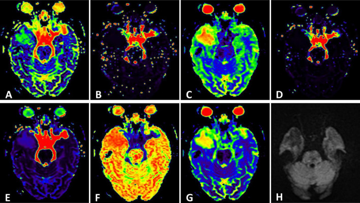
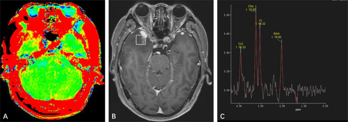
Background: Cerebral syphilitic gumma is a rare intracranial infectious disorder. Without a clear history of syphilis and comprehensive serological examinations, it's challenging to diagnose it accurately prior to surgery through routine magnetic resonance imaging (MRI). Advanced neuroimaging techniques have been widely used in diagnosing brain tumors, yet there's limited report on their application in cerebral syphilitic gumma. This report presents a case of an elderly male patient with cerebral syphilitic gumma and analyzes its characteristics of advanced neuroimaging.
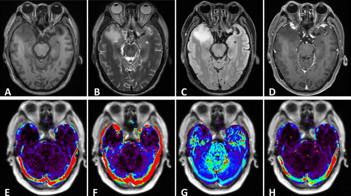
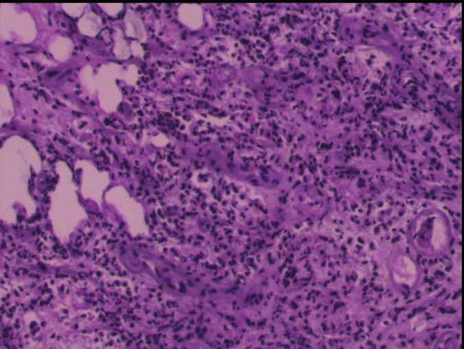
Case presentation: A 68-year-old male patient was admitted to our institution presenting with bilateral hearing loss complicated with continuing headaches without obvious cause. Laboratory tests indicated positive treponema pallidum. Conventional MRI showed nodules closely related to the adjacent meninges in bilateral temporal lobes. The patient underwent surgical resection of the nodule in the right temporal lobe due to the mass effect and the final pathological diagnosis revealed cerebral syphilitic gumma.
Conclusions: With the return of syphilis in recent years, accurate diagnosis of cerebral syphilitic gumma is a matter of great urgency. Advanced neuro-MRI can serve as a significant complement to conventional MRI examination.
Keywords: Advanced neuroimaging; case report; cerebral syphilitic gumma; intracranial neoplasm; magnetic resonance imaging.
Copyright © 2024 Shen, Zhu, Li, Zhang, Zhang and Zhang.
Conflict of interest statement
The authors declare that the research was conducted in the absence of any commercial or financial relationships that could be construed as a potential conflict of interest.
Figures
Similar articles
-
Cerebral syphilitic gumma misdiagnosed as brain abscess: A case report.World J Clin Cases. 2024 Jan 26;12(3):650-656. doi: 10.12998/wjcc.v12.i3.650. World J Clin Cases. 2024. PMID: 38322467 Free PMC article.
-
Neuroimaging findings of cerebral syphilitic gumma.Exp Ther Med. 2019 Dec;18(6):4185-4192. doi: 10.3892/etm.2019.8089. Epub 2019 Oct 8. Exp Ther Med. 2019. PMID: 31772624 Free PMC article.
-
The Application of MR Spectroscopy and MR Perfusion in Cerebral Syphilitic Gumma: A Case Report.Front Neurosci. 2020 Oct 21;14:544802. doi: 10.3389/fnins.2020.544802. eCollection 2020. Front Neurosci. 2020. PMID: 33192243 Free PMC article.
-
Cerebral syphilitic Gumma in the modern era: a report of an unusual case and brief review of recent published reports.Br J Neurosurg. 2024 Dec;38(6):1283-1288. doi: 10.1080/02688697.2022.2159923. Epub 2022 Dec 23. Br J Neurosurg. 2024. PMID: 36564899 Review.
-
Spinal Cord Syphilitic Gumma Presenting with Brown-Séquard Syndrome: A Case Report and Literature Review.Ann Clin Lab Sci. 2019 Mar;49(2):265-270. Ann Clin Lab Sci. 2019. PMID: 31028074 Review.
References
-
- Callot V., Galanaud D., Le Fur Y., Confort-Gouny S., Ranjeva J. P., Cozzone P. J. (2008). (1)H MR spectroscopy of human brain tumours: A practical approach. Eur. J. Radiol. 67 268–274. - PubMed
Publication types
LinkOut - more resources
Full Text Sources