. 2024 Oct;11(40):e2402671.
doi: 10.1002/advs.202402671. Epub 2024 Aug 29.

FOXO1-NCOA4 Axis Contributes to Cisplatin-Induced Cochlea Spiral Ganglion Neuron Ferroptosis via Ferritinophagy

Affiliations

FOXO1-NCOA4 Axis Contributes to Cisplatin-Induced Cochlea Spiral Ganglion Neuron Ferroptosis via Ferritinophagy

Xue Wang et al. Adv Sci (Weinh). 2024 Oct.

Abstract

Mammalian cochlea spiral ganglion neurons (SGNs) are crucial for sound transmission, they can be damaged by chemotherapy drug cisplatin and lead to irreversible sensorineural hearing loss (SNHL), while such damage can also render cochlear implants ineffective. However, the mechanisms underlying cisplatin-induced SGNs damage and subsequent SNHL are still under debate and there is no currently effective clinical treatment. Here, this study demonstrates that ferroptosis is triggered in SGNs following exposure to cisplatin. Inhibiting ferroptosis protects against cisplatin-induced SGNs damage and hearing loss, while inducing ferroptosis intensifies these effects. Furthermore, cisplatin prompts nuclear receptor coactivator 4 (NCOA4)-mediated ferritinophagy in SGNs, while knocking down NCOA4 mitigates cisplatin-induced ferroptosis and hearing loss. Notably, the upstream regulator of NCOA4 is identified and transcription factor forkhead box O1 (FOXO1) is shown to directly suppress NCOA4 expression in SGNs. The knocking down of FOXO1 amplifies NCOA4-mediated ferritinophagy, increases ferroptosis and lipid peroxidation, while disrupting the interaction between FOXO1 and NCOA4 in NCOA4 knock out mice prevents the cisplatin-induced SGN ferroptosis and hearing loss. Collectively, this study highlights the critical role of the FOXO1-NCOA4 axis in regulating ferritinophagy and ferroptosis in cisplatin-induced SGNs damage, offering promising therapeutic targets for SNHL mitigation.

Keywords: cisplatin; ferritinophagy; ferroptosis; forkhead box transcription factor O1; hearing loss; nuclear receptor coactivator 4; spiral ganglion neuron.


Conflict of interest statement

The authors declare no conflict of interest.

Figures
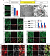
Figure 1
Figure 1
Cisplatin induces ferroptosis in cochlear SGNs in vitro. A) Cultured SGNs from P3 WT mice were exposed to 50 µM cisplatin for 48 h. B) Immunostaining and cell counting indicated that cisplatin treatment resulted in ≈64.02 ± 4.49% of SGNs remaining after treatment compared to the normal control group, n = 6 samples for each group. Scale bar = 25 µm. C) TEM showed that cisplatin led to mitochondrial shrinkage, a decrease or loss of mitochondrial cristae, and an increase in mitochondrial membrane density. The damaged mitochondria are indicated by the red arrows. n = 3 samples for each group. Scale bar = 500 nm. D) Immunostaining demonstrated that the fluorescence intensity of 4‐HNE was significantly higher in SGNs treated with cisplatin than those in the control group, n = 6 samples for each group. Scale bar = 25 µm. E) Western blot analysis showed that the protein levels of SLC7A11, FSP1 and GPX4 in SGNs were markedly decreased after cisplatin treatment, n = 3 samples. F–H) Immunofluorescence staining revealed stronger TfR1 F), FerroOrange G) and Mito‐FerroGreen H) fluorescences in the cisplatin‐treated group than those in the control group, n = 4 samples for each group. F, G: scale bar = 25 µm. H: scale bar = 5 µm. I) The TMRM fluorescence intensity was significantly decreased in cisplatin‐treated SGNs, n = 4 samples. Scale bar = 5 µm. The data are presented as the mean ± S.D. *p < 0.05, **p < 0.01, ***p < 0.001, two‐tailed, unpaired Student's t‐tests.
Figure 2
Figure 2
Cisplatin induces ferroptosis in cochlear SGNs in vivo. A) WT C57BL/6 mice were treated with 3 mg/kg cisplatin for 7 consecutive days by intraperitoneal (i.p.) injection beginning at P30. B,C) Cisplatin administration led to significant increases in the ABR threshold B) and CAP threshold C) at all tested frequencies, n = 6 mice. D) Cisplatin administration reduced in the CAP amplitude at 90 dB SPL across all tested frequencies, n = 6 mice. E) Immunostaining showed that cisplatin caused significant SGN loss in the cochlea, n = 6 samples. Scale bar = 25 µm. F,G) Cisplatin administration significantly upregulated 4‐HNE F) and TfR1 G), n = 4 samples for each group. Scale bar = 25 µm. H) Western blot analysis revealed that cisplatin treatment downregulated the expressions of SLC7A11, FSP1 and GPX4 in cochlear SGNs in vivo, n = 3 samples. The data are presented as the mean ± S.D. *p < 0.05, **p < 0.01, ***p < 0.001, two‐tailed, unpaired Student's t‐tests or two‐way ANOVA with a post‐hoc Student Newman‐Keuls test.
Figure 3
Figure 3
Inhibition of ferroptosis prevents cisplatin‐induced SGN loss and hearing loss. A) Cultured SGNs from P3 WT mice were treated with 50 µM cisplatin for 48 h in the presence or absence of the ferroptosis activator erastin (25 µM) or the ferroptosis inhibitor DFO (80 µM). B) Western blot analysis showed that cisplatin treatment caused a reduction of FSP1 and GPX4 in SGNs compared to the control group. Pretreatment with erastin in the Cis + erastin group significantly exacerbated these reductions, while pretreatment with DFO in the Cis + DFO group migrated them compared to the cisplatin‐only group. n = 3 samples. C) Increased SGN loss was observed following erastin pretreatment and cisplatin injury compared to the cisplatin treatment alone group, but this loss was attenuated by pretreatment with DFO. n = 6 samples for each group. Scale bar = 25 µm. D) C57BL/6 mice were i.p. injected with 3 mg/kg cisplatin daily for 7 days with or without erastin or DFO co‐treatment starting at P30. E,F) The ABR threshold shift E) and CAP threshold shift F) were much larger in the Cis + erastin group, whereas they were smaller in the Cis + DFO group than in the cisplatin only group. n = 6 mice. G) DFO alleviated the reduction in the CAP amplitude in cisplatin‐treated mice, whereas erastin administration had the opposite effect. n = 6 mice. H) Cisplatin administration in mice induced downregulations of FSP1 and GPX4 in SGNs compared with the control mice, and the expressions of FSP1 and GPX4 were further decreased in the Cis + erastin group, while they were increased in the Cis + DFO group, compared to the cisplatin only group. n = 3 samples. I) Cell counting revealed fewer surviving SGNs in the Cis + erastin group but significantly more SGNs in the Cis + DFO group than in the cisplatin alone group. n = 6 samples for each group. Scale bar = 25 µm. The data are presented as the mean ± S.D. *p < 0.05, **p < 0.01, ***p < 0.001, one‐way ANOVA with Tukey's post‐hoc test or two‐way ANOVA with a post‐hoc Student Newman‐Keuls test. DFO, deferoxamine.
Figure 4
Figure 4
Ferritinophagy is activated in cochlear SGNs after cisplatin treatment. A) Cultured SGNs from P3 WT mice or Bhlhb5‐cre/Rosa26‐tdTomato mice were exposed to 50 µM cisplatin for 48 h. B) Western blot analysis showed an increase in NCOA4 expression and a decrease in FTH1 expression in cultured SGNs after cisplatin treatment. n = 3 samples. C) Immunofluorescence showed colocalization of NCOA4 and FTH1 in cisplatin‐treated SGNs. n = 4 samples for each group. Scale bar = 5 µm. D) The colocalization of FTH1 and LAMP2 was increased in SGNs after cisplatin injury. n = 4 samples for each group. Scale bar = 5 µm. E) Cultured SGNs from P3 WT mice were treated with 50 µM cisplatin for 48 h alone or with the autophagy activator RAP (0.1 µM) or the autophagy inhibitor Baf (100 nM). F,G) RAP significantly upregulated NCOA4 protein expression and downregulated FTH1 expression, resulting in increased accumulation of intracellular iron and reductions in FSP1 and GPX4 protein levels in SGNs treated with cisplatin. Baf co‐treatment had the opposite effect. n = 3 samples for each group. Scale bar = 25 µm. The data are presented as the mean ± S.D. *p < 0.05, **p < 0.01, ***p < 0.001, two‐tailed, unpaired Student's t‐tests or one‐way ANOVA with Tukey's post‐hoc test. RAP, rapamycin; Baf, bafilomycin A1.
Figure 5
Figure 5
NCOA4 knockdown rescues cisplatin‐induced SGN loss and ferroptosis in cultured SGNs. A) Cultured SGNs from P3 WT mice were incubated with 4 × 10¹⁰ GC mL−1 Anc80‐Ncoa4 shRNA for 24 h. Then, the medium was replaced with normal medium, and the SGNs were further incubated with 50 µM cisplatin for 48 h. B) Western blot analysis revealed that the increase in NCOA4 expression and decrease in FTH1 expression in SGNs treated with cisplatin were significantly alleviated by pretreatment with Anc80‐Ncoa4 shRNA. n = 3 samples. C) More surviving SGNs were found in the Cis + Anc80‐Ncoa4‐shRNA group than in the cisplatin only group. n = 6 samples for each group. Scale bar = 25 µm. D,E) NCOA4 knockdown significantly reduced the expression of TfR1 D) and the FerroOrange intensity E) in cisplatin‐treated SGNs. n = 4 samples for each group. Scale bar = 25 µm. F,G) NCOA4 knockdown increased the TMRM signal intensity F) and the protein levels of FSP1 and GPX4 G) in cisplatin‐treated SGNs compared with SGNs in the cisplatin only group. n = 3 samples for each group. Scale bar = 5 µm. The data are presented as the mean ± S.D. *p < 0.05, **p < 0.01, ***p < 0.001, one‐way ANOVA with Tukey's post‐hoc test.
Figure 6
Figure 6
NCOA4 knockdown alleviates cisplatin‐induced hearing loss, SGN loss and ferroptosis in mice. A) WT mice were injected with 1 × 10¹⁰ GCs of Anc80‐Ncoa4 shRNA via the RWM at P5 and then administered 3 mg kg−1 cisplatin by i.p. injection for 7 consecutive days beginning at P30. B–D) The shifts in the ABR threshold B) and CAP threshold C) in the Cis + Anc80‐Ncoa4 shRNA group were significantly inhibited compared to those in the cisplatin only group, whereas the change in the CAP amplitude D) was increased in the Cis + Anc80‐Ncoa4 shRNA group. n = 6 mice. E) Cell counting revealed more surviving SGNs in the Cis+Anc80‐Ncoa4 shRNA group than in the cisplatin only group. n = 6 samples for each group. Scale bar = 25 µm. F) The protein levels of FSP1 and GPX4 in mice cotreated with cisplatin and Anc80‐Ncoa4 shRNA were increased compared to those in mice treated with cisplatin alone. n = 3 samples. G,H) The increase in TfR1 G) and 4‐HNE H) fluorescence intensity in cisplatin‐treated SGNs was markedly inhibited by pretreatment with Anc80‐Ncoa4 shRNA. n = 4 samples for each group. Scale bar = 25 µm. The data are presented as the mean ± S.D. *p < 0.05, **p < 0.01, ***p < 0.001, one‐way ANOVA with Tukey's post‐hoc test or two‐way ANOVA with a post‐hoc Student Newman‐Keuls test.
Figure 7
Figure 7
FOXO1 binds to the promoter of NCOA4, suppresses NCOA4 expression, and inhibits ferritinophagy and ferroptosis in cisplatin‐treated SGNs. A) Two TF prediction websites (JASPAR and Animal TFDB) were used to conduct bioinformatics analysis and predict the potential TFs that bind to the promoter sequence of Ncoa4. B,C) Immunofluorescence staining B) and Western blot C) revealed that the expression level of FOXO1 was significantly decreased in cisplatin‐treated cultured SGNs. n = 3 samples for each group. Scale bar = 25 µm. D) As predicted by the JASPAR database, there were two potential FOXO1 binding sites in the NCOA4 promoter. E,F) ChIP‒PCR showed that FOXO1 bound directly to the NCOA4 promoter. n = 3 samples. G) Cultured SGNs from P3 WT mice were incubated with 4 × 10¹⁰ GC mL−1 Anc80‐Foxo1 shRNA for 24 h. Then, the medium was replaced with normal medium, and the SGNs were further incubated with 50 µM cisplatin for 48 h. H) Western blot analysis revealed that the expression of FOXO1 was further reduced, the expression of NCOA4 was increased, and the expression of FTH1 was decreased in the Cis + Anc80‐Foxo1 shRNA group SGNs compared to SGNs in the cisplatin only group. n = 3 samples. I–L) FOXO1 deficiency increased intracellular iron accumulation I), aggravated SGN loss J), increased 4‐HNE protein level and further reduced FSP1 and GPX4 protein levels K,L) in the Cis + Anc80‐Foxo1 shRNA group than in the cisplatin only group. n = 3 samples for each group. Scale bar = 25 µm. The data are presented as the mean ± S.D. *p < 0.05, **p < 0.01, ***p < 0.001, two‐tailed, unpaired Student's t‐tests or one‐way ANOVA with Tukey's post‐hoc test.
Figure 8
Figure 8
Disruption of the FOXO1‐NCOA4 axis rescues cisplatin‐induced SGN loss and ferroptosis in cultured SGNs. A) Cultured SGNs from P3 WT mice or Ncoa4 −/− mice were incubated with 4 × 10¹⁰ GC mL−1 Anc80‐Foxo1 shRNA for 24 h, after which the medium was replaced with normal medium. The SGNs were then further incubated with 50 µM cisplatin for 48 h. B,C) Genetic knockout of NCOA4 significantly increased FTH1 expression, decreased intracellular iron accumulation, and upregulated FSP1 and GPX4 protein expression in Ncoa4 −/− mouse SGNs after cisplatin treatment. NCOA4 deficiency inhibited the accumulation of intracellular iron and the reduction in FSP1 and GPX4 levels in the Cis + Ncoa4 −/− + Anc80‐Foxo1 shRNA group compared with the Cis + Anc80‐Foxo1 shRNA group. n = 3 samples for each group. Scale bar = 25 µm. D) The number of surviving SGNs was significantly greater in the Cis + Ncoa4 −/− group than in the cisplatin only group. NCOA4 deficiency increased the number of surviving SGNs in the Cis + Ncoa4 −/− + Anc80‐Foxo1 shRNA group compared with the Cis + Anc80‐Foxo1 shRNA group. n = 4 samples for each group. Scale bar = 25 µm. The data are presented as the mean ± S.D. *p < 0.05, **p < 0.01, ***p < 0.001, one‐way ANOVA with Tukey's post‐hoc test.
Figure 9
Figure 9
Disruption of the FOXO1‐NCOA4 axis rescues cisplatin‐induced SGN ferroptosis and hearing loss in mice. A) WT mice or Ncoa4 −/− mice were injected with 1 × 10¹⁰ GCs of Anc80‐Foxo1 shRNA via the RWM at P5, followed by administration of 3 mg/kg cisplatin via i.p. injection for 7 consecutive days beginning at P30. B–D) Increases in the ABR thresholds B) and CAP thresholds C), along with a decrease in CAP amplitude D), were observed in the Cis + Anc80‐Foxo1 shRNA group compared with the cisplatin‐only group. Hearing function was improved in the Cis + Ncoa4 −/− + Anc80‐Foxo1 shRNA mice relative to the Cis + Anc80‐Foxo1 shRNA mice. n = 6 mice. E,F) FOXO1 knockdown significantly increased TfR1 expression E), and decreased the protein levels of FSP1 and GPX4 F) in mice treated with cisplatin. These protein changes were reversed in the Cis + Ncoa4 −/− + Anc80‐Foxo1 shRNA group compared to the Cis + Anc80‐Foxo1 shRNA group. n = 3 samples for each group. Scale bar = 25 µm. G) FOXO1 knockdown reduced the number of surviving SGNs in WT mice treated with cisplatin, while significantly more SGNs were observed in the Cis + Ncoa4 −/− + Anc80‐Foxo1 shRNA group than in the Cis + Anc80‐Foxo1 shRNA group. n = 4 samples for each group. Scale bar = 25 µm. Data are presented as the mean ± S.D. *p < 0.05, **p < 0.01, ***p < 0.001, one‐way ANOVA with Tukey's post‐hoc test or two‐way ANOVA with a post‐hoc Student Newman‐Keuls test.
Figure 10
Figure 10
A graphic illustration for the role of FOXO1‐NCOA4 axis in cisplatin‐induced SGNs ferroptosis and hearing loss. Cisplatin exposure induces ROS production and triggers ferroptosis in SGNs. The TF FOXO1, which is downregulated in response to cisplatin exposure, binds to the NCOA4 promoter enhancing its expression. Elevated NCOA4 levels increase ferritinophagy, thereby releasing free iron and driving Fenton reaction, ultimately leading to ferroptosis in SGNs. This diagram highlights the potential intervention points where inhibiting FOXO1 or NCOA4 could mitigate ferroptosis and thus reducing hearing loss associated with cisplatin treatment. IHC, inner hair cell; SGN, spiral ganglion neuron; FOXO1, forkhead box transcription factor O1; NCOA4, nuclear receptor coactivator 4; ROS, reactive oxygen species.

References

    1. Chadha S., Kamenov K., Cieza A., Bull W. H O. 2021, 99, 242. - PMC - PubMed
    1. Géléoc G. S., Holt J. R., Science (New York, N.Y.) 2014, 344, 1241062. - PMC - PubMed
    1. Nayagam B. A., Muniak M. A., Ryugo D. K., Hearing Research 2011, 278, 2. - PMC - PubMed
    1. Wang M., Xu L., Han Y., Wang X., Chen F., Lu J., Wang H., Liu W., Front. Mol. Neurosci. 2021, 14, 829564. - PMC - PubMed
    1. Lu J., Wang M., Meng Y., An W., Wang X., Sun G., Wang H., Liu W., Front. Neurosci. 2023, 17, 1334162. - PMC - PubMed

MeSH terms

LinkOut - more resources