. 2024 Aug 29;9(20):e179525.
doi: 10.1172/jci.insight.179525.

Mutant induced neurons and humanized mice enable identification of Niemann-Pick type C1 proteostatic therapies

Affiliations

Mutant induced neurons and humanized mice enable identification of Niemann-Pick type C1 proteostatic therapies

Ruth D Azaria et al. JCI Insight. .

Abstract

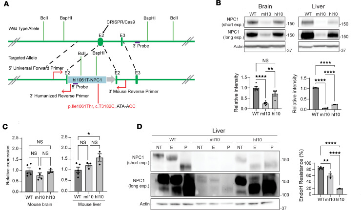
Therapeutics that rescue folding, trafficking, and function of disease-causing missense mutants are sought for a host of human diseases, but efforts to leverage model systems to test emerging strategies have met with limited success. Such is the case for Niemann-Pick type C1 disease, a lysosomal disorder characterized by impaired intracellular cholesterol trafficking, progressive neurodegeneration, and early death. NPC1, a multipass transmembrane glycoprotein, is synthesized in the endoplasmic reticulum and traffics to late endosomes/lysosomes, but this process is often disrupted in disease. We sought to identify small molecules that promote folding and enable lysosomal localization and functional recovery of mutant NPC1. We leveraged a panel of isogenic human induced neurons expressing distinct NPC1 missense mutations. We used this panel to rescreen compounds that were reported previously to correct NPC1 folding and trafficking. We established mo56-hydroxycholesterol (mo56Hc) as a potent pharmacological chaperone for several NPC1 mutants. Furthermore, we generated mice expressing human I1061T NPC1, a common mutation in patients. We demonstrated that this model exhibited disease phenotypes and recapitulated the protein trafficking defects, lipid storage, and response to mo56Hc exhibited by human cells expressing I1061T NPC1. These tools established a paradigm for testing and validation of proteostatic therapeutics as an important step toward the development of disease-modifying therapies.

Keywords: Lysosomes; Neurological disorders; Neuroscience; Protein misfolding.


Figures

Figure 1
Figure 1. mo56Hc increases Endo H resistance of I1061T NPC1 in iNeurons.
(A and B) Differentiated WT and I1061T NPC1 iNeurons were treated with vehicle (NT), 0.2 μM vorinostat (Vori.), 0.2 μM CI-994, 0.005 μM onalespib (Onal.), 100 μM 4-phenylbutyric acid (4-PBA), 10 μM quinestrol (Quin.), 10 μM abiraterone (Abir.), 1 μM 25-hydroxycholesterol (25Hc), 1 μM mo56-hydroxycholesterol (mo56Hc), or 400 μM arimoclomol (Ari.) for 48 hours. Lysates were digested with Endo H and analyzed by Western blot (values on right indicate ladder standard weights in kilodaltons). Quantified at right. (C and D) Differentiated I1061T NPC1 iNeurons were treated (C) for 48 hours with the indicated concentrations of mo56Hc or (D) with 1 µM mo56Hc for the indicated times.” Both experiments utilize mo56Hc treatment. Lysates were digested with Endo H and analyzed by Western blot. Quantified at right. All data are mean ± SEM from indicated number of independent experiments. *P ≤ 0.05, ***P ≤ 0.001, ****P ≤ 0.0001 by (A, C, and D) 1-way ANOVA with Tukey’s post hoc test or (B) t test. (A) n = 5 WT, 9 I1061T NPC1 NT, 3 I1061T NPC1 plus each drug; (B) n = 3 I1061T NPC1 NT, 3 I1061T NPC1 Ari.; (C) n = 3 I1061T NPC1 plus mo56Hc at each concentration; (D) n = 3 I1061T NPC1 plus mo56Hc for each treatment duration.
Figure 2
Figure 2. mo56Hc increases Endo H resistance of NPC1 trafficking mutants in iNeurons.
(A and B) Differentiated WT and R934L NPC1 iNeurons were treated with vehicle (NT), 0.2 μM vorinostat (Vori.), 0.2 μM CI-994, 0.005 μM onalespib (Onal.), 100 μM 4-phenylbutyric acid (4-PBA), 10 μM quinestrol (Quin.), 10 μM abiraterone (Abir.), 1 μM 25-hydroxycholesterol (25Hc), 1 μM mo56-hydroxycholesterol (mo56Hc), or 400 μM arimoclomol (Ari.) for 48 hours. Lysates were digested with Endo H and analyzed by Western blot. Quantified at right. (C and D) Differentiated R1186H and P1007A iNeurons were treated with vehicle (NT), 1 μM 25Hc, or 1 μM mo56Hc for 48 hours. Lysates were digested with Endo H and subjected to Western blot. Quantified at right. All data are mean ± SEM from indicated number of independent experiments. **P ≤ 0.01, ***P ≤ 0.001, ****P ≤ 0.0001 by (A, C, and D) 1-way ANOVA with Tukey’s post hoc test or (B) t test. (A) n = 3 WT, 7 R934L NPC1 NT, 3 R934L NPC1 plus each drug; (B) n = 3 R934L NPC1 NT, 3 R934L NPC1 Ari.; (C) n = 3 R1186H NPC1 NT, 3 R1186H NPC1 plus each drug; (D) n = 3 P1007A NPC1 NT, 5 P1007A NPC1 plus each drug.
Figure 3
Figure 3. m56Hc increases thermal stability of NPC1 protein.
Purified WT NPC1 protein was treated with 50 μM mo56Hc (blue lines and text), 50 μM 25Hc (red lines and text), or 50 μM mo56Hc + 50 μM 25Hc (purple lines and text) before thermal stability analysis was performed via monitoring of inherent tryptophan fluorescence using nano-differential scanning fluorimetry. First derivative analysis of the 330 nm emission wavelength is shown. The stabilization of NPC1 by mo56Hc is apparent in a shift in Tm as well as an increase in slope of the thermal transition.
Figure 4
Figure 4. Generation of humanized I1061T NPC1 mice.
(A) Depiction of the editing strategy used to create the humanized I1061T NPC1 mouse. The human NPC1 cDNA sequence encoding the I1061T NPC1 mutation was inserted into exon 2 of mouse Npc1, downstream of the signal peptide sequence, by CRISPR/Cas9 homology-directed repair. Restriction enzymes (BcII, BspHI) and Southern blot (5′ and 3′ probes) were used to determine proper insertion and PCR (5′ universal forward primer, 3′ humanized reverse primer, 3′ mouse reverse primer) were designed for mouse genotyping. (B) Brain and liver tissue from 6- to 8-week-old WT, knockin mouse I1061T NPC1 (mI10), and humanized I1061T NPC1 (hI10) were analyzed for total NPC1 protein by Western blot. Quantified below. (C) Brain and liver tissue from WT, mI10, and hI10 mice were analyzed for NPC1 mRNA by qPCR. (D) Liver lysates from WT, mI10, and hI10 mice were digested with Endo H and subjected to Western blot. Quantified at right. NT, not treated; E, Endo H digested; P, PNGase F digested. All data are mean ± SEM from the indicated number of independent experiments. *P ≤ 0.05, **P ≤ 0.01, ****P ≤ 0.0001 by (BD) 1-way ANOVA with Tukey’s post hoc test. (B) For brain, n = 5 WT, 5 mI10, 4 hI10; for liver, n = 4 WT, 4 mI10, 3 hI10. (C) For brain, n = 5 WT, 4 mI10, 3 hI10; for liver, n = 5 WT, 3 mI10, 3 hI10; (D) n = 5 WT, 4 mI10, 3 hI10.
Figure 5
Figure 5. Humanized I1061T NPC1 mice exhibit age-dependent motor impairment, diminished body weight, and decreased survival.
(A) Mice were tested for their average time to cross a balance beam at the indicated weeks of age. (B) Mice were tested for their average time spent running on an accelerating rotarod at the indicated weeks of age. (C) Body weights were measured at the indicated weeks of age. Males, left; females, right. (D) Survival probability of tested mice was calculated over time. P = 0.0003 by log-rank test. (AD) Npc1+/– in black; hI10/– in red. All data are shown as mean ± SEM. *P ≤ 0.05, **P ≤ 0.01, ****P ≤ 0.0001 by (AD) 2-way ANOVA with Bonferroni’s post hoc test. (A, B, and D) n = 8 Npc1+/–, 12 h10/–; (C) n = 4–5 males/group, n = 3–6 females/group.
Figure 6
Figure 6. Age-dependent neurodegeneration in humanized I1061T NPC1 mice.
(A) Midline sagittal sections of cerebellar lobules V–VI were stained with H&E and imaged. Top, Npc1+/–; bottom, hI10/–. Scale bar = 50 μm. (B) Purkinje cell count and molecular layer thickness in cerebellar lobules V–VI were quantified in Npc1+/– and hI10/– mice at 16 and 52 weeks of age. (C) The cerebellums of 16-week-old mice were stained for GFAP (green). Nuclei stain by DAPI (blue). Scale bar = 100 μm. (D) Purkinje neurons in the cerebellum of 52-week-old mice were stained for calbindin (red) and LAMP1 (green). LAMP1-positive area quantified at right. Scale bar = 10 μm. (E) Cerebellar lysates from 52-week-old Npc1+/–(+/-) and hI10+ (h/-) mice were probed for calbindin and GFAP. Quantified below. (F and G) Mass spectrometry lipidomic analysis of hindbrain from Npc1+/– and hI10/– at 52 weeks showed a significant (F) decrease of phosphatidylinositol (PI 18:0/20:4) and (G) increase of ganglioside GM3 (d18:1/20:0) in mutant mice. B, D, and E are shown as mean ± SEM. F and G are shown as mean ± SD. *P ≤ 0.05, **P ≤ 0.01, ***P ≤ 0.001, ****P ≤ 0.0001 by (B) 2-way ANOVA with Bonferroni’s post hoc test or (DG) t test. (B) At 16 weeks n = 5/group; at 52 weeks, n = 4–5/group; (D) n = 4 mice/genotype; (EG) n = 3/group.
Figure 7
Figure 7. Liver pathology in humanized I1061T NPC1 mice.
(A) Serum AST and ALT were measured in mice at 52 weeks. U/L, units/liter. (B) Liver from 52-week-old Npc1+/– and hI10/– mice were stained with H&E and imaged. Arrows highlight clusters of foamy macrophages. Scale bar = 50 μm. (CG) Mass spectrometry lipidomic analysis of liver tissue from Npc1+/– and hI10/– at 52 weeks showed significant increases of (C) free cholesterol, (D) sphingomyelins (SM), (E) hexosylceramides (HexCer), (F) ceramides (Cer), and (G) bis(monoacylglycero)phosphate (BMP) in mutant mice. (DG) Npc1+/– in black; hI10/– in red. (A) Mean ± SEM. (CG) Mean ± SD. *P ≤ 0.05, **P ≤ 0.01, ***P ≤ 0.001 by (A and CG) 2-tailed t test. (A) n = 6/group, (CG) n = 3/group. I.S., internal standard.
Figure 8
Figure 8. mo56Hc rescues humanized I1061T NPC1 trafficking, localization, and function.
(A and B) Human fibroblasts (hFB) expressing WT or I1061T NPC1 (I10) and mouse fibroblasts (MEFs) expressing mouse I1061T NPC1 (mI10) or human I1061T NPC1 (hI10) were treated with 1 μM mo56Hc for 48 hours. (A) Total NPC1 was analyzed by Western blot. Quantified at right. (B) Lysates were digested with Endo H and subjected to Western blot. Quantified at right. (C) MEFs expressing mouse WT or human I1061T NPC1 were treated with vehicle or 1 μM mo56Hc for 48 hours and subjected to qPCR for NPC1 gene expression. (D) MEFs expressing mouse WT or human I1061T NPC1 were treated with vehicle or 1 μM mo56Hc for 48 hours. Cells were stained for NPC1 (red) and LAMP1 (green). Nuclei were stained with DAPI (blue). Scale bar = 10 μm. Colocalization of NPC1 and LAMP1 is quantified to right. (E) MEFs expressing mouse WT or human I1061T NPC1 were treated with vehicle or varying doses of mo56Hc for 48 hours. Unesterified cholesterol was labeled with filipin and quantified. All data are mean ± SEM from the indicated number of independent experiments. *P ≤ 0.05, **P ≤ 0.001, ****P ≤ 0.0001 by (AE) 1-way ANOVA with Tukey’s post hoc test. (AC) n = 4 for all groups. (D) The box encompasses the 25th to 75th percentile of each data set, the horizontal bar indicates the data set median, and the whiskers represent min and max values for the data set. n = 33 WT MEF images, 32 hI10 MEF Veh images, 32 hI10 MEF mo56Hc images. (E) n = 4 for all groups, with each n representing an average of 8 images per data point.

References

    1. Ellgaard L, Helenius A. Quality control in the endoplasmic reticulum. Nat Rev Mol Cell Biol. 2003;4(3):181–191. doi: 10.1038/nrm1052. - DOI - PubMed
    1. Brodsky JL, Skach WR. Protein folding and quality control in the endoplasmic reticulum: recent lessons from yeast and mammalian cell systems. Curr Opin Cell Biol. 2011;23(4):464–475. doi: 10.1016/j.ceb.2011.05.004. - DOI - PMC - PubMed
    1. Pentchev PG, et al. A defect in cholesterol esterification in Niemann-Pick disease (type C) patients. Proc Natl Acad Sci U S A. 1985;82(23):8247–8251. doi: 10.1073/pnas.82.23.8247. - DOI - PMC - PubMed
    1. Higgins JJ, et al. A clinical staging classification for type C Niemann-Pick disease. Neurology. 1992;42(12):2286–2290. doi: 10.1212/WNL.42.12.2286. - DOI - PubMed
    1. Carstea ED, et al. Niemann-Pick C1 disease gene: homology to mediators of cholesterol homeostasis. Science. 1997;277(5323):228–231. doi: 10.1126/science.277.5323.228. - DOI - PubMed

MeSH terms