. 2024 Aug 29;134(20):e180904.
doi: 10.1172/JCI180904.

Retinal perivascular macrophages regulate immune cell infiltration during neuroinflammation in mouse models of ocular disease

Affiliations

Retinal perivascular macrophages regulate immune cell infiltration during neuroinflammation in mouse models of ocular disease

Jacob K Sterling et al. J Clin Invest. .

Abstract

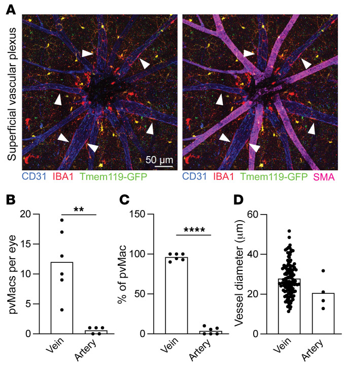
The blood-retina barrier (BRB), which is disrupted in diabetic retinopathy (DR) and uveitis, is an important anatomical characteristic of the retina, regulating nutrient, waste, water, protein, and immune cell flux. The BRB is composed of endothelial cell tight junctions, pericytes, astrocyte end feet, a collagen basement membrane, and perivascular macrophages. Despite the importance of the BRB, retinal perivascular macrophage function remains unknown. We found that retinal perivascular macrophages resided on postcapillary venules in the superficial vascular plexus and expressed MHC class II. Using single-cell RNA-Seq, we found that perivascular macrophages expressed a prochemotactic transcriptome and identified platelet factor 4 (Pf4, also known as CXCL4) as a perivascular macrophage marker. We used Pf4Cre mice to specifically deplete perivascular macrophages. To model retinal inflammation, we performed intraocular CCL2 injections. Ly6C+ monocytes crossed the BRB proximal to perivascular macrophages. Depletion of perivascular macrophages severely hampered Ly6C+ monocyte infiltration. These data suggest that retinal perivascular macrophages orchestrate immune cell migration across the BRB, with implications for inflammatory ocular diseases including DR and uveitis.

Keywords: Macrophages; Monocytes; Ophthalmology; Retinopathy.


Figures

Figure 1
Figure 1. Flow cytometric identification of retinal macrophage heterogeneity.
(A) Flow cytometric gating strategy. Top left panel: Live cells were identified from singlets. Bottom left panel: CD45+ cells were identified from live, singlet cells. Top middle panel: Lineage (CD4+, CD8+ [T cells], B220 [B cells], Ly6G [neutrophils], NK1.1 [NK cells], SiglecF [eosinophils]) versus CD11b plot to gate forward CD11b+Linneg mononuclear phagocytes. Bottom middle panel: CD64+ macrophages were identified. Top right panel: Tmem119GFP versus Cx3cr1 plot to delineate Cx3cr1hiTmem119GFP+ microglia. Bottom right panel: Nonmicroglia were plotted on a CD206 versus CD169 plot to identify CD206hiCD169+ hyalocytes and CD206intCD169neg perivascular macrophages. FSC-A, forward scatter area. (B) Modal frequency histogram for CD206 expression. (C) Hyalocytes expressed the most CD206, while perivascular macrophages expressed intermediate CD206 levels. (D) Modal frequency histogram for CD169 expression. (E) Hyalocytes expressed the most CD169, whereas perivascular macrophages expressed low levels of CD169. (F) Microglia were the most abundant retinal macrophages, followed by perivascular macrophages and hyalocytes. Data are presented as the mean ± SEM. ***P < 0.001 and ****P < 0.0001, by repeated-measures, 1-way ANOVA followed by Tukey’s multiple-comparison test. n = 10 per group. pvMac, perivascular macrophages.
Figure 2
Figure 2. Perivascular macrophages only reside in the superficial vascular plexus.
(AC) In the representative images of superficial (A), intermediate (B), and deep (C) vascular plexuses, white arrowheads identify perivascular macrophages, orange arrowheads show microglia, and red arrowheads highlight hyalocytes. Scale bar: 50 μm. (D) Zoomed-in representative image of each cell type. Scale bars: 50 μm. (EG) Counts of microglia (E), periventricular macrophages (F), and hyalocytes (G) in each vascular plexus. Microglia were in all 3 plexuses, while perivascular macrophages and hyalocytes were only found in the superficial vascular plexus. **P < 0.01, ***P < 0.001, and ****P < 0.0001, by repeated-measures, 1-way ANOVA followed by Tukey’s multiple-comparison test. (HK) MotiQ quantitative morphometry showing total area, spanned area, tree length, and ramification index. ****P < 0.0001, by 1-way ANOVA followed by Tukey’s multiple-comparison test. n = 11 per group (EG); n = 25–50 cells per group (HK). Data are presented as the mean ± SEM. ONH, optic nerve head.
Figure 3
Figure 3. Perivascular macrophages reside on venules.
(A) Representative images of the superficial vascular plexus. White arrowheads identify perivascular macrophages on SMAneg venules. Scale bar: 50 μm. (B and C) Number and percentage of perivascular macrophages on venules versus arterioles. (D) Perivascular macrophages reside on vessels with an average diameter of 25–30 μm. Data are presented as the mean ± SEM. **P < 0.01 and ****P < 0.0001, by 2-tailed t test. n = 5–6 mice per group (B and C). n = 129 vein and n = 4 artery perivascular macrophages (D).
Figure 4
Figure 4. Perivascular macrophages express Pf4.
(A) Uniform manifold approximation and projection (UMAP) dimension plot. (B and C) Microglia (MG) express higher levels of P2ry12 and Tmem119 than do non-MG. Mrc1 and Pf4 are more highly expressed in non-MG retinal macrophages. (D) Perivascular macrophages and hyalocytes were targeted by Pf4Cre to a significantly greater degree than microglia. **P < 0.01 and ***P < 0.001, by repeated-measures, 1-way ANOVA followed by Tukey’s multiple-comparison test. (E) Representative image of the superficial vascular plexus. Orange arrowheads delineate CD169+ hyalocytes; white arrowheads identify perivascular macrophages. Scale bar: 200 μm. (FH) Representative zoomed-in images of Pf4-zsGreen perivascular macrophages and hyalocytes stained for CD169 (FH) and CD206 (IK). n = 4 per group for D. Images in FK are representative of 3 mice. Scale bars: 50 μm. Data are presented as the mean ± SEM.
Figure 5
Figure 5. Pf4+ cells are MHCII+ and prochemotactic.
(A) UMAP dimension plot. (B) DotPlot of cluster-identifying genes showing 2 Pf4+ clusters (red arrows): Pf4-A and Pf4-B. (C) Modal frequency histogram of MHCII MFI from multiparameter flow cytometry. (D) Hyalocytes and perivascular macrophages expressed significantly higher levels of MHCII than did microglia, in agreement with the scRNA-Seq data. (E) Representative images of MHCII immunofluorescence in the superficial vascular plexus. White arrowheads identify MHCII+ perivascular macrophages, and orange arrowheads show MHCII hyalocytes. Scale bar: 50 μm. (F) Perivascular macrophages expressed significantly higher levels of MHCII than did microglia and hyalocytes. (G) GO enrichment for Pf4-A and Pf4-B showing increased expression of chemotaxis GO terms. (H) DotPlot of pro-chemotaxis genes differentially expressed in Pf4-A or Pf4-B. Size of dot indicates percentage of cells expressing the gene. Darkness of dot shows average expression with blue > purple. Data are presented as the mean ± SEM. *P < 0.05, **P < 0.01, and ***P < 0.001, by repeated-measures, 1-way ANOVA followed by Tukey’s multiple-comparison test (D and F). n = 6 per group.
Figure 6
Figure 6. Perivascular macrophages correlate with Ly6C+ inflammatory cell infiltration.
(A) Schematic showing approximate location for intravitreal injections of CCL2. (B) Frequency histogram of each cell type and their distance from the optic nerve. Significant overlap was found between the distribution of perivascular macrophages and Ly6C+ cells using a transformation of the Kolmogorov-Smirnov test. n = 2,998 cells. (C) Overview representative image of Ly6C+ cell infiltration at the superficial vascular plexus. Scale bar: 50 μm. (DF) Zoomed-in representative images identifying Ly6C+ cells close to the optic nerve head where most perivascular macrophages (white arrowheads) resided in Tmem119GFP mice. Scale bar: 200 μm. (G and H) Zoomed-in representative images of cells from Pf4-zsGreen mice showing close association between Ly6C+ monocytes (red cell, white arrowhead) and Pf4+ perivascular macrophages. Scale bar: 50 μm.
Figure 7
Figure 7. Pf4-DTR mice deplete perivascular macrophages and block immune cell infiltration.
(A and B) Representative immunofluorescence images of the superficial vascular plexus from PBS- or DT-treated Pf4-DTR mice. White arrows identify DTR+ perivascular macrophages. Scale bar: 100 μm. (C) DT treatment reduced the number of perivascular macrophages without affecting hyalocytes. **P < 0.01, by 2-way ANOVA followed by Šidák’s multiple-comparison test. n = 7 per group. (D and E) Representative immunofluorescence images of the superficial vascular plexus from PBS- and DT-treated Pf4Cre:R:26CAG-LSL-DTR mice after intravitreal CCL2 injection. White arrows identify Ly6C+ infiltrating immune cells. Red dash-outlined boxes indicate enlarged areas in G and H. Scale bars: 100 μm. (F) DT treatment decreased the density of Ly6C+ monocytes after CCL2 injection. **P < 0.01, by Mann-Whitney U test because the distribution was nonparametric on the Shapiro-Wilk test. n = 9 per group. (G and H) Enlarged images from D and E separated by lectin and Ly6C channels for PBS (G) and DT (H) treatment. Data are presented as the mean ± SEM.

Similar articles

Cited by

References

    1. Ehlers JP, et al. Quantitative ultra-widefield angiography and diabetic retinopathy severity: an assessment of panretinal leakage index, ischemic index and microaneurysm count. Ophthalmology. 2019;126(11):1527–1532. doi: 10.1016/j.ophtha.2019.05.034. - DOI - PMC - PubMed
    1. Pecen PE, et al. Peripheral findings and retinal vascular leakage on ultra-widefield fluorescein angiography in patients with uveitis. Ophthalmol Retina. 2017;1(5):428–434. doi: 10.1016/j.oret.2017.01.016. - DOI - PubMed
    1. Faraco G, et al. Perivascular macrophages mediate the neurovascular and cognitive dysfunction associated with hypertension. J Clin Invest. 2016;126(12):4674–4689. doi: 10.1172/JCI86950. - DOI - PMC - PubMed
    1. Wolter JR. Perivascular glia of the blood vessels of the human retina. Am J Ophthalmol. 1957;44(6):766–773. doi: 10.1016/0002-9394(76)90779-0. - DOI - PubMed
    1. Mendes-Jorge L, et al. Scavenger function of resident autofluorescent perivascular macrophages and their contribution to the maintenance of the blood-retinal barrier. Invest Ophthalmol Vis Sci. 2009;50(12):5997–6005. doi: 10.1167/iovs.09-3515. - DOI - PubMed

MeSH terms

Substances