. 2024 Aug 29;134(20):e177384.
doi: 10.1172/JCI177384.

B cell-based therapy produces antibodies that inhibit glioblastoma growth

Affiliations

B cell-based therapy produces antibodies that inhibit glioblastoma growth

Si Wang et al. J Clin Invest. .

Abstract

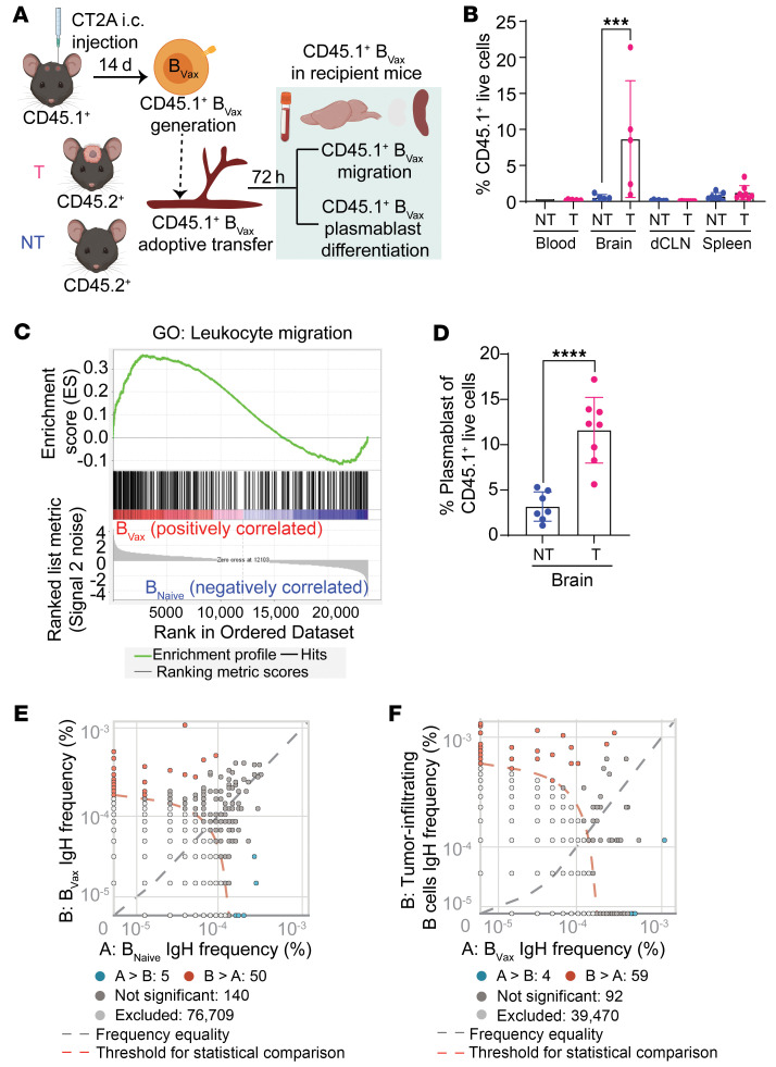
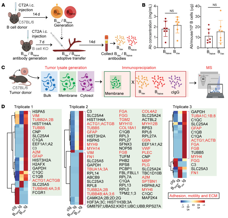
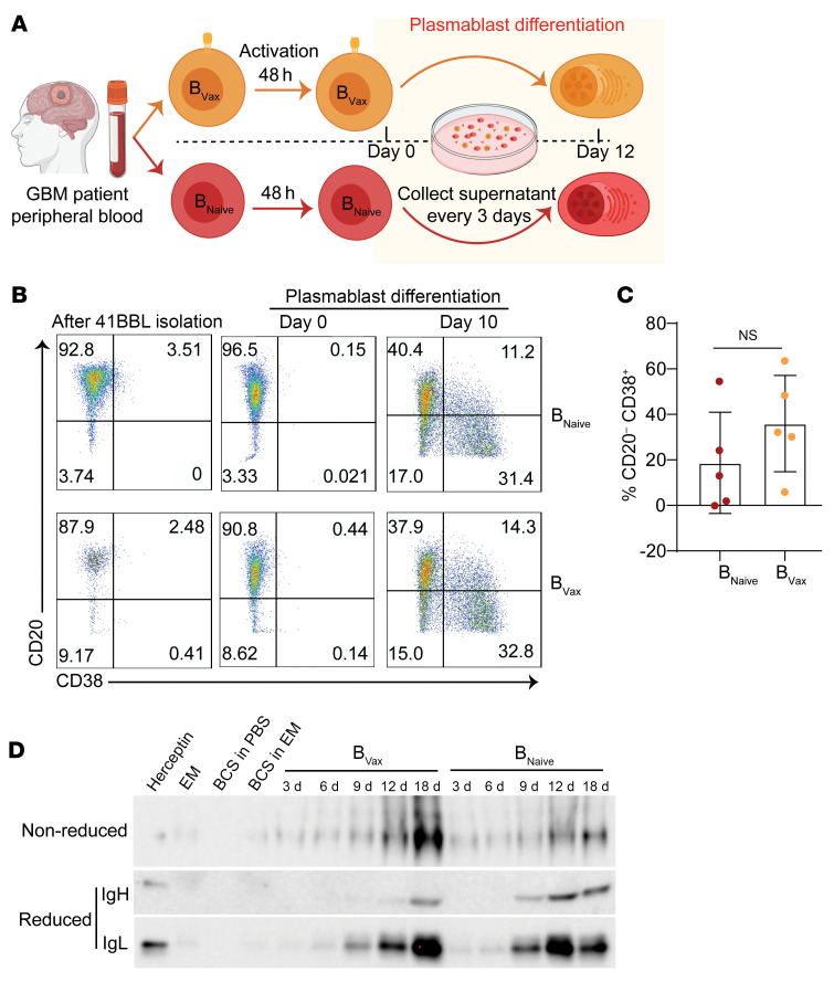
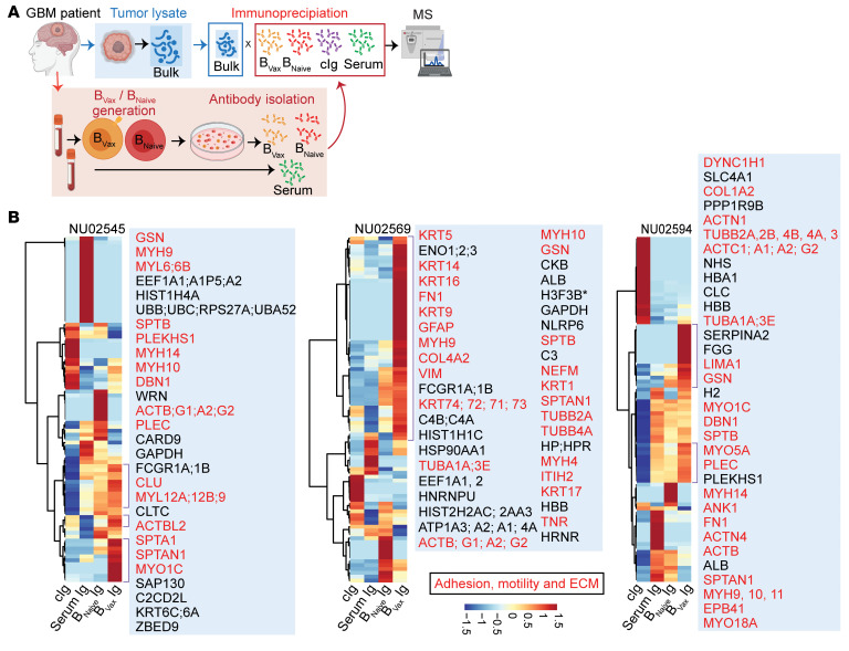
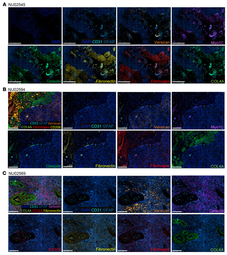
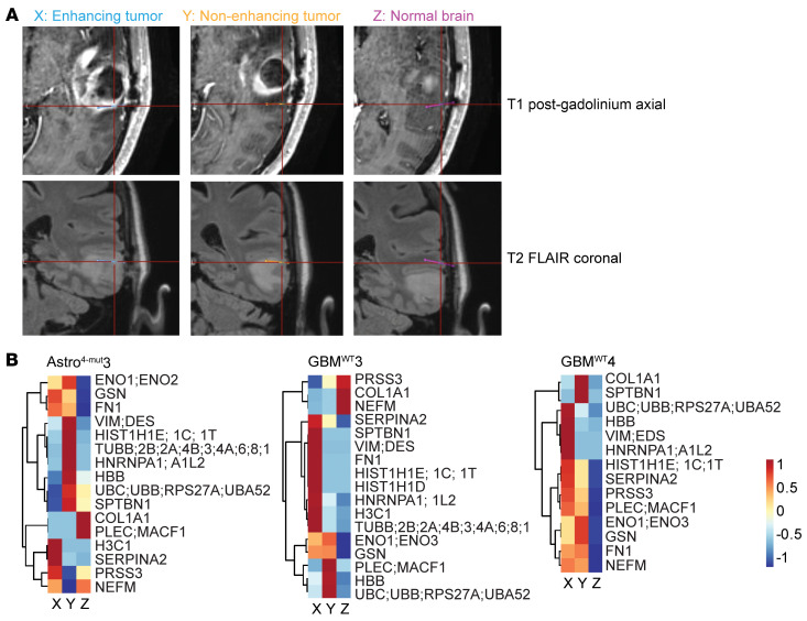
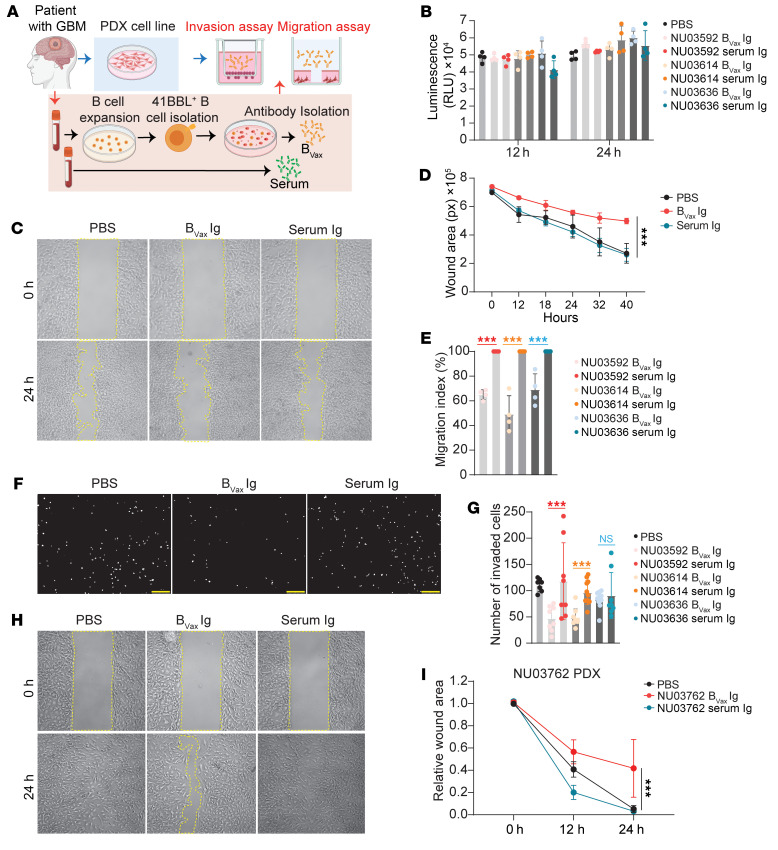
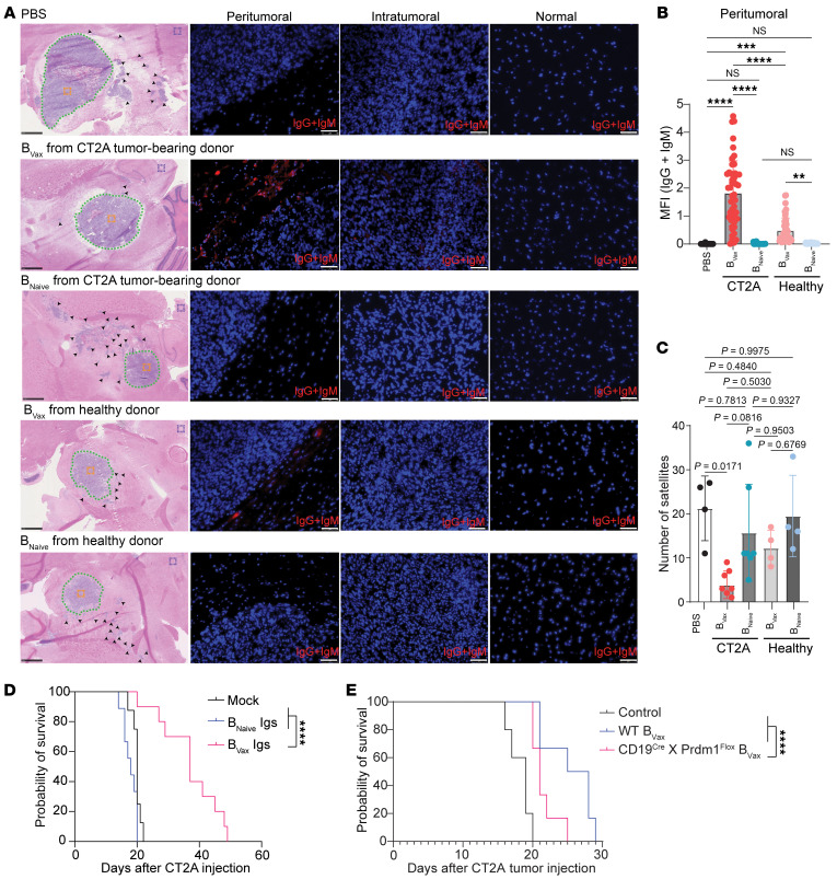
Glioblastoma (GBM) is a highly aggressive and malignant brain tumor with limited therapeutic options and a poor prognosis. Despite current treatments, the invasive nature of GBM often leads to recurrence. A promising alternative strategy is to harness the potential of the immune system against tumor cells. Our previous data showed that the BVax (B cell-based vaccine) can induce therapeutic responses in preclinical models of GBM. In this study, we aimed to characterize the antigenic reactivity of BVax-derived Abs and evaluate their therapeutic potential. We performed immunoproteomics and functional assays in murine models and samples from patients with GBM. Our investigations revealed that BVax distributed throughout the GBM tumor microenvironment and then differentiated into Ab-producing plasmablasts. Proteomics analyses indicated that the Abs produced by BVax had unique reactivity, predominantly targeting factors associated with cell motility and the extracellular matrix. Crucially, these Abs inhibited critical processes such as GBM cell migration and invasion. These findings provide valuable insights into the therapeutic potential of BVax-derived Abs for patients with GBM, pointing toward a novel direction for GBM immunotherapy.

Keywords: Brain cancer; Cancer immunotherapy; Extracellular matrix; Immunology; Oncology.


Figures

Figure 1
Figure 1. BVax differentiates into plasmablasts and generates potentially tumor-reactive B cell Igs.
(A) Schema illustrating the experimental design to investigate the potential of BVax to migrate to the glioma and differentiate into Ab-producing cells (plasmablasts). i.c., intracranial; T, tumor; NT, nontumor. (B) Percentage of BVax (CD45.1+) cells in various tissues (blood, brain, deep cervical lymph node [dCLN], and spleen) of intracranial tumor–bearing and nontumor-bearing mice via flow cytometry (n = 5 for each group). (C) GSEA of the indicated datasets comparing the transcriptional profile between BVax and BNaive. Data were pooled from 3 independent experiments. (D) Percentage of plasmablasts (CD19+CD20CD38+) within the BVax population of the brain via flow cytometry (n = 8 for each group). (E) Representative dot plot of BCR clones from BCR IgH sequencing comparing murine BVax and BNaive. Unique clones in BVax are shown in red; unique clones in BNaive are shown in blue (n = 3 for each group). (F) Representative dot plot of BCR clones from BCR IgH sequencing comparing murine TIB cells (n = 2) and BVax (n = 3). Unique clones in TIB cells are shown in red; unique clones in BVax are shown in blue. Data are the mean ± SD. ***P < 0.001 and ****P < 0.0001, by 1-way ANOVA. NT, nontumor; T, tumor.
Figure 2
Figure 2. Characterization of murine BVax-derived Ig reactivity.
(A) Schema demonstrating how in vivo BVax-derived Igs are produced from mice bearing CT2A gliomas. (B) Amount of BVax-derived Igs generated from mice bearing GBM tumors ( n = 7 for each group). (C) Schema depicting the protocol for the murine IP-MS experiments used to identify tumor-specific antigens recognized by BVax-derived Igs. (D) Heatmap revealing hierarchical clustering of GBM tumor antigens recognized by BVax-derived Igs. Each triplicate corresponds to an independent IP-MS experiment. In triplicate 1, BVax-derived Igs were pooled from 10 mice, and BNaive-derived Igs were pooled from 11 mice. In triplicate 2, BVax-derived Igs were pooled from 11 mice, and BNaive-derived Igs were pooled from 10 mice. Triplicate 3 involved BVax-derived Igs pooled from 10 mice and BNaive-derived Igs pooled from 12 mice. Data in B are the mean ± SD and were analyzed by 2-tailed Student’s t test.
Figure 3
Figure 3. Production of BVax-derived Igs in patients with GBM.
(A) Schema of the ex vivo generation of GBM patient BVax-derived Abs. (B) Dot plots of flow cytometric analysis of CD20 and CD38 expression during different stages in the BVax activation protocol to generate GBM patient–derived Abs ex vivo. (C) Box-and-whisker plot of the percentage of plasmablasts generated at day 10 of BVax/BNaive activation in patients with GBM (n = 5 for each group). (D) Western blot confirming the presence of Abs in the media during various stages of the ex vivo BVax activation protocol for patients with GBM. EM, expansion medium; BCS, B cell supplement. Data in C are the mean ± SD and were analyzed by 2-tailed Student’s t test.
Figure 4
Figure 4. Characterization of BVax-derived Ig reactivity in patients with GBM.
(A) Schema depicting the protocol for the human IP-MS experiments used to identify tumor-specific antigens recognized by BVax-derived Abs. (B) Heatmap revealing hierarchical clustering of GBM tumor antigens recognized by BVax-derived Igs (n = 3). Targets related to adhesion, motility, or the ECM are shown in red.
Figure 5
Figure 5. BVax-derived, Ig-recognized antigens are part of ECM.
Representative spatial multiplex IF images generated using the COMET system (Lunaphore Technologies) from paired GBM patients (n = 3) showing that BVax-derived, Ig-recognized antigens are part of the GBM ECM (including versican, fibronectin, and COL4A), ECM modulators (gelsolin), and proteins involved in cell adhesion and motility (MYO1C and fibrinogen). (A) Images for patient NU02545. Scale bars: 2 mm. (B) Images for patient NU02594. Scale bars: 200 μm. (C) Images for patient NU02569. Scale bars: 200 μm. CD31 (endothelial cells), GFAP (glioma tumor cells and astrocytes), CD163 (macrophage scavenger receptor), CD206 (immunosuppressive macrophages).
Figure 6
Figure 6. BVax-derived, Ig-recognized antigens are detected in the extracellular fluid with brain microdialysis.
(A) T1 post-gadolinium axial and T2 fluid-attenuated inversion recovery (FLAIR) coronal MRIs demonstrating the stereotactic target location of each catheter in enhancing tumor, nonenhancing tumor, and normal brain (MRI for patient GBMWT3 is shown). (B) Heatmap revealed the relative intensity of high-grade glioma antigens recognized by BVax-derived Igs in the micro dialysate. Samples were collected from 3 distinct patients, different from those in Figure 5.
Figure 7
Figure 7. BVax-derived Igs inhibit tumor invasion and migration.
(A) Schema depicting the protocol for the ex vivo functional assay. (B) Cell viability of GBM43 cells after treated with serum- or BVax-derived Igs from patients with GBM (NU03592, NU03614, and NU03636). n = 3. (C) Representative images of wound areas (marked by yellow lines) on confluent monolayers of GBM43 cells at 0 hours and 24 hours; cells were treated with serum- or BVax-derived Igs from a patient with GBM (NU03592). Original magnification, ×4 (C, F, and H). (D) Quantification of the wound area at different time points of GBM43 cells treated with serum- or BVax-derived Igs from a patient with GBM (NU03592). (E) Quantification of the migration index of GBM43 cells at 24 hours that were treated with serum- or BVax-derived Igs from patients with GBM (NU03592, NU03614, and NU03636). n = 3. (F) Representative images and (G) quantification of invading GBM43 cells (DAPI+) at 24 hours; cells had been treated with serum- or BVax-derived Igs from patients with GBM (NU03592, NU03614, and NU03636). n = 3. Each white dot represents a single invaded cell. Scale bars: 250 μm. (H) Representative images of wound areas (marked by yellow lines) on confluent monolayers of PDX cells at 0 hours and 24 hours; cells had been treated with serum- or BVax-derived Igs from the same patient (NU03762). (I) Quantification of the wound area of PDX cells at different time points; cells had been treated with serum- or BVax-derived Igs from the same patient (NU03762). Data are the mean ± SD. ***P < 0.001, by 1-way ANOVA (B, E, and G) or 2-way ANOVA (D and I).
Figure 8
Figure 8. BVax cells from tumor-bearing mice have a superior ability to produce Abs that localize to the peritumoral region and promote survival of GBM-bearing mice survival.
(A) Representative images of H&E and IF staining for anti–mouse IgG and IgM to assess the presence and localization of BVax-derived Igs. BVax or BNaive cells from healthy or CT2A tumor–bearing C57BL/6 mice were adoptively transferred into CT2A tumor–bearing muMT mice. Following treatment, brain tissues were harvested from recipient mice and stained for anti–mouse IgG and IgM (red). H&E-stained images show the organization of the tumors and the locations where the IF images were taken: peritumoral region (dotted green line), intratumoral region (orange box), and relatively normal brain (purple box). (B) Quantification of the relative intensity of BVax-Igs in the peritumoral region. A total of 10–15 images were taken around the peritumoral region in each mouse (dotted green line). The MFI of anti–mouse IgG and IgM (red) in each image was quantified using ImageJ as described previously (68, 69). PBS group: n = 3; BVaxCT2A group: n = 5; BNaiveCT2A group: n = 5; BVaxhealthy group: n = 3; BNaivehealthy group: n = 3. Data are representative of 2 independent experiments. (C) Quantification of satellites (black arrowhead) away from the CT2A tumor core based on H&E images from each mouse. PBS group: n = 3; BVax-CT2A group: n = 5; BNaiveCT2A group: n = 5; BVax healthy group: n = 3; BNaivehealthy group: n = 3. The data are representative of 2 independent experiments. (D) Survival of CT2A tumor–bearing mice was evaluated in 3 groups: mock-treated (n = 8), BNaive Ig–treated (n = 9), and BVax Ig–treated (n = 10). (E) Survival of CT2A tumor–bearing muMT mice was assessed according to the 3 treatment groups: PBS control (n = 5), WT BVax (n = 6), and Prdm1-deficient BVax (n = 6). Data are presented as the mean ± SD. **P < 0.01, ***P < 0.001, and ****P < 0.0001, by 1-way ANOVA (B and C) or log-rank test (D and E).

References

    1. Laug D, et al. A glial blueprint for gliomagenesis. Nat Rev Neurosci. 2018;19(7):393–403. doi: 10.1038/s41583-018-0014-3. - DOI - PMC - PubMed
    1. Schaff LR, Mellinghoff IK. Glioblastoma and other primary brain malignancies in adults: a review. JAMA. 2023;329(7):574–587. doi: 10.1001/jama.2023.0023. - DOI - PMC - PubMed
    1. Hambardzumyan D, Bergers G. Glioblastoma: defining tumor niches. Trends Cancer. 2015;1(4):252–265. doi: 10.1016/j.trecan.2015.10.009. - DOI - PMC - PubMed
    1. Zhang Y, Zhang Z. The history and advances in cancer immunotherapy: understanding the characteristics of tumor-infiltrating immune cells and their therapeutic implications. Cell Mol Immunol. 2020;17(8):807–821. doi: 10.1038/s41423-020-0488-6. - DOI - PMC - PubMed
    1. Petitprez F, et al. B cells are associated with survival and immunotherapy response in sarcoma. Nature. 2020;577(7791):556–560. doi: 10.1038/s41586-019-1906-8. - DOI - PubMed

MeSH terms

LinkOut - more resources