Review
. 2024 Sep 1;80(Pt 9):647-660.
doi: 10.1107/S2059798324007794. Epub 2024 Aug 29.

Post-translational modifications in the Protein Data Bank

Affiliations
Review

Post-translational modifications in the Protein Data Bank

Lucy C Schofield et al. Acta Crystallogr D Struct Biol. .

Abstract

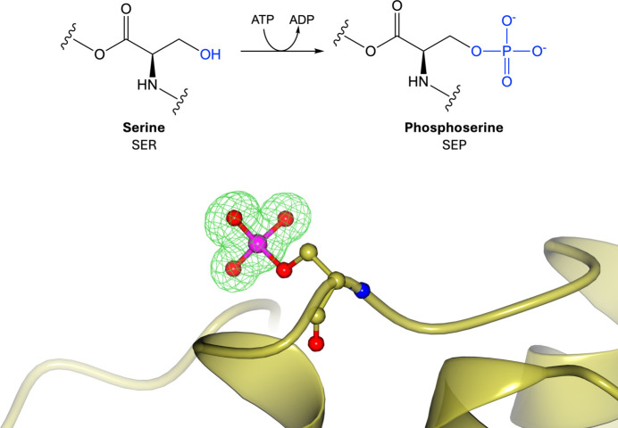
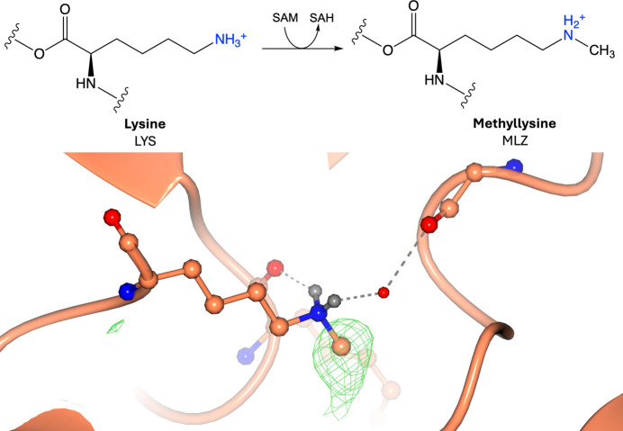
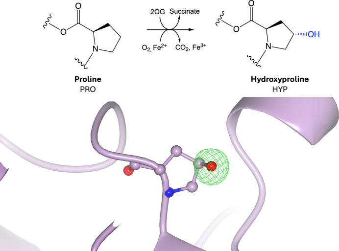
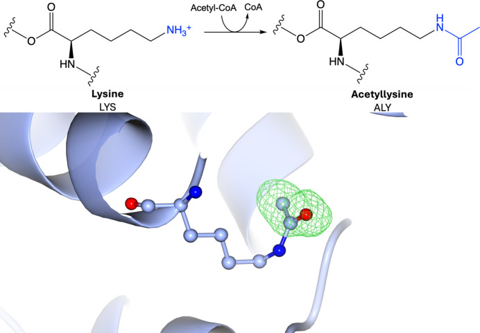
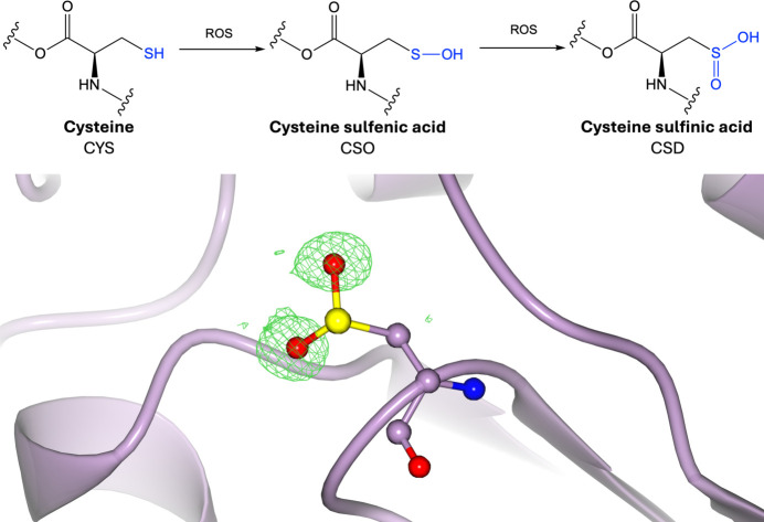
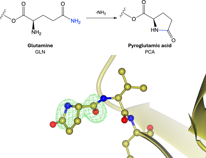
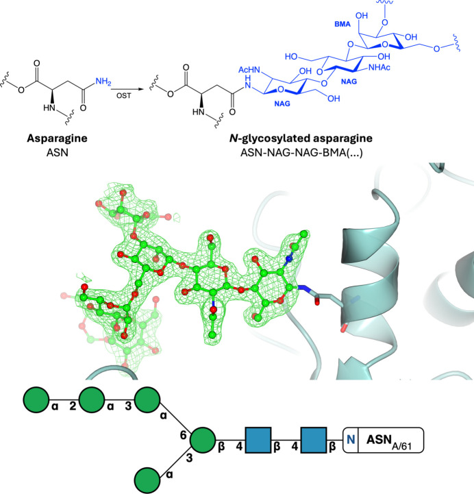
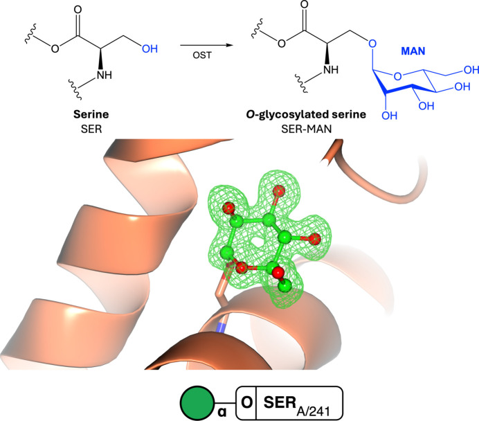
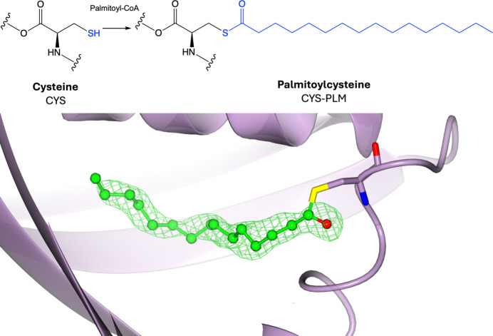
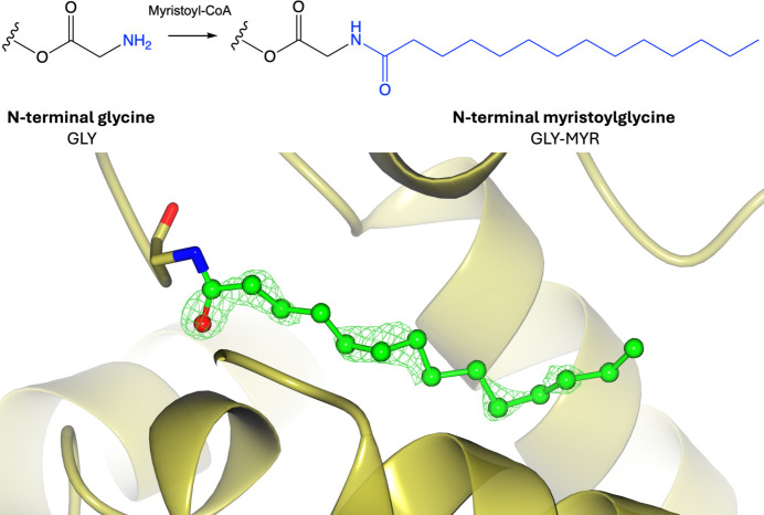
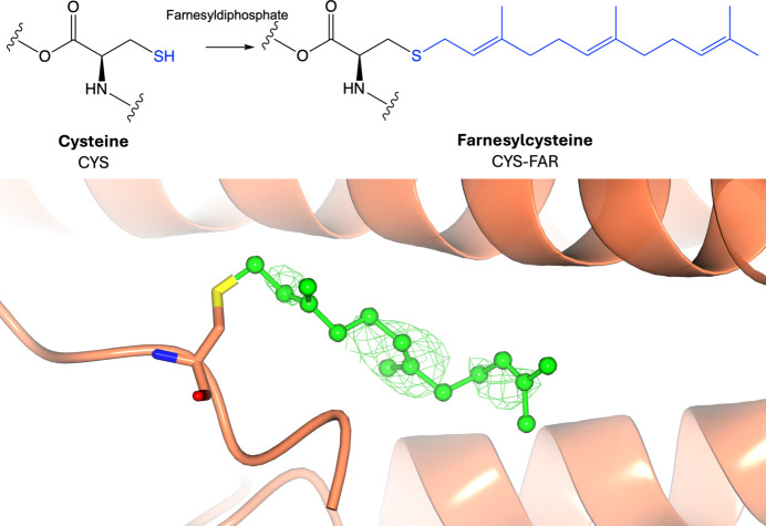
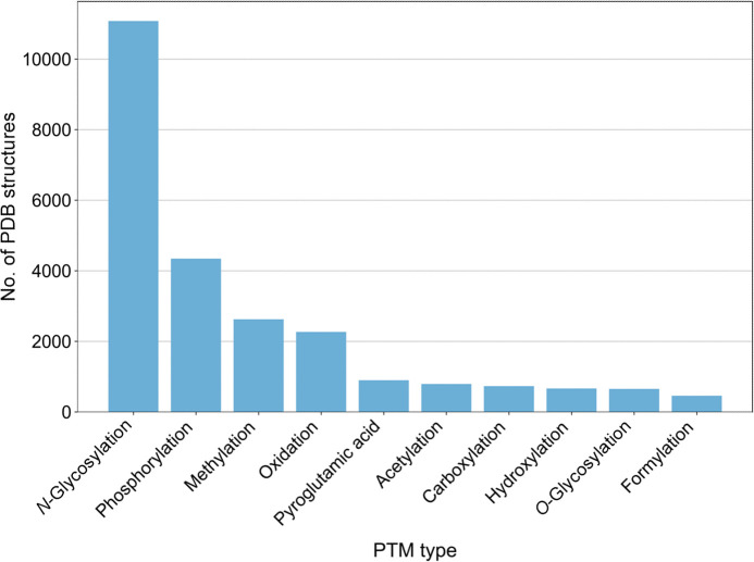
Proteins frequently undergo covalent modification at the post-translational level, which involves the covalent attachment of chemical groups onto amino acids. This can entail the singular or multiple addition of small groups, such as phosphorylation; long-chain modifications, such as glycosylation; small proteins, such as ubiquitination; as well as the interconversion of chemical groups, such as the formation of pyroglutamic acid. These post-translational modifications (PTMs) are essential for the normal functioning of cells, as they can alter the physicochemical properties of amino acids and therefore influence enzymatic activity, protein localization, protein-protein interactions and protein stability. Despite their inherent importance, accurately depicting PTMs in experimental studies of protein structures often poses a challenge. This review highlights the role of PTMs in protein structures, as well as the prevalence of PTMs in the Protein Data Bank, directing the reader to accurately built examples suitable for use as a modelling reference.

Keywords: Protein Data Bank; acetylation; glycosylation; phosphorylation; post-translational modifications.


Figures

Figure 1
Figure 1
Phosphorylation. Top: phosphorylation of serine involves the addition of a phosphate group donated by ATP to the side-chain hydroxyl group. Bottom: phosphoserine (PDB entry 5n3h; Sadowsky et al., 2011; CCD code SEP). Positive omit density is shown in green at 3σ for the modified residue. The rest of the protein chain is represented by a yellow ribbon model.
Figure 2
Figure 2
Methylation. Top: methylation of lysine involves the addition of a methyl group donated by S-adenosylmethionine (SAM) to the side-chain amino group. Bottom: methyllysine (PDB entry 3kmt; Wei & Zhou, 2010; CCD code MLZ). Positive omit density is shown in green at 3σ for the modified residue. The rest of the protein chain is represented by an orange ribbon model. The environment surrounding the modification is shown, indicating that the NZ atom is protonated (hydrogenation of the NZ atom was performed using Coot; Emsley et al., 2010 ▸). Hydrogen bonds are displayed as grey dashed lines.
Figure 3
Figure 3
Hydroxylation. Top: hydroxylation of proline involves the addition of a hydroxyl group donated by 2-oxoglutarate (2OG) to the side-chain pyrrolidine ring. Bottom: hydroxyproline (PDB entry 1gk8; Taylor et al., 2001; CCD code HYP). Positive omit density is shown in green at 3σ for the modified residue. The rest of the protein chain is represented by a purple ribbon model.
Figure 4
Figure 4
Acetylation. Top: acetylation of lysine involves the addition of an acetyl group donated by acetyl-CoA to the side-chain amino group. Bottom: acetyllysine (PDB entry 5e2f; Y. Kim, G. Joachimiak, M. Endres, G. Babnigg & A. Joachimiak, unpublished work; CCD code ALY). Positive omit density is shown in green at 3σ for the modified residue. The rest of the protein chain is represented by a blue ribbon model.
Figure 5
Figure 5
Oxidation. Top: oxidation of cysteine involves the reaction between the side-chain thiol group of cysteine and reactive oxygen species (ROS) to form cysteine sulfenic, then cysteine sulfinic acid and cysteine sulfonic acid (Supplementary Fig. S5). Bottom: cysteine sulfinic acid (PDB entry 1soa; Canet-Avilés et al., 2004; CCD code CSD). Positive omit density is shown in green at 3σ for the modified residue. The rest of the protein chain is represented by a purple ribbon model.
Figure 6
Figure 6
Pyroglutamic acid. Top: formation of pyroglutamic acid involves the cyclization of an N-terminal glutamine or glutamic acid. Bottom: pyroglutamic acid (PDB entry 8ojt; Davies et al., 2023; CCD code PCA). The first three N-terminal residues are shown. Positive omit density is shown in green at 3σ for the modified residue. The rest of the protein chain is represented by a yellow ribbon model.
Figure 7
Figure 7
N-Glycosylation. Top: N-glycosylation of asparagine involves the addition of an N-glycan to the side-chain amino group via oligosaccharide transferase (OST). Middle: N-linked glycosylation (PDB entry 5fji; Agirre et al., 2016 ▸). Positive omit density is shown in green at 3σ for the modified residue. The rest of the protein chain is represented by a blue ribbon model. Bottom: the symbol nomenclature for glycans (SNFG) representation is shown and was generated using the Privateer Web App (Dialpuri, Bagdonas, Schofield, Pham, Holland, Bond et al., 2024 ▸).
Figure 8
Figure 8
O-Glycosylation. Top: O-glycosylation of serine or threonine involves the addition of an O-glycan to the side-chain hydroxyl group via oligosaccharide transferase (OST). Middle: O-linked glycosylation (PDB entry 2ciw; Kühnel et al., 2006 ▸). Omit density is shown in green at 3σ for the modified residue. The rest of the protein chain is represented by an orange ribbon model. Bottom: the symbol nomenclature for glycans (SNFG) representation is shown and was generated using the Privateer Web App (Dialpuri, Bagdonas, Schofield, Pham, Holland, Bond et al., 2024 ▸).
Figure 9
Figure 9
Palmitoylation. Top: palmitoylation of cysteine involves the addition of a palmitoyl group donated by palmitoyl-CoA to the side-chain thiol group. Bottom: palmitoylcysteine (PDB entry 2w3y; Quevillon-Cheruel et al., 2009; CCD code PLM). Positive omit density is shown in green at 3σ for the modified residue. The rest of the protein chain is represented by a purple ribbon model.
Figure 10
Figure 10
Myristoylation. Top: myristoylation of N-terminal glycine involves the addition of a myristoyl group donated by myristoyl-CoA to the N-terminal amino group. Bottom: myristoylglycine (PDB entry 4zv5; Doležal et al., 2016; CCD code MYR). Positive omit density is shown in green at 3σ for the modified residue. The rest of the protein chain is represented by a yellow ribbon model.
Figure 11
Figure 11
Prenylation. Top: prenylation of cysteine involves the addition of a farnesyl group (or geranylgeranyl group; Supplementary Fig. S9) donated by farnesyl diphosphate (or geranylgeranyl diphosphate) to the side-chain thiol group. Bottom: farnesylcysteine (PDB entry 6k1z; Ji et al., 2019; CCD code FAR). Positive omit density is shown in green at 3σ for the modified residue. The rest of the protein chain is represented by an orange ribbon model.
Figure 12
Figure 12
Most common types of PTM in the PDB. The top ten PTM types identified in the PDB are shown. Data were obtained by searching the RCSB PDB Search API using identified CCD codes corresponding to PTMs. Glycosylation data were obtained from the Privateer Database (Dialpuri, Bagdonas, Schofield, Pham, Holland, Bond et al., 2024; Dialpuri, Bagdonas, Schofield, Pham, Holland & Agirre, 2024 ▸).
Figure 13
Figure 13
Most common small-molecule post-translationally modified residues in the PDB. The top 15 small-molecule PTMs identified in the PDB are shown. Data were obtained by searching the RCSB PDB Search API using identified CCD codes corresponding to PTMs. These data include CCD codes which are located in the polymeric sequence.
Figure 14
Figure 14
Glycosylation types in the PDB. Bars show the number of PDB structures containing each glycosylation type. The y axis is shown in log count. Data were obtained from the Privateer database (Dialpuri, Bagdonas, Schofield, Pham, Holland & Agirre, 2024; Dialpuri, Bagdonas, Schofield, Pham, Holland, Bond et al., 2024 ▸).
Figure 15
Figure 15
Lipidation types in the PDB. Bars show the number of PDB structures containing each detected lipidation type. The y axis is shown in log count. Data were obtained by searching the RCSB PDB Search API using identified CCD codes corresponding to lipid PTMs.
Figure 16
Figure 16
Unmodelled PTMs in the PDB. Density for protein modifications may be present but left unmodelled. (a) Lys84 has positive difference density surrounding the side-chain amino group (PDB entry 2jc7; Santillana et al., 2007 ▸). (b) Addition of a carboxyl group to the NZ atom of Lys84 [using Coot (Emsley et al., 2010 ▸) and AceDRG (Long et al., 2017 ▸)], followed by refinement with REFMAC (Murshudov et al., 2011 ▸). 2mFoDFc electron density is shown in blue at 1σ for the residue. Positive difference density (mFoDFc) is shown in green contoured to 4σ and clipped within 4 Å of Lys84. The rest of the protein chain is represented by a yellow ribbon model.

Similar articles

Cited by

References

    1. Adam, R. M., Mukhopadhyay, N. K., Kim, J., Di Vizio, D., Cinar, B., Boucher, K., Solomon, K. R. & Freeman, M. R. (2007). Cancer Res.67, 6238–6246. - PubMed
    1. Agirre, J. (2017). Acta Cryst. D73, 171–186. - PMC - PubMed
    1. Agirre, J., Ariza, A., Offen, W. A., Turkenburg, J. P., Roberts, S. M., McNicholas, S., Harris, P. V., McBrayer, B., Dohnalek, J., Cowtan, K. D., Davies, G. J. & Wilson, K. S. (2016). Acta Cryst. D72, 254–265. - PMC - PubMed
    1. Agirre, J., Atanasova, M., Bagdonas, H., Ballard, C. B., Baslé, A., Beilsten-Edmands, J., Borges, R. J., Brown, D. G., Burgos-Mármol, J. J., Berrisford, J. M., Bond, P. S., Caballero, I., Catapano, L., Chojnowski, G., Cook, A. G., Cowtan, K. D., Croll, T. I., Debreczeni, J. É., Devenish, N. E., Dodson, E. J., Drevon, T. R., Emsley, P., Evans, G., Evans, P. R., Fando, M., Foadi, J., Fuentes-Montero, L., Garman, E. F., Gerstel, M., Gildea, R. J., Hatti, K., Hekkelman, M. L., Heuser, P., Hoh, S. W., Hough, M. A., Jenkins, H. T., Jiménez, E., Joosten, R. P., Keegan, R. M., Keep, N., Krissinel, E. B., Kolenko, P., Kovalevskiy, O., Lamzin, V. S., Lawson, D. M., Lebedev, A. A., Leslie, A. G. W., Lohkamp, B., Long, F., Malý, M., McCoy, A. J., McNicholas, S. J., Medina, A., Millán, C., Murray, J. W., Murshudov, G. N., Nicholls, R. A., Noble, M. E. M., Oeffner, R., Pannu, N. S., Parkhurst, J. M., Pearce, N., Pereira, J., Perrakis, A., Powell, H. R., Read, R. J., Rigden, D. J., Rochira, W., Sammito, M., Sánchez Rodríguez, F., Sheldrick, G. M., Shelley, K. L., Simkovic, F., Simpkin, A. J., Skubak, P., Sobolev, E., Steiner, R. A., Stevenson, K., Tews, I., Thomas, J. M. H., Thorn, A., Valls, J. T., Uski, V., Usón, I., Vagin, A., Velankar, S., Vollmar, M., Walden, H., Waterman, D., Wilson, K. S., Winn, M. D., Winter, G., Wojdyr, M. & Yamashita, K. (2023). Acta Cryst. D79, 449–461.
    1. Agirre, J., Davies, G. J., Wilson, K. S. & Cowtan, K. D. (2017). Curr. Opin. Struct. Biol.44, 39–47. - PubMed