Review
. 2024 Oct 14;25(10):6357-6366.
doi: 10.1021/acs.biomac.4c00995. Epub 2024 Aug 29.

How Many Glucan Chains Form Plant Cellulose Microfibrils? A Mini Review

Affiliations
Review

How Many Glucan Chains Form Plant Cellulose Microfibrils? A Mini Review

Daniel J Cosgrove et al. Biomacromolecules. .

Abstract

Assessing the number of glucan chains in cellulose microfibrils (CMFs) is crucial for understanding their structure-property relationships and interactions within plant cell walls. This Review examines the conclusions and limitations of the major experimental techniques that have provided insights into this question. Small-angle X-ray and neutron scattering data predominantly support an 18-chain model, although analysis is complicated by factors such as fibril coalescence and matrix polysaccharide associations. Solid-state nuclear magnetic resonance (NMR) spectroscopy allows the estimation of the CMF width from the ratio of interior to surface glucose residues. However, there is uncertainty in the assignment of NMR spectral peaks to surface or interior chains. Freeze-fracture transmission electron microscopy images show cellulose synthase complexes to be "rosettes" of six lobes each consistent with a trimer of cellulose synthase enzymes, consistent with the synthesis of 18 parallel glucan chains in the CMF. Nevertheless, the number of chains in CMFs remains to be conclusively demonstrated.


Conflict of interest statement

The authors declare no competing financial interest.

Figures
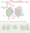
Figure 1
Figure 1
Features of the cellulose microfibril (CMF) structure and organization. (a) CMFs are relatively stiff rod-like structures with high order (crystallinity) and perhaps some disordered (noncrystalline) regions, but the location and proportion of any disordered cellulose within CMFs is uncertain. Surface chains are more mobile than core chains. CMFs have multiple faces with distinct hydrophilic and hydrophobic properties. (b) Possible models of CMFs made of 18 glucan chains are illustrated in cross sections with hexagonal and approximately rectangular habits (shapes). Other habits may also be possible. The habit determines the proportion of the different CMF surfaces that are relatively hydrophilic vs hydrophobic. The distance between the “sheets” of cellulose chains is ∼0.4 nm and between chains within a sheet the distance is ∼0.8 nm. Xyloglucan (XyG; blue) is hypothesized to bind to the hydrophobic surfaces, whereas xylan is hypothesized to bind to the hydrophilic surfaces. The internal core chains in the left fibril are shaded green. (c) Hypothetical arrangements of CMFs and the matrix in an abbreviated cross section of a single cell wall lamella. In primary cell walls, CMF regions may be found as “singletons” (ii) or in bundles of two or more CMFs laterally bound to each other, without (i) or with (iii) intermediary xyloglucan. In this illustration, CMF–CMF binding occurs via the hydrophobic surfaces, but other studies suggest hydrophilic surfaces also mediate bundling. In secondary cell walls, xyloglucan is absent, and bundling into macrofibrils with several CMFs may involve xylan and lignin interactions. Images a, b, and d were adapted with permission from ref (2). Copyright 2018 Elsevier.
Figure 2
Figure 2
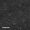
Hexameric rosette CSCs in the plasma membrane of a cell synthesizing a secondary cell wall. A sample containing differentiating tracheary elements of Zinnia elegans was frozen, fractured, shadowed, and replicated prior to TEM imaging. The resolved transmembrane regions of oligomerized CesAs are organized into six lobes in each CSC and visualized top-down. The cytosolic (catalytic) parts of the CSCs were below the plasma membrane and were not visible. The scale bar is 100 nm. (Image provided by R.L. Blanton, NC State University.)
Figure 3
Figure 3
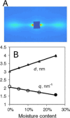
(A) SANS data from spruce wood showing two equatorial peaks corresponding to the spacing between CMFs. (B) Spacing from SANS as a function of the moisture content. The increase in spacing (d) with water absorption suggests that there are hydrophilic chains in between CMFs, placing an upper bound of the CMF diameter to less than 3 nm. Reproduced with permission from ref (41). Copyright 2011 PNAS.
Figure 4
Figure 4
Illustration of the three conformations of the C6 hydroxymethyl group of glucose residues in cellulose. The tg conformation has a C4 chemical shift around 89 ppm (D1), whereas the gg and gt conformation glucose residues have a C4 chemical shift around 84 ppm (D2).
Figure 5
Figure 5
Comparison of an image average of a CSC structure with a theoretical model assembled from CesA trimers. Left: The image average is reprinted with permission under a Creative Commons license CC BY 4.0 from ref (17). Middle: Envelope of the transmembrane region of a theoretical CSC assembled from CesA trimers (PDB: 6WLB). Right: The semitransparent envelope is shown together with a cartoon representation of the transmembrane segments of the assembled CSC.
Figure 6
Figure 6
Correlation between CSC structure and predicted CMF structure. (A) Cartoon of a rosette CSC with 3 CesAs within each of 6 lobes. Each CesA is assumed to be active, synthesizing one glucan chain. (B) Cross-section of an 18-chain CMF model with a 2–3–4–4–3–2 habit (note the number of glucan chains in each horizontal layer). The overall arrangement of glucan chains (within six boxes with dashed borders) mimics the arrangement of CesAs within the CSC. Reproduced with permission from ref (44). Copyright 2020 Springer Nature.

Similar articles

Cited by

References

    1. Nishiyama Y. Structure and properties of the cellulose microfibril. J. Wood Sci. 2009, 55, 241–9. 10.1007/s10086-009-1029-1. - DOI
    1. Cosgrove D. J. Nanoscale structure, mechanics and growth of epidermal cell walls. Curr. Opin Plant Biol. 2018, 46, 77–86. 10.1016/j.pbi.2018.07.016. - DOI - PubMed
    1. Jarvis M. C. Structure of native cellulose microfibrils, the starting point for nanocellulose manufacture. Philos. Trans A Math Phys. Eng. Sci. 2018, 376, 20170045.10.1098/rsta.2017.0045. - DOI - PubMed
    1. Nishiyama Y.; Sugiyama J.; Chanzy H.; Langan P. Crystal structure and hydrogen bonding system in cellulose I(alpha) from synchrotron X-ray and neutron fiber diffraction. J. Am. Chem. Soc. 2003, 125, 14300–6. 10.1021/ja037055w. - DOI - PubMed
    1. Nishiyama Y.; Langan P.; Chanzy H. Crystal structure and hydrogen-bonding system in cellulose Ibeta from synchrotron X-ray and neutron fiber diffraction. J. Am. Chem. Soc. 2002, 124, 9074–82. 10.1021/ja0257319. - DOI - PubMed

Publication types

LinkOut - more resources