. 2024 Dec 6;42(12):1040-1054.
doi: 10.1093/stmcls/sxae053.

Immunomodulatory potential of cytokine-licensed human bone marrow-derived mesenchymal stromal cells correlates with potency marker expression profile

Affiliations

Immunomodulatory potential of cytokine-licensed human bone marrow-derived mesenchymal stromal cells correlates with potency marker expression profile

Jiemin Wang et al. Stem Cells. .

Abstract

Cytokine(s) pre-activation/licensing is an effective way to enhance the immunomodulatory potency of mesenchymal stromal cells (MSCs). Currently, IFN-γ licensing received the most attention in comparison with other cytokines. After licensing human bone marrow-derived MSCs with pro-/anti-inflammatory cytokines IFN-γ, IL-1β, TNF-α, TGF-β1 alone or in combination, the in vitro immunomodulatory potency of these MSCs was studied by incubating with allogeneic T cells and macrophage-like THP-1 cells. In addition, immunomodulation-related molecules filtered by bioinformatics, complement 1 subcomponent (C1s), and interferon-induced GTP-binding protein Mx2 (MX2), were studied to verify whether to reflect the immunomodulatory potency. Herein, we reported that different cytokines cause different effects on the function of MSC. While TGF-β1 licensing enhances the capacity of MSCs to induce T cells with an immunosuppressive phenotype, IFN-γ-licensing strengthens the inhibitory effect of MSC on T cell proliferation. Both TGF-β1 and IFN-γ licensing can enhance the effect of MSC on reducing the expression of pro-inflammatory cytokines by M1 macrophage-like THP-1 cells. Interestingly, IFN-γ upregulates potential potency markers extracellular C1s and kynurenine (KYN) and intracellular MX2. These 3 molecules have the potential to reflect mesenchymal stromal cell immunomodulatory potency. In addition, we reported that there is a synergistic effect of TGF-β1 and IFN-γ in immunomodulation.

Keywords: IFN-γ; TGF-β1; immunomodulation; licensing; mesenchymal stromal cells; potency assay.


Conflict of interest statement

The authors declared no potential conflicts of interest.

Figures

Graphical Abstract
Graphical Abstract
Figure 1.
Figure 1.
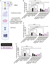
The inhibitory effect of (licensed) mesenchymal stromal cell (MSC) on T-cell proliferation. A. Schematic depicting the experimental set-up of MSC/peripheral blood mononuclear cell (PBMC) co-cultures. B-D. Percentage of proliferated CD3+, CD3+/CD4+ and CD3+/CD8+ T cells. All groups, except the unstimulated control, were stimulated with Human T-Activator CD3/CD28 Dynabeads. Flow cytometry was used to assess the proliferation. The proliferated ratio of each group is shown. Representative results of 3 independent experiments are shown ±SD and analyzed by 1-way ANOVA with Tukey’s multiple comparisons test. *P < .05; **P < 0.01; ***P < 0.001.
Figure 2.
Figure 2.
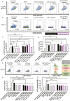
The T-cell phenotype is altered after coculturing with (licensed) mesenchymal stromal cell (MSC). A-B. The representative pseudocolor plots of FOXP3/CD25+ cells (regulatory T cells, Tregs), CD69+ T cells. C-D. The percentage of Tregs and CD69+ T cells. E. The representative pseudocolor plots of CD73+ T cells. F. The representative histograms of CD73 expression. G. The percentage of CD73+ T cells. H. The expression of CD73 (MFI, median fluorescence intensity). All groups, except the unstimulated control, were stimulated with Human T-Activator CD3/CD28 Dynabeads. Flow cytometry was used to assess the protein expression. Representative results of 3 independent experiments are shown ±SD and analyzed by 1-way ANOVA with Tukey’s multiple comparisons test. *P < .05; **P < .01; ***P < .001.
Figure 3.
Figure 3.
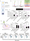
Both TGF-β1 and IFN-γ licensing enhances the effect of mesenchymal stromal cells (MSCs) on reducing the TNF-α and IL-1β expression of M1φ THP-1 cells. A. Schematic illustrating the experimental overview of MSC/THP-1 coculturing assay. B. The representative histograms IL-1β and TNF-α by THP-1 cells. C. The expression of IL-1β and TNF-α by THP-1 cells (MFI, median fluorescence intensity). D. Schematic illustrating the experimental overview of MSC/THP-1 coculturing assay to focus on their crosstalk. E. The percentage of THP-1 cells crosstalk with MSCs (Q2 in Figure 3G). F. The percentage of MSCs which has not crosstalked with THP-1 cells(Q1 in Figure 3G). G. The representative pseudocolor plots of MSC/THP-1 crosstalk assay. Flow cytometry was used to assess the protein and crosstalk. Representative results of 3 independent experiments are shown ± SD and analyzed by one-way ANOVA with Tukey’s multiple comparisons test. *P < 0.05; **P < 0.01; ***P < 0.001.
Figure 4.
Figure 4.
The bioinformatics analysis of strong immunomodulatory mesenchymal stromal cells (MSCs). A. The volcano plots of each datasets. B. The Venn diagram of significant upregulated genes in each dataset. Complement 1 subcomponent (C1s) and interferon-induced GTP-binding protein Mx2 (MX2) are upregulated in all the datasets. C. The fold change and adjust P value of C1s and MX2 in each dataset.
Figure 5.
Figure 5.
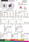
The gene and protein alteration of IFN-γ-licensed mesenchymal stromal cells (MSCs). A. PD-L1 expression in RNA and protein. B. The representative histograms of PD-L1. C. The representative dots of MX2, GAPDH, and IDO1 via dot-blotting in each group. D. IDO1 expression in RNA and protein. E. MX2 expression in RNA and protein. F. C1s expression in RNA and protein. The IDO1 and MX2 protein expressions are assessed by dot-blotting. The PD-L1 protein expression is assessed by flow cytometry. The C1s protein concentration is assessed by ELISA. All of the gene quantification is assessed by qPCR. Representative results of 3 independent experiments are shown ±SD and analyzed by one-way ANOVA with Tukey’s multiple comparisons test. *P < .05; **P < .01; ***P < 0.001.
Figure 6.
Figure 6.
IFN-γ licensing increases IDO1-dependent kynurenine (KYN) concentration and KYN has the strongest negative correlation with the proliferated T-cell ratio. A. KYN concentration in each 24-hour licensing (or not) group. B. Schematic depicting the experimental set-up of KYN IDO1-dependence assay. C-D. KYN concentration corresponds to different time points. E. Time-dependence of KYN concentration. F. The correlation study between selected potency markers and immunomodulatory properties. The slope and P-value were labeled. A was analyzed by 1-way ANOVA with Tukey’s multiple comparisons test. C-E were done by analysis of 2-way ANOVA with Sidak’s multiple comparisons test. *P < .05; **P < .01; ***P < 0.001. F was analyzed by univariate linear regression models.

References

    1. Castro LL, Kitoko JZ, Xisto DG, et al.Multiple doses of adipose tissue-derived mesenchymal stromal cells induce immunosuppression in experimental asthma. Stem Cells Transl Med. 2020;9(2):250-260. https://doi.org/10.1002/sctm.19-0120 - DOI
    1. Poggi A, Varesano S, Zocchi MR.. How to hit mesenchymal stromal cells and make the tumor microenvironment immunostimulant rather than immunosuppressive. Front Immunol. 2018;9:262. - PMC - PubMed
    1. Sukho P, Hesselink JW, Kops N, et al.Human mesenchymal stromal cell sheets induce macrophages predominantly to an anti-inflammatory phenotype. Stem Cells Dev. 2018;27(13):922-934. https://doi.org/10.1089/scd.2017.0275 - DOI - PubMed
    1. Ge XH, Shi K, Hou J, et al.Galectin-1 secreted by bone marrow-derived mesenchymal stem cells mediates anti-inflammatory responses in acute airway disease. Exp Cell Res. 2021;407(1):112788. https://doi.org/10.1016/j.yexcr.2021.112788 - DOI - PubMed
    1. Ortiz-Virumbrales M, Menta R, Pérez LM, et al.Human adipose mesenchymal stem cells modulate myeloid cells toward an anti-inflammatory and reparative phenotype: role of IL-6 and PGE2. Stem Cell Res Therapy. 2020;11(1):462. https://doi.org/10.1186/s13287-020-01975-2 - DOI - PMC - PubMed

Publication types

MeSH terms