. 2024 Oct 3;187(20):5604-5619.e14.
doi: 10.1016/j.cell.2024.07.053. Epub 2024 Aug 28.

Cryo-EM-based discovery of a pathogenic parvovirus causing epidemic mortality by black wasting disease in farmed beetles

Affiliations

Cryo-EM-based discovery of a pathogenic parvovirus causing epidemic mortality by black wasting disease in farmed beetles

Judit J Penzes et al. Cell. .

Abstract

We use cryoelectron microscopy (cryo-EM) as a sequence- and culture-independent diagnostic tool to identify the etiological agent of an agricultural pandemic. For the past 4 years, American insect-rearing facilities have experienced a distinctive larval pathology and colony collapse of farmed Zophobas morio (superworm). By means of cryo-EM, we discovered the causative agent: a densovirus that we named Zophobas morio black wasting virus (ZmBWV). We confirmed the etiology of disease by fulfilling Koch's postulates and characterizing strains from across the United States. ZmBWV is a member of the family Parvoviridae with a 5,542 nt genome, and we describe intersubunit interactions explaining its expanded internal volume relative to human parvoviruses. Cryo-EM structures at resolutions up to 2.1 Å revealed single-strand DNA (ssDNA) ordering at the capsid inner surface pinned by base-binding pockets in the capsid inner surface. Also, we demonstrated the prophylactic potential of non-pathogenic strains to provide cross-protection in vivo.

Keywords: Zophobas morio; capsid structure; capsid-DNA interactions; cryo-EM; densovirus; diagnostic virology; emerging infectious diseases; entomology; parvovirus; superworm.


Conflict of interest statement

Declaration of interests Rutgers University filed US Provisional Application 63/591,484 “Method for inhibiting Z. morio black wasting disease” on behalf of J.J.P. and J.T.K. S.A.Y. is employed by REGENXBIO Inc. and holds shares in the company. J.T.K. receives a research grant from REGENXBIO Inc.

Figures

Figure 1
Figure 1
Discovery of a pathogenic parvovirus via cryo-electron microscopy (CryoEM). (A) Top: deceased superworms (Zophobas morio) typifying the blackening in Zophobas morio black wasting disease (ZmBWD). Bottom: progression of ZmBWD in 4-week-old Z. morio larvae. Larva source is presented in Table S3. (B) Sucrose step gradient from blackened Z. morio larvae displaying two fractions (arrows) at the 20% and 30% sucrose interfaces, respectively, which appear blue in fluorescent light due to the presence of viral particles. (C) Electron micrographs displaying the particles from those fractions of ~28nm. Insets emphasize the absence of genome inside particles of the 20% band and presence inside particles of the 30% band. (D) Isosurface rendering (left) and central slice through (right) 3D reconstructions of the particles purified from diseased larvae. The gold line separates reconstructions of the 20% band (upper right) and 30% band (lower left). The protein structure is almost identical between the two bands but only the 30% band contains genome. The initial polyalanine chain is displayed as a ribbon diagram, with each T=1-related subunit in a different color. Results of searches based on this map are presented in Supplemental Tables 1-2. (E) Comparison of the capsid surface of the densovirus described herein associated with ZmBWD and consequently designated as Zophobas morio black wasting virus (ZmBWV) with those of other members of the Parvoviridae family: Subfamilies Densovirinae (Galleria mellonella densovirus [GmDV)] Acheta domesticus densovirus [AdDV] and Bombyx mori densovirus [BmDV]), Hamaparvovirinae (Penaeus stylirostris densovirus [PstDV]), and Parvovirinae (adeno-associated virus 2 [AAV2])., colored by radial distance. (F) Discovery of an iflavirus at low concentration alongside NJ2-molitor. Left: extract of a micrograph showing an iflavirus virion (arrow) alongside a ZmBWV virion. Right: polyalanine chain trace through the reconstructed density, used for structure-based identification.
Figure 2
Figure 2
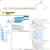
Characterization of the genome, proteins and phylogenetic relationships of the densovirus, ZmBWV. (A) The I-shaped ssDNA secondary structure, flanking the ZmBWV genome. The presented DNA region represents the entire left inverted terminal repeat of the genome and the bases forming the terminal hairpin are highlighted in green. (B) Top, diagram of the ZmBWV genome showing ITRs (rectanges) and ORF locations (arrows) The pink and teal boxes indicate the respective size and position of the superfamily 3 helicase (SF3) and the phospholipase A2 (PLA2) domains, respectively. Bottom, diagram showing the putative expression pattern of the structural proteins (VPs) from cap1 and cap2 based on the tandem mass spectrometry data (Fig S1A). (C) SDS-PAGE gel, showing the four structural proteins of ZmBWV, present in both the empty and virion fractions. Mass spectrometry is presented in Figure S1. (D) Inferred phylogeny of the complete coding sequences of all ZmBWV strains, identified in this study and shown in bold, plus members of the species Blattambidensovirus incertum1, derived mostly from metagenomic studies. The tree is outgroup-rooted to the closest related species of the same genus.
Figure 3
Figure 3
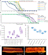
The pathogenesis of the Zophobas morio black wasting virus (ZmBWV) type strain, UT-morio. (A) Line diagrams showing the distribution of symptoms and the survival rate of Z. morio larvae inoculated by ZmBWV UT-morio as direct fat body injections. Mock-infected larvae were inoculated by sterile phosphate buffered saline; negative control specimens received no injection. Inoculation titers are shown in the panel legend. (B) Line diagrams representing the manifestation of symptoms and the survival rate of Z. morio larvae after introduction of ZmBWV strain UT-morio by adding blackened carcasses of ZmBWV-killed larvae, which were ingested by the subjects (purple); or by dripping a suspension of virus in PBS onto the larval cuticle (gray and green). Controls as in panel A. (C) Box and whiskers plots showing the qPCR viral titers of the larvae from panels A and B, sampled at 12 d.p.i. (halfway through the experiment). Each plot summarizes the results of three biological replicates. (D) Viral yield of infected Z. morio specimens at various life stages. The box and whiskers plots summarize the qPCR results of three infected colonies, located in New York, Arkansas and Georgia. (E) MicroCT reconstructions of the midgut of a healthy (left) and freshly-deceased (right) 4-week-old Z. morio larvae. The specimen in the right panel, fixed immediately upon becoming non-responsive, had been inoculated with ZmBWV strain UT-morio by ingestion of an infected carcass and showed the typical pathology (including blackening) associated with ZmBWV. Both images show the cross section of the midgut, tilted obliquely, revealing both the lumen and outer surface of the organ.
Figure 4
Figure 4
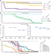
Cross-species pathogenesis of Zophobas morio black wasting virus (ZmBWV). (A) Line diagrams of the survival rate and the prevalence of symptoms in case of 4-week-old Z. morio larvae inoculated by direct fat body injections of Tenebrio molitor-derived NJ2-molitor strain. Figure S2B presents the control experiment to this study, inoculating the negative larvae of the same stock using the pathogenic representative UT-morio ZmBWV strain. Negative control received no injection. Mock-infection was with sterile PBS. (B) Line diagrams comparing the survival rate and prevalence of symptoms in ≈4-week-old Z. morio larvae inoculated by UT-morio or NJ2-molitor ZmBWV strains via feeding. Controls as panel A. (C) Box and whisker plots showing viral titers of Z. morio larvae following inoculation by the strain NJ1-molitor. Titer values were determined by qPCR. T.p.i.: time post-inoculation. (D) Box and whiskers plots showing the ZmBWV titer in the tenebrionid beetle Alphitobius diaprenius and the blattodean roach Blaptica duibia. Both were obtained from a facility in New York that experienced Z. morio colony collapse due to ZmBWV. * indicates uninfected larva. (E) Line diagrams of the survival rate and symptom prevalence in double-inoculated 4-week-old Z. morio larvae. See the panel legend for the exact order and type of strains used in each treatment group. An earlier double-inoculation experiment is shown in Figure S2C.
Figure 5
Figure 5
Intersubunit interactions of the ZmBWV capsid. (A) Ribbon diagrams of the atomic model of one subunit of the capsid. Colored arrows mark the first ordered residue of each model. The β-strands comprising the jelly roll core are labeled. The fivefold symmetry axis is labeled by a pentagon, the threefold axis by a triangle and the twofold axis is marked by the ellipsoid. (B) Ribbon diagram of adjacent subunits. Note the domain-swapping conformation of the N-termini, composing an additional, inter-subunit β-sheet (cyan). In this conformation, the βA strand can interact with the βF strand of the threefold neighboring subunit (red box). This interaction only forms in the presence of a packaged genome (see inset). Comparison to homologs is shown in Figure S5A. (C) Difference map between empty capsids and full virions highlights the externalization of the capsid N-termini in empty particles. Top-down (left) and cross-sectional side views (right) contain the empty capsid density map (gray) with excess density of the virions (red) and of the empty capsids (green). In the cross-sectional view, one copy of the DE loop has been made translucent and a ribbon diagram of the full virion is shown; the DE loop is ordered only in full virions. Protease susceptibility is shown in Figure S4. (D) The ZmBWV β-annulus, located at the threefold symmetry axis as shown in the magnified left panel, coordinates an ion. Distances to nearby heteroatoms are labeled in Ångströms. At right, the threefold axis is highlighted in gold on the B-factor map of ZmBWV and of GmDV (cutaway at upper right). The B-factor is low (blue) in this region in ZmBWV but high in GmDV, whose crystal structure lacks an ion. (E) An electron micrograph of full ZmBWV UT-morio virions dialyzed into Tris-buffered saline evinced spontaneous loss of encapsidated DNA and increased prevalence of disrupted or partial capsids.
Figure 6
Figure 6
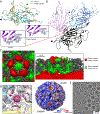
Non-protein densities in the cryoEM map of ZmBWV. (A) Excess density (magenta) not accounted for by protein (cyan) in the ZmBWV icosahedral map is concentrated at one location per ASU. (B) NJ2-molitor map filtered to 5Å resolution showing capsid residue Asn235 (blue), other capsid density (gray), and non-protein density (orange). (C) No ordered density for the unidentified moiety is seen adjacent to Asn235 in the map of OR-molitor filtered to 5Å (colored as above). (D) Virions of the NJ2-molitor strain include 17 ordered nucleotides per subunit, surrounding the threefold symmetry axis. Inter-chain hydrogen bonds in yellow. Nucleotide properties are described in Table S5. (E) A column of stacked bases including a single base from the 9-mer under three bases from the 5-mer. (F-H) Three base-binding pockets on the capsid interior surface: a pyrimidine (modeled as cytosine) of the 3-mer apposes two lysine residues (F); an adenosine of the 9-mer is base-flipped into a pocket (G); and a cytosine of the 5-mer base-flips to π-stack against Tyr404 while oriented to form a hydrogen bond with Glu493 (H). In panels F-H, density attributable to DNA is translucent pink (3-mer), purple (9-mer), or dark purple (5-mer), protein density is gray, and water density is red; in B-C and E-H protein carbons are displayed in cyan, DNA carbons in pink/purple/dark purple (as for the DNA density) and heteroatoms as per standard conventions.

References

    1. Bishop RF, Davidson GP, Holmes IH, and Ruck BJ (1974). Detection of a New Virus By Electron Microscopy of Fæcal Extracts From Children With Acute Gastroenteritis. The Lancet. - PubMed
    1. Ksiazek TG, Erdman D, Goldsmith CS, Zaki SR, Peret T, Emery S, Tong S, Urbani C, Comer JA, Lim W, et al. (2003). A novel coronavirus associated with severe acute respiratory syndrome. N Engl J Med 348, 1953–1966. 10.1056/NEJMoa030781. - DOI - PubMed
    1. Blumberg BS (1977). Australia antigen and the biology of hepatitis B. Science 197, 17–25. 10.1126/science.325649. - DOI - PubMed
    1. Choo QL, Kuo G, Weiner AJ, Overby LR, Bradley DW, and Houghton M (1989). Isolation of a cDNA clone derived from a blood-borne non-A, non-B viral hepatitis genome. Science. - PubMed
    1. Feng H, Shuda M, Chang Y, and Moore PS (2008). Clonal integration of a polyomavirus in human Merkel cell carcinoma. Science 319, 1096–1100. 10.1126/science.1152586. - DOI - PMC - PubMed

MeSH terms

LinkOut - more resources