Review
. 2024 Oct 31;64(4):2401095.
doi: 10.1183/13993003.01095-2024. Print 2024 Oct.

Pathology and pathobiology of pulmonary hypertension: current insights and future directions

Affiliations
Review

Pathology and pathobiology of pulmonary hypertension: current insights and future directions

Christophe Guignabert et al. Eur Respir J. .

Abstract

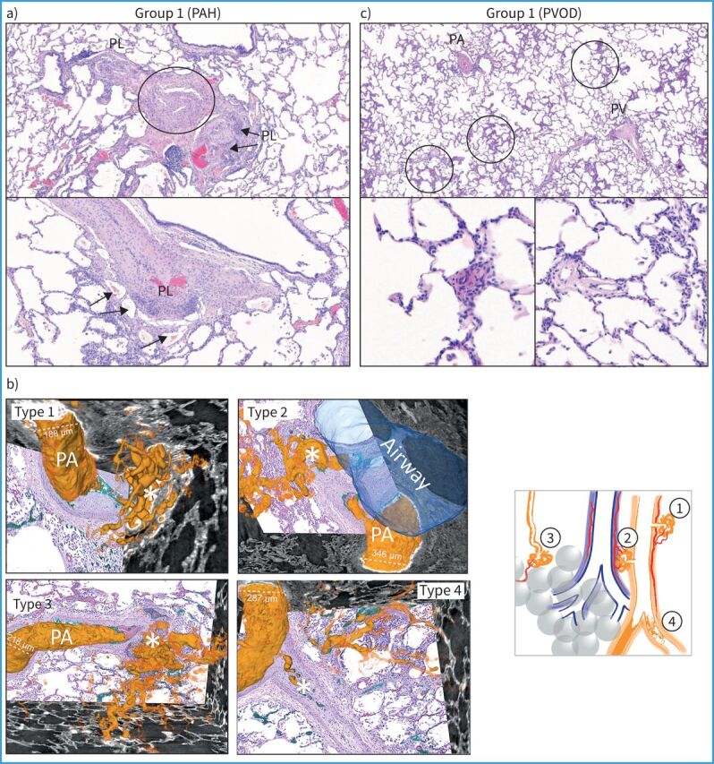
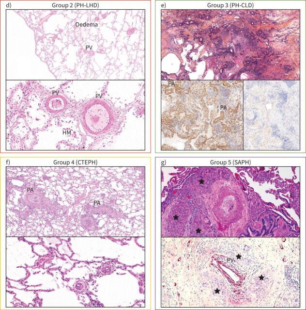
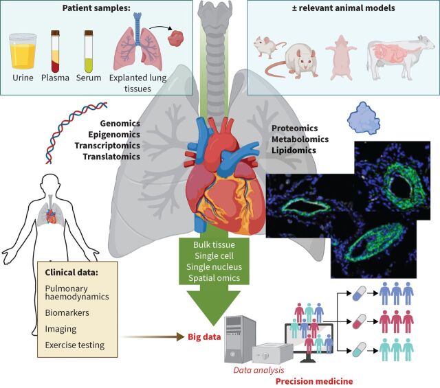
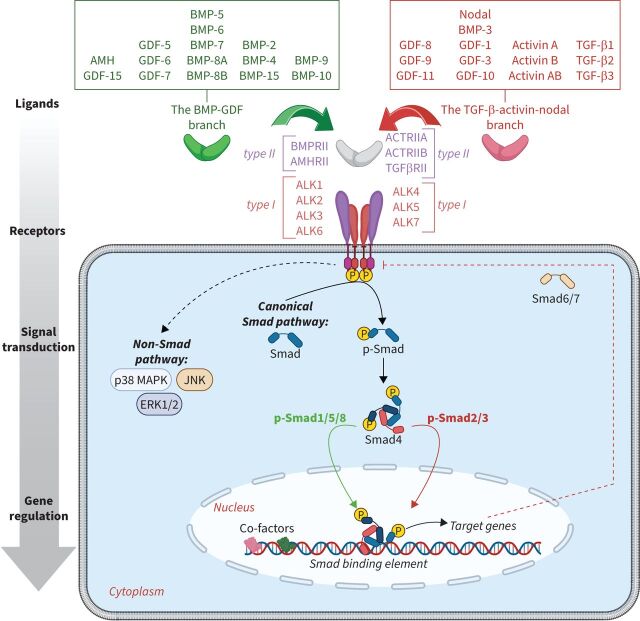
In recent years, major advances have been made in the understanding of the cellular and molecular mechanisms driving pulmonary vascular remodelling in various forms of pulmonary hypertension, including pulmonary arterial hypertension, pulmonary hypertension associated with left heart disease, pulmonary hypertension associated with chronic lung disease and hypoxia, and chronic thromboembolic pulmonary hypertension. However, the survival rates for these different forms of pulmonary hypertension remain unsatisfactory, underscoring the crucial need to more effectively translate innovative scientific knowledge into healthcare interventions. In these proceedings of the 7th World Symposium on Pulmonary Hypertension, we delve into recent developments in the field of pathology and pathophysiology, prioritising them while questioning their relevance to different subsets of pulmonary hypertension. In addition, we explore how the latest omics and other technological advances can help us better and more rapidly understand the myriad basic mechanisms contributing to the initiation and progression of pulmonary vascular remodelling. Finally, we discuss strategies aimed at improving patient care, optimising drug development, and providing essential support to advance research in this field.


Conflict of interest statement

Conflict of interest: C. Guignabert reports grants from Acceleron Pharma, MSD, Corteria Pharmaceuticals, Structure Therapeutics (ex-ShouTi) and Gossamer Bio, payment or honoraria for lectures, presentations, manuscript writing or educational events from MSD, and patents planned, issued or pending (WO/2024/023139, WO/2018/011376). J. Aman has no potential conflicts of interest to disclose. S. Bonnet reports grants from Morphic Therapeutic, Sunshine Bio and Janssen, consultancy fees from Morphic Therapeutic and Chiesi, and participation on a data safety monitoring board or advisory board with Morphic Therapeutic and Allienaire. P. Dorfmüller reports payment or honoraria for lectures, presentations, manuscript writing or educational events from AstraZeneca. A.J. Olschewski reports grants from Austrian Science Fund (FWF) (10.55776/I6299 and 10.55776/KLI1153), payment or honoraria for lectures, presentations, manuscript writing or educational events from MSD, patents pending (PCT/EP2017/055440), and stock (or stock options) with Bayer. S. Pullamsetti reports grants and consultancy fees from Gossamer Bio. M. Rabinovitch reports consultancy fees from Pfizer, Amgen, Merck and Tiakis, payment or honoraria for lectures, presentations, manuscript writing or educational events from NIH, patents planned, issued or pending (FK506: tacrolimus in pulmonary hypertension), participation on a data safety monitoring board or advisory board with Amgen and NIH, is associate editor for JACC BTS, receipt of equipment, materials, drugs, medical writing, gifts or other services from Tiakis (tiprelestat). R.T. Schermuly reports grants from Chiesi and Attgeno, and consultancy fees from Gossamer, Attgeno and Chiesi. M. Humbert reports grants from Gossamer and Merck, consultancy fees from 35 Pharma, Aerovate, AOP Orphan, Chiesi, Ferrer, Gossamer, Janssen, Keros, Liquidia, Merck, Novartis, Respira, Roivant and United Therapeutics, payment or honoraria for lectures, presentations, manuscript writing or educational events from Janssen and Merck, and participation on a data safety monitoring board or advisory board with 35 Pharma, Aerovate, Janssen, Keros, Merck, Novartis and United Therapeutics. K.R. Stenmark reports grants from NIH/NHLBI and DoD, and a leadership role with PVRI.

Figures

FIGURE 1
FIGURE 1
Spectrum of pulmonary vascular diseases in pulmonary hypertension (PH): histology of lung samples from the five groups of the clinical classification. a) In pulmonary arterial hypertension (PAH), plexiform lesions (PL) are a hallmark of the disease; the upper photo is centred on a pulmonary artery (PA, circled) which shows dichotomous branching and development of PL on both branches, to the left and to the right. Note that the plexiform core of the right-sided lesion appears to involve the outer layer (adventitia) of the PA (arrows). The lower image highlights this adventitial involvement and depicts the PL's vicinity to vasa vasorum (arrows). b) Schematic illustration of four types of plexiform lesions (*) identified with synchrotron-based imaging [14]. Type 1 are present in supernumerary arteries and typically connect to the vasa vasorum and type 2 connect pulmonary arteries and peribronchial vessels. Connections to the bronchial circulation from those two types of lesions may relieve suprasystemic pressure and thereby protect the right ventricle, but shunting will cause desaturation. Type 3 lesions are present at abrupt ends of distal pulmonary arteries with runoff through dilated pulmonary venules, and type 4 are obstructed pulmonary arteries with recanalisation. c) Lungs from patients with pulmonary veno-occlusive disease (PVOD) show remodelling of pulmonary veins (PV), PAs, microvessels <70 µm in diameter and capillaries. Note the patchy distribution of pulmonary capillary haemangiomatosis (upper image, circled), which corresponds to characteristic centrilobular ground-glass opacities on high-resolution computed tomography of the chest. The lower left image depicts a muscularised arteriole (<70 µm diameter); the lower right image a venule with intimal fibrosis of the same patient. (Continues on following page.)
FIGURE 1
FIGURE 1
Continued. d) Lungs from a patient with PH associated with heart failure with preserved ejection fraction (group 2) showing lung oedema, hemosiderin-laden macrophages (HM) and important muscularisation of PV and venules that may reach the degree of arterialisation on PVs. e) In PH associated with idiopathic pulmonary fibrosis (IPF), lung parenchyma shows extensive collagen-rich fibrosis, destruction of the elastic fibres and distortion of the alveoloseptal architecture (upper image; collagen is orange, elastic fibres are black;Elastica–van Gieson–saffron staining). Smooth muscle actin staining highlights the increase in interstitial smooth muscle cells and myofibroblasts, as well as heavily remodelled PA in this area (lower left image); note that interstitial and PA remodelling are often associated in IPF. Staining with collagen 1A1 highlights typical fibroblastic foci in this same area, which are frequently located in the interstitial space of small airways and considered to be the activity hotspots of IPF. Note their vicinity to remodelled PA. f) Lungs from chronic thromboembolic pulmonary hypertension (CTEPH) patients typically show PA with thrombotic lesions that are partially organised/recanalised (upper image). PV may show remodelling, too, which is probably due to functional bronchopulmonary anastomoses and increase in venous drainage of systemic blood from bronchial arteries and vasa vasorum, in analogy to PAH (see earlier). Of note, pre- and post-capillary microvessels (arterioles and venules, lower image) show secondary remodelling that is not due to embolism or thrombosis, but to increase of shear stress and pressure. g) An example of PH group 5 (sarcoidosis-associated PH (SAPH)). Several mechanisms are responsible for the increase of pulmonary vascular resistance in this condition, including hypoxaemia, interstitial fibrosis, vascular wall remodelling resembling PAH and PVOD, but also compression of pulmonary vessels by chronic granulomatous inflammation, as shown. Note at least three granulomas with epithelioid histiocytes and Langhans giant cells (★) surrounding the PA, which displays medial thickening and near-occlusive intimal fibrosis. The lower image depicts an endothelial staining focusing on a PV visibly compressed by granulomas. All photographed cases were collected from different institutions and with courtesy of Anton Vonk-Nordegraaf (University of Amsterdam, Amsterdam, the Netherlands), Marc Humbert (University Paris-Saclay, Le Kremlin-Bicetre, France) and Werner Seeger (University of Giessen, Giessen, Germany), and their pathology departments. LHD: left heart disease; CLD: chronic lung disease.
FIGURE 2
FIGURE 2
Paving the way for precision medicine in pulmonary hypertension. Schematic illustration underscoring the imperative of bridging omics data, be it genomic, epigenomic, transcriptomic, proteomic, metabolomic or lipidomic, with comprehensive clinical and demographic information. This approach, whether conducted in bulk, at single-cell, single-nuclei or spatial resolution, enhances data readability and interpretation, steering medicine closer to precise and individualised practices.
FIGURE 3
FIGURE 3
Signalling by the bone morphogenetic proteins (BMP) and transforming growth factor (TGF)-β signalling. GDF: growth differentiation factor; AMH: anti-Müllerian hormone; BMPRII: BMP receptor type II; AMHRII: AMH receptor type II; ACTRII: activin receptor type II; TGFβRII: TGF-β receptor type II; ALK: activin receptor-like kinase; Smad: small mothers against decapentaplegic; MAPK: mitogen-activated protein kinase; JNK: C-Jun N-terminal kinase; ERK: extracellular signal-regulated kinase. Reproduced and modified with permission [61].

Comment in

References

    1. Humbert M, Guignabert C, Bonnet S, et al. . Pathology and pathobiology of pulmonary hypertension: state of the art and research perspectives. Eur Respir J 2019; 53: 1801887. doi:10.1183/13993003.01887-2018 - DOI - PMC - PubMed
    1. Ghigna MR, Guignabert C, Montani D, et al. . BMPR2 mutation status influences bronchial vascular changes in pulmonary arterial hypertension. Eur Respir J 2016; 48: 1668–1681. doi:10.1183/13993003.00464-2016 - DOI - PubMed
    1. Tuder RM, Archer SL, Dorfmuller P, et al. . Relevant issues in the pathology and pathobiology of pulmonary hypertension. J Am Coll Cardiol 2013; 62: D4–D12. doi:10.1016/j.jacc.2013.10.025 - DOI - PMC - PubMed
    1. Gu S, Goel K, Forbes LM, et al. . Tensions in taxonomies: current understanding and future directions in the pathobiologic basis and treatment of group 1 and group 3 pulmonary hypertension. Compr Physiol 2023; 13: 4295–4319. doi:10.1002/cphy.c220010 - DOI - PMC - PubMed
    1. Huertas A, Perros F, Tu L, et al. . Immune dysregulation and endothelial dysfunction in pulmonary arterial hypertension: a complex interplay. Circulation 2014; 129: 1332–1340. doi:10.1161/CIRCULATIONAHA.113.004555 - DOI - PubMed

LinkOut - more resources