Review
. 2024 Oct 31;64(4):2401321.
doi: 10.1183/13993003.01321-2024. Print 2024 Oct.

Pathophysiology of the right ventricle and its pulmonary vascular interaction

Affiliations
Review

Pathophysiology of the right ventricle and its pulmonary vascular interaction

Anna R Hemnes et al. Eur Respir J. .

Abstract

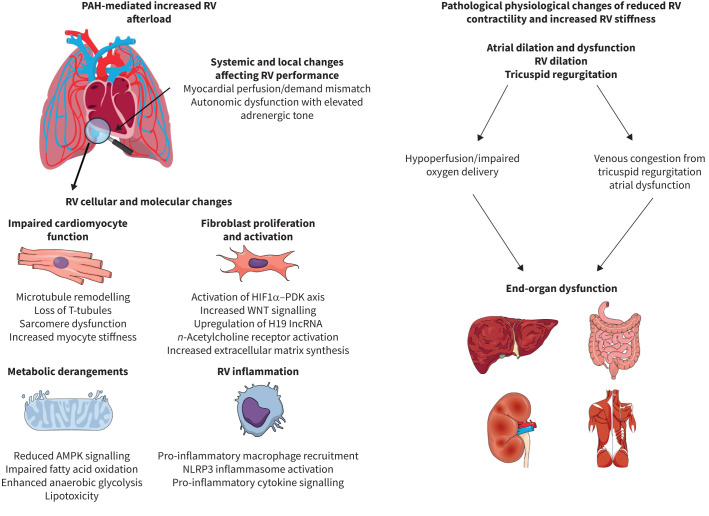
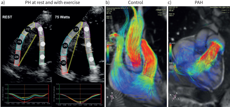
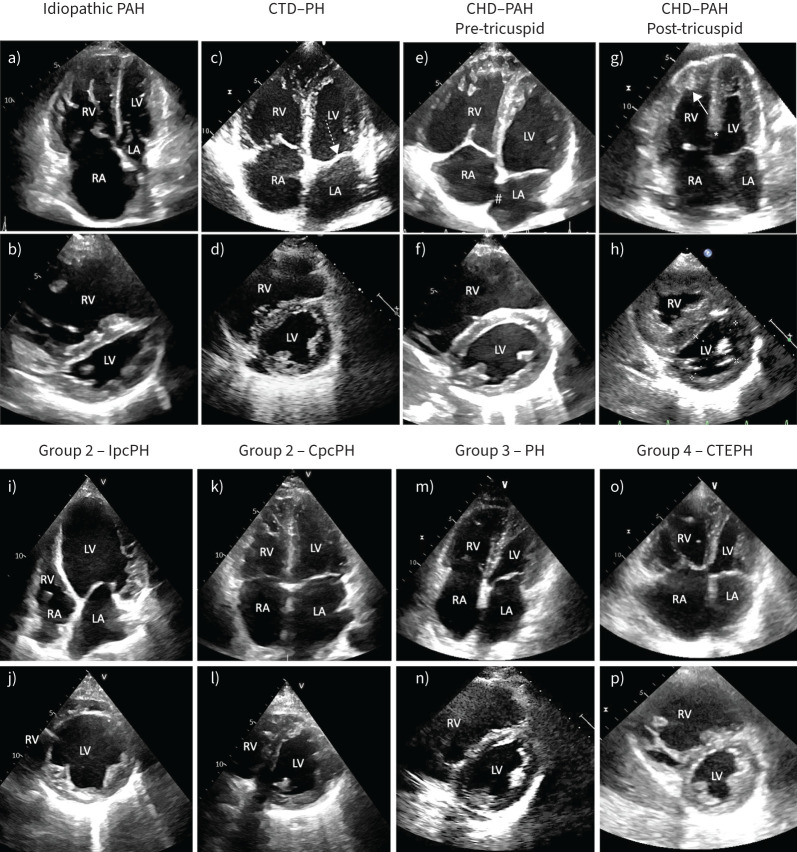
The right ventricle and its stress response is perhaps the most important arbiter of survival in patients with pulmonary hypertension of many causes. The physiology of the cardiopulmonary unit and definition of right heart failure proposed in the 2018 World Symposium on Pulmonary Hypertension have proven useful constructs in subsequent years. Here, we review updated knowledge of basic mechanisms that drive right ventricular function in health and disease, and which may be useful for therapeutic intervention in the future. We further contextualise new knowledge on assessment of right ventricular function with a focus on metrics readily available to clinicians and updated understanding of the roles of the right atrium and tricuspid regurgitation. Typical right ventricular phenotypes in relevant forms of pulmonary vascular disease are reviewed and recent studies of pharmacological interventions on chronic right ventricular failure are discussed. Finally, unanswered questions and future directions are proposed.


Conflict of interest statement

Conflict of interest: A.R. Hemnes reports grants from NIH/NHLBI, consultancy fees from Bayer, Gossamer Bio, Merck, Janssen, United Therapeutics and Tenax, participation on a data safety monitoring board or advisory board with NIH/NHLBI, leadership roles with Nashville Ballet and Pulmonary Vascular Research Institute, and stock (or stock options) with Tenax Therapeutics. D.S. Celermajer has no potential conflicts of interest to disclose. M. D'Alto reports consultancy fees and payment or honoraria for lectures, presentations, manuscript writing or educational events from Merck Sharp and Dhome, Dompé, AOP and Janssen, and support for attending meetings from Dompé, AOP and Janssen. F. Haddad reports grants from Johnson & Johnson, and consultancy fees from Merck. P.M. Hassoun reports grants from NIH/NHLBI (R01 R01HL114910), and participation on a data safety monitoring board or advisory board with MSD and ARIA-CV. K.W. Prins reports grants from NHLBI (R01 HL158795 and 162927) and Bayer (PHAB grant), and consultancy fees from Edwards. R. Naeije reports consultancy fees from AOP Orphan Pharma, Johnson & Johnson, United Therapeutics and Lung Biotechnology, payment or honoraria for lectures, presentations, manuscript writing or educational events from AOP Orphan Pharma, support for attending meetings from AOP Orphan Pharma, and participation on a data safety monitoring board or advisory board with Johnson & Johnson, AOP Orphan Pharma and United Therapeutics. A. Vonk Noordegraaf reports payment or honoraria for lectures, presentations, manuscript writing or educational events from Actelion and Johnson & Johnson.

Figures

FIGURE 1
FIGURE 1
The systemic, organ, cellular and molecular causes and subsequent physiological consequences of right ventricular (RV) dysfunction in pulmonary arterial hypertension (PAH). Increased afterload paired with derangements in autonomic tone and myocardial perfusion/demand mismatch lead to pathogenic changes in cardiomyocyte structure and function driven in part by mitochondrial metabolic dysfunction, heightened fibrosis due to fibroblast proliferation and activation and pathological inflammation. These cellular and organ level alterations increase RV stiffness and impair contractility that leads to right atrial dilation/dysfunction, RV dilation and tricuspid regurgitation. This ultimately results in end-organ hypoperfusion and venous congestion causing hepatic and renal dysfunction. HIF: hypoxia-inducible factor; PDK: pyruvate dehydrogenase kinase; WNT: wingless/int; lncRNA: long noncoding RNA; n-acetylcholine: nicotinic acetylcholine; AMPK: AMP kinase; NLRP: NACHT-, LRR-, and pyrine domain-containing protein.
FIGURE 2
FIGURE 2
Novel right ventricular (RV) imaging in pulmonary hypertension. a) RV longitudinal strain patters demonstrate regional heterogeneity. A 65-year-old female with scleroderma presenting with undifferentiated dyspnoea underwent supine bicycle echocardiography. Rest images demonstrate a heterogeneous pattern of strain-derived regional contractility [70], tricuspid annular plane systolic excursion (TAPSE) 2.12 cm, normal RV systolic pressure (RVSP). With 75 W of exercise, the basal segment does not augment while there is increase in regional contractility of the midventricular and apical segments, TAPSE 1.53 cm, associated with chamber dilatation (increase in RV end-diastolic area and end-systolic area, increase midventricular dimension) and RVSP 58 mmHg [71]. Echocardiography-based strain imaging on exercise is technically difficult and not at this stage recommended for standard clinical use. Magnetic resonance imaging four-dimensional flow measurements in b) a healthy control and c) a patient with pulmonary arterial hypertension (PAH). In the healthy person only forward flow and the RV serves as a bellow in which all parts of the RV contribute to the flow movement. In PAH the contribution of the apical region to forward flow is minimal, indicating the heterogeneous contribution of the RV to power output. In addition, the flow is not linear in the atrium and vortex flow can be observed in the pulmonary artery.
FIGURE 3
FIGURE 3
a–h) While distinguishing types of pulmonary hypertension (PH) based on echocardiography is challenging and depends on the stage of disease, presented here are example images of echocardiographic features of PH subtypes. a) and b) Severe idiopathic pulmonary arterial hypertension (PAH) with a) marked dilation of the right chambers and b) flattening of the interventricular septum, with a squeezed left ventricle (LV) (eccentricity index >1.5). Due to the prominence of the right chambers, this may be described as “right phenotype”. c) and d) PH associated with connective tissue disease (CTD). Patients show prominence of the right chambers, but may have thickening of the mitral (dashed arrow) or the aortic valve and a certain degree of LV diastolic dysfunction. e) and f) PAH associated with an atrial septal defect (#) demonstrating enlarged right atrium (RA) and right ventricle (RV). g) and h) PAH associated with a ventricular septal defect (*) in natural history (Eisenmenger syndrome). Typically, these patients show severe right ventricular hypertrophy (arrow) with a normal or only mildly dilated right ventricle. i–p) Typical echocardiographic features of group 2, 3 and 4 PH. i) and j) Typical remodelling of a patient with heart failure and reduced ejection fraction with isolated post-capillary PH (IpcPH) (“left phenotype”). There is a marked prevalence of the left chambers (g) and a round-shaped left ventricle (eccentricity index 1). k) and l) Patients with combined post- and pre-capillary PH (CpcPH) show an intermediate phenotype (“neither right nor left”) with i) bi-atrial dilation and left ventricle prevalence, but j) initial flattening of the interventricular septum. m) and n) Patients with PH associated with lung disease have usually a prevalence of the right chambers (“right phenotype”), but they are often older than PAH patients and may have a component of left-heart involvement. o) and p) Patients with chronic thromboembolic PH (CTEPH) have a “right phenotype” indistinguishable from PAH patients. Nevertheless, some may have by chance a left-heart disease and features of left heart involvement. CHD: congenital heart disease; LA: left atrium.

Comment in

References

    1. Vonk Noordegraaf A, Westerhof BE, Westerhof N. The relationship between the right ventricle and its load in pulmonary hypertension. J Am Coll Cardiol 2017; 69: 236–243. doi: 10.1016/j.jacc.2016.10.047 - DOI - PubMed
    1. Lahm T, Douglas IS, Archer SL, et al. Assessment of right ventricular function in the research setting: knowledge gaps and pathways forward. An Official American Thoracic Society Research Statement. Am J Respir Crit Care Med 2018; 198: e15–e43. doi: 10.1164/rccm.201806-1160ST - DOI - PMC - PubMed
    1. Sanz J, Sánchez-Quintana D, Bossone E, et al. Anatomy, function, and dysfunction of the right ventricle: JACC state-of-the-art review. J Am Coll Cardiol 2019; 73: 1463–1482. doi: 10.1016/j.jacc.2018.12.076 - DOI - PubMed
    1. Leopold JA, Kawut SM, Aldred MA, et al. Diagnosis and treatment of right heart failure in pulmonary vascular diseases: a National Heart, Lung, and Blood Institute Workshop. Circ Heart Fail 2021; 14: e007975. doi: 10.1161/CIRCHEARTFAILURE.120.007975 - DOI - PMC - PubMed
    1. Naeije R, Richter MJ, Rubin LJ. The physiological basis of pulmonary arterial hypertension. Eur Respir J 2022; 59: 2102334. doi: 10.1183/13993003.02334-2021 - DOI - PMC - PubMed

MeSH terms

LinkOut - more resources