Editorial

Spinal CSF Leaks: The Neuroradiologist Transforming Care

Mark D Mamlouk et al. AJNR Am J Neuroradiol. .

Abstract

Spinal CSF leak care has evolved during the past several years due to pivotal advances in its diagnosis and treatment. To the reader of the American Journal of Neuroradiology (AJNR), it has been impossible to miss the exponential increase in groundbreaking research on spinal CSF leaks and spontaneous intracranial hypotension (SIH). While many clinical specialties have contributed to these successes, the neuroradiologist has been instrumental in driving this transformation due to innovations in noninvasive imaging, novel myelographic techniques, and image-guided therapies. In this editorial, we will delve into the exciting advancements in spinal CSF leak diagnosis and treatment and celebrate the vital role of the neuroradiologist at the forefront of this revolution, with particular attention paid to CSF leak-related work published in the AJNR.


Figures

FIG 1.
FIG 1.
Number of spinal CSF leak publications in AJNR from 2000 to June 2024.
FIG 2.
FIG 2.
A ventral dural tear on a dynamic CT myelogram in a 34-year-old woman showing the direct outflow of the contrast medium (open arrow, A and B) within seconds into the ventral epidural space at the T3–T4 level. The contrast medium then flows cranially within the epidural space (solid arrows, A). The underlying cause is a calcified disc at the T3–T4 level (open arrowhead, A and B).
FIG 3.
FIG 3.
Digital DSM and conebeam CT of a lateral dural tear in a 36-year-old man. DSM with the patient in the right lateral decubitus position in an anterior-posterior projection suggests a small epidural contrast medium egress, which is unchanged in the subsequent single radiograph (white solid arrows, A and B). C, conebeam CT that follows a few minutes later confirms the epidural accumulation of contrast medium in the coronal view (white solid arrows) at the right T12–L1 level. In addition, a cyst-like structure can be seen within the contrast collection, corresponding to an arachnoid layer (dashed black arrow, C) herniating through a lateral dural tear in the axilla of the exiting nerve root sleeve (black arrowhead, C), which was later confirmed by surgery.
FIG 4.
FIG 4.
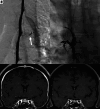
CVF on a DSM (A) in a 47-year-old man with a right T5 CVF (arrow) that was treated with Onyx (Medtronic) embolization (B). The pre-embolization brain MRI (C) demonstrates dural enhancement that nearly normalized 1 month after embolization (D).
FIG 5.
FIG 5.
Benefit of conebeam CT for detection of a right T6 CVF. Select unsubtracted (A) and subtracted (B) images from a right lateral decubitus DSM show faint linear paraspinal venous opacification (A, arrow). The finding is extremely difficult to appreciate on the unsubtracted image and essentially not seen on the subtracted image (B) due to a combination of pulmonary markings and respiratory motion. Axial (C) and coronal (D) MIP images from a conebeam CT performed minutes later during active contrast injection show a clear right T6 CVF involving several lateral branch veins. In cases such as this, conebeam CT serves as an excellent adjunct to DSM.
FIG 6.
FIG 6.
CVF occlusion with targeted fibrin glue patching. A, Axial right decubitus CTM shows a right T7 CVF with a paravertebral segmental vein (arrows). B, Axial CT treatment image demonstrates injected fibrin glue within the neural foramen and paravertebral space that matches the CVF drainage course, which is an important feature for treatment success. Posttreatment axial right decubitus CTM (C) shows resolution of the CVF. The pretreatment brain MRI (D) demonstrates dural enhancement (arrows) that resolved 1 month after patching (E).
FIG 7.
FIG 7.
The advantage of high spatial resolution to detect a right T10 CVF on photon-counting CTM. Axial and sagittal 0.2-mm images (A and B) from a right decubitus photon-counting CTM reconstructed using a relatively smoother Br56 kernel, demonstrate a right T10 CVF involving the ventral and dorsal internal epidural venous plexus (A and B, arrows). Axial 0.2-mm images at the same level, reconstructed using both a smoother Br56 kernel (C) and a sharper Qr89 kernel with denoising (D), show involvement of the intervertebral vein that is only evident on the sharper Qr89 kernel. In some cases, maximizing spatial resolution using a sharper kernel with denoising is necessary to appreciate the full extent of venous opacification.
FIG 8.
FIG 8.
Use of denoised sharp kernel images on photon-counting CTM for localization of a T5 ventral dural tear. Axial images from a prone dynamic photon-counting CTM, all at the same slice, time point, and window/level setting, demonstrate a ventral dural tear just to the right of midline (A–C, arrows). The precise location of the ventral leak is demonstrated with greater resolution when using a sharper Qr89 kernel (B and C) compared with a smoother Br56 kernel (A). Because the Qr89 kernel introduced noise into the image, a denoising algorithm was applied to permit the use of a sharper kernel while retaining an acceptable SNR (C).

References

    1. Schievink WI. Spontaneous intracranial hypotension. N Engl J Med 2021;385:2173–78 10.1056/NEJMra2101561 - DOI - PubMed
    1. Schievink WI, Maya MM, Jean-Pierre S, et al. A classification system of spontaneous spinal CSF leaks. Neurology 2016;87:673–79 10.1212/WNL.0000000000002986 - DOI - PubMed
    1. Farb RI, Nicholson PJ, Peng PW, et al. Spontaneous intracranial hypotension: a systematic imaging approach for CSF leak localization and management based on MRI and digital subtraction myelography. AJNR Am J Neuroradiol 2019;40:745–53 10.3174/ajnr.A6016 - DOI - PMC - PubMed
    1. Schievink WI, Maya MM, Tay AS-MS, et al. Lateral spinal CSF leaks in patients with spontaneous intracranial hypotension: radiologic-anatomic study of different variants. AJNR Am J Neuroradiol 2024;45:951–56 10.3174/ajnr.A8261 - DOI - PMC - PubMed
    1. Madhavan AA, Farb RI, Brinjikji W, et al. Expounding on the distinction between lateral dural tears and leaking meningeal diverticula in spontaneous intracranial hypotension. AJNR Am J Neuroradiol 2024;45:E28–29 10.3174/ajnr.A8308 - DOI - PMC - PubMed

Publication types

MeSH terms

LinkOut - more resources