. 2024 Aug 29;15(1):7514.
doi: 10.1038/s41467-024-51804-w.

Helical ultrastructure of the L-ENA spore aggregation factor of a Bacillus paranthracis foodborne outbreak strain

Affiliations

Helical ultrastructure of the L-ENA spore aggregation factor of a Bacillus paranthracis foodborne outbreak strain

Mike Sleutel et al. Nat Commun. .

Abstract

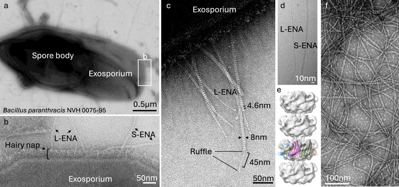
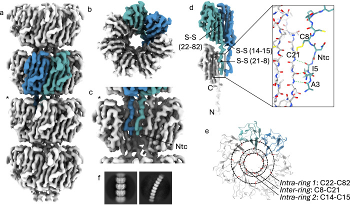
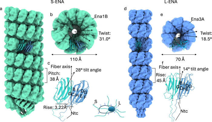
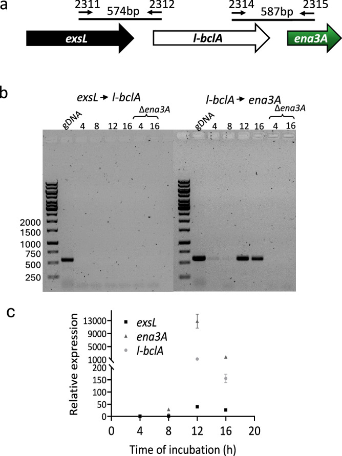
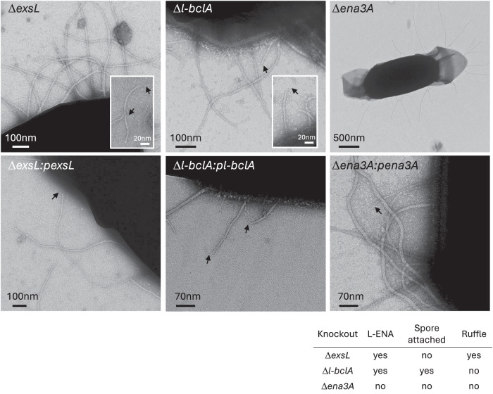
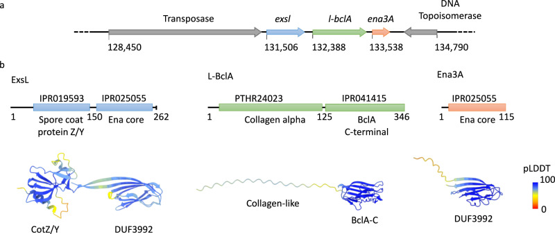
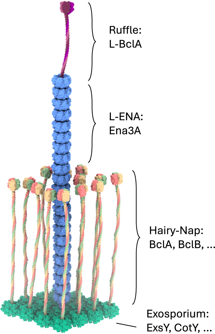
In pathogenic Bacillota, spores can form an infectious particle and can take up a central role in the environmental persistence and dissemination of disease. A poorly understood aspect of spore-mediated infection is the fibrous structures or 'endospore appendages' (ENAs) that have been seen to decorate the spores of pathogenic Bacilli and Clostridia. Current methodological approaches are opening a window on these long enigmatic structures. Using cryoID, Alphafold modelling and genetic approaches we identify a sub-class of robust ENAs in a Bacillus paranthracis foodborne outbreak strain. We demonstrate that L-ENA are encoded by a rare three-gene cluster (ena3) that contains all components for the self-assembly of ladder-like protein nanofibers of stacked heptameric rings, their anchoring to the exosporium, and their termination in a trimeric 'ruffle' made of a complement C1Q-like BclA paralogue. The role of ENA fibers in spore-spore interaction and the distribution of L-ENA operon as mobile genetic elements in B. cereus s.l. strains suggest that L-ENA fibers may increase the survival, spread and virulence of these strains.


Conflict of interest statement

The authors declare no competing interests.

Figures

Fig. 1
Fig. 1. Negative stain transmission electron microscopy images of the endospores of the food-poisoning outbreak strain B. paranthracis NVH 0075-95.
a Composite montage of a single endospore with a central spore body and the surrounding exosporial sack, b High-magnification image of the exosporium decorated with the hairy nap and two types of endospore appendages (L- and S-ENA), c L-ENA fibers anchored to the exosporium, exhibit a ‘ladder-like’ pattern (4.6 nm intervals), are terminally decorated with single tip fibrillae, i.e., ruffles. Ruffles consist of a 45 nm long stalk that terminates into a globular head domain, d cryoEM image of isolated ENA fibers, e reconstructed cryoEM-volume of L-ENA fibers (5.8 Å at FSC = 0.143) with rigid body docking of 7 copies of Ena1B19-117, f nsTEM image of recEna3A produced in and purified from the cytoplasm of E. coli. The data shown are representative of experiments made independently in triplicate.
Fig. 2
Fig. 2. Cryo-EM volume of recEna3A L-ENA fibers.
a Helical ultrastructure of L-ENA determined at 3.32 Å global resolution (cut-off criterion at FSC = 0.143 Å1) revealing an axial stacking of heptameric Ena3A rings that rotate 18.5° clockwise relative to each other. Two neighboring Ena3A monomers are colored in blue and cyan, b top-view of a single ring with a highlight of the β-sheet augmentation between the blue and cyan subunit, c zoom-in of a single ring - for clarity, two subunits were removed from the ring highlighted with an asterisk in (a) - showing the docking of the N-terminal connectors that covalently tether the ring docked above, d Highlight of the lateral intra-ring contacts: two types of disulfide bridges (Cys14-Cys15; Cys22-Cys82) exist within two neighboring subunits. In turn, each subunit connects to a subunit in the ring below via their Ntc (Cys8-Cys21; inset in stick representation), e On-axis top-view of the L-ENA fiber model in cartoon with disulfide bridges shown in red; f 2D class averages of straight and curved L-ENA segments covering 4 and 9 rings, respectively.
Fig. 3
Fig. 3. Structural comparison between the S- and L-ENA fiber architectures.
a Side and b on-axis view of an S-ENA fiber (pdb-id: 7A02) composed of Ena1B subunits. Helical parameters: Rise: 3.22 Å, Twist: 31.0°, Pitch: 38 Å, c Dimeric contacts of Ena1B subunits via β-sheet augmentation at the interface between the G and C strands. Subunits are tilted 28° with respect to the fiber axis. This out-of-plane interaction leads to a helical stacking of Ena1B monomers, d Side and e on-axis view of an L-ENA fiber (pdb-id: 8PDZ) composed of Ena3A subunits. Helical parameters: Rise = pitch: 45 Å, Twist: 18.5°, f Dimeric contacts of Ena3A subunits via β-sheet augmentation at the interface between the G and C strands. Although subunits are tilted 14° with respect to the fiber axis, their contacts remain in-plane yielding a heptameric ring.
Fig. 4
Fig. 4. Expression of L-ENA genes is concomitant with sporulation.
a Chromosomal organization of exsL, l-bclA, and ena3A, and primers used in standard PCR using cDNA template; b The L-ENA genes l-bclA and ena3A form an operon. Agarose gel electrophoresis (1%) of PCR products conducted on cDNA from NVH 0075-95 culture (4, 8, 12, and 16 h). Primer combinations and expected product sizes are shown in (a). Genomic DNA (gDNA) from NVH 0075-95 and cDNA prepared from its isogenic Δena1ABCena3A mutant were used as positive and negative controls for the PCR, respectively; c Expression of L-ENA genes relative to rpoB during vegetative growth and sporulation. Data are presented as mean values with error bars representing the standard deviation of three independent experiments, each with three technical replicates. Source data are provided as a Source Data file.
Fig. 5
Fig. 5. Phenotypic read-out of L-ENA mutants.
nsTEM micrographs of the respective L-ENA operon deletion mutants. The data shown are representative of experiments made independently in triplicate.
Fig. 6
Fig. 6. Genetic organization of the L-ENA gene cluster.
a the ena3A gene is embedded in a three-gene cluster, preceded on the CP116205.1 plasmid by two genes here annotated as exsL and l-bclA, b Domain organization of the L-ENA proteins as identified by Interpro (10.1093/nar/gkac993). ExsL: consists of an N-terminal spore coat protein Z/Y domain and a C-terminal Ena-core domain (DUF3992), L-BclA: consists of an N-terminal collagen-like domain and a C-terminal BclA-C domain, Ena3A: consists of a single Ena-core domain (DUF3992). AlphaFold2 predictions are shown in cartoon representation, color-coded according to the pLDDT score.
Fig. 7
Fig. 7. Mesoscale model of an L-ENA fiber projecting from the exosporium.
L-ENA fibers are helical, protein ultrastructures composed of heptameric Ena3A rings that stack axially. At the tip that is distal from the spore, the L-ENA fiber terminates into a bacterial collagen-like ruffle protein, i.e., L-BclA. The L-ENA fiber protrudes through the hairy nap layer while being tethered to the exosporium via a dedicated anchoring protein ExsL. ExsL is expected to couple to or be integrated in the 2D crystal lattice of the exosporium. Note that for clarity, the L-ENA fibers are depicted shorter here.

References

    1. McKenney, P. T., Driks, A. & Eichenberger, P. The Bacillus subtilis endospore: assembly and functions of the multilayered coat. Nat. Rev. Microbiol.11, 33–44 (2013). 10.1038/nrmicro2921 - DOI - PMC - PubMed
    1. Gao, Y. et al. Bacterial spore germination receptors are nutrient-gated ion channels. Science380, 387–391 (2023). 10.1126/science.adg9829 - DOI - PMC - PubMed
    1. Henriques, A. O., Charles, J. & Moran, P. Structure, assembly, and function of the spore surface layers. Annu. Rev. Microbiol.61, 555–588 (2007). 10.1146/annurev.micro.61.080706.093224 - DOI - PubMed
    1. Rodenburg, C. M., McPherson, S. A., Turnbough, C. L. Jr. & Dokland, T. Cryo-EM analysis of the organization of BclA and BxpB in the Bacillus anthracis exosporium. J. Struct. Biol.186, 181–187 (2014). 10.1016/j.jsb.2014.02.018 - DOI - PMC - PubMed
    1. Panessa-Warren, B. J., Tortora, G. T. & Warren, J. B. High resolution FESEM and TEM reveal bacterial spore attachment. Microsc. Microanal.13, 251–266 (2007). 10.1017/S1431927607070651 - DOI - PubMed

MeSH terms

Substances

LinkOut - more resources