. 2024 Oct;26(10):1691-1699.
doi: 10.1038/s41556-024-01488-7. Epub 2024 Aug 29.

Nuclear proteasomes buffer cytoplasmic proteins during autophagy compromise

Affiliations

Nuclear proteasomes buffer cytoplasmic proteins during autophagy compromise

So Jung Park et al. Nat Cell Biol. 2024 Oct.

Abstract

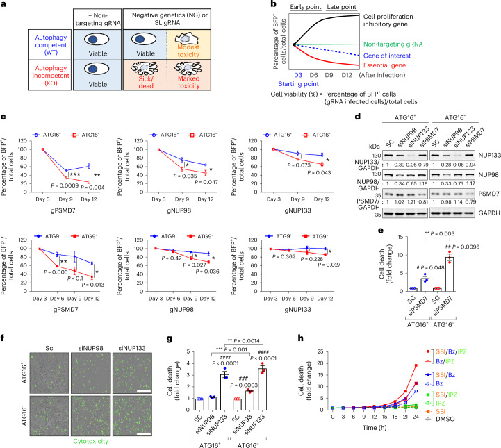
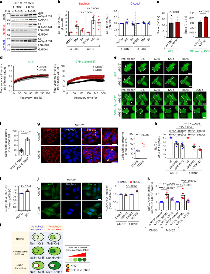
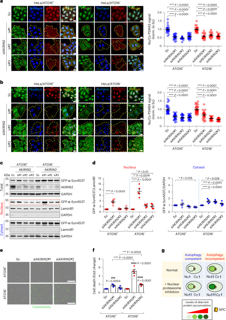
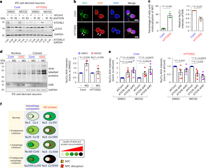
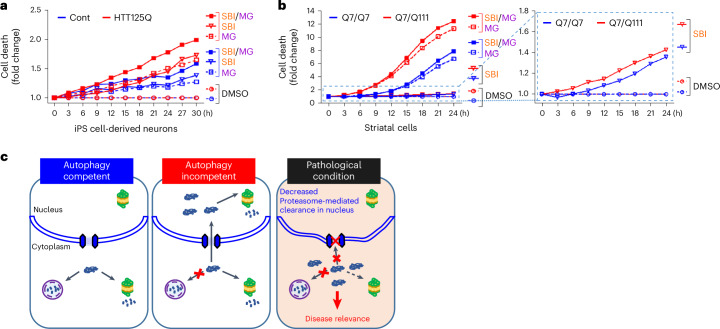
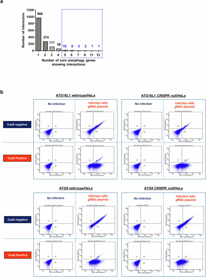
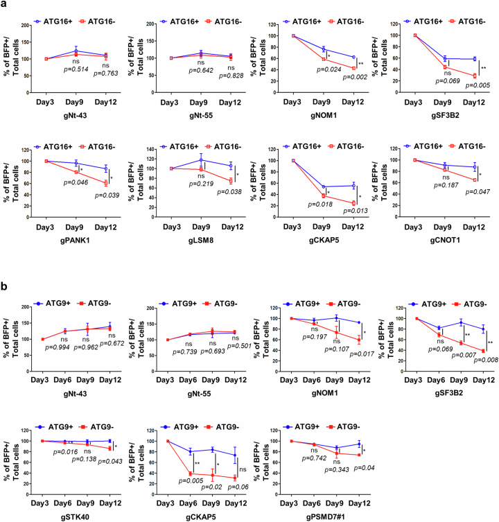
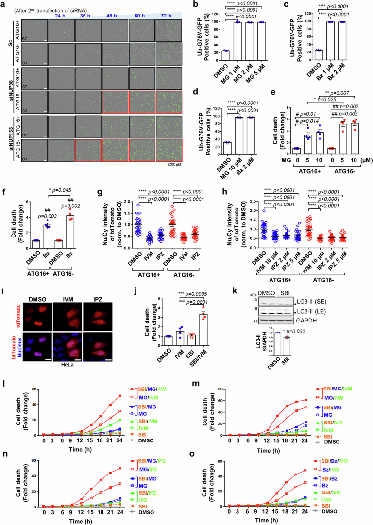
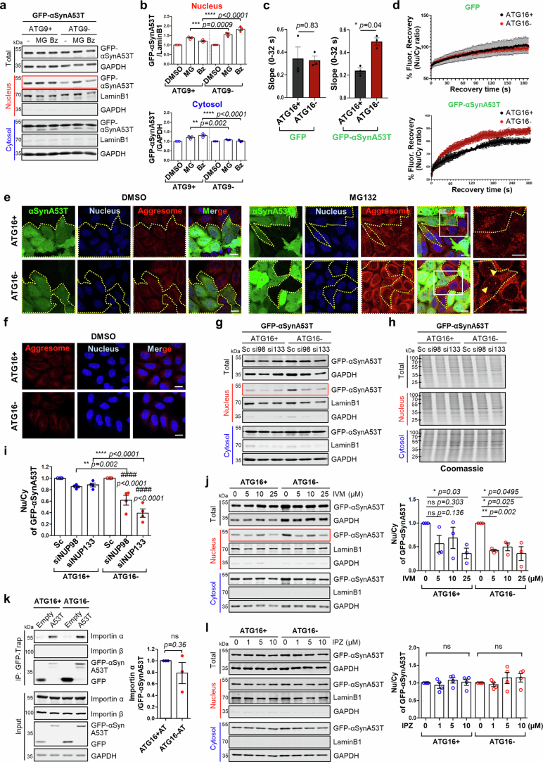
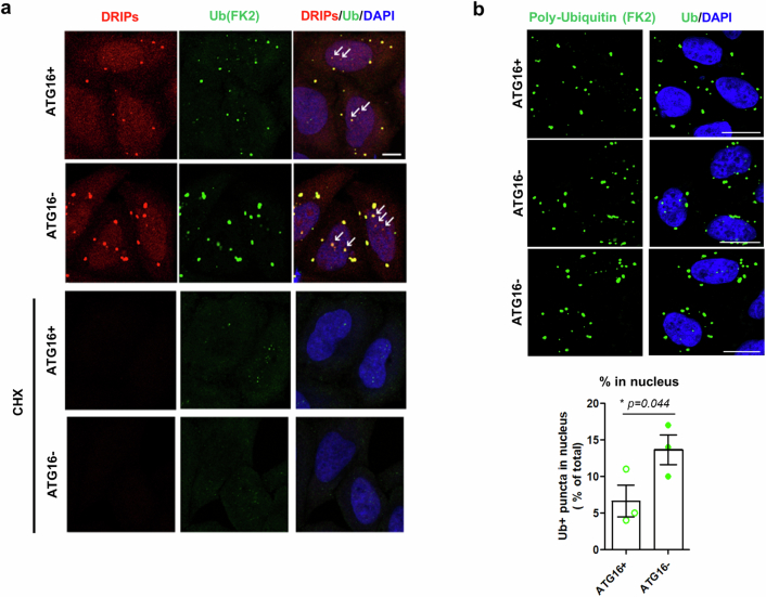
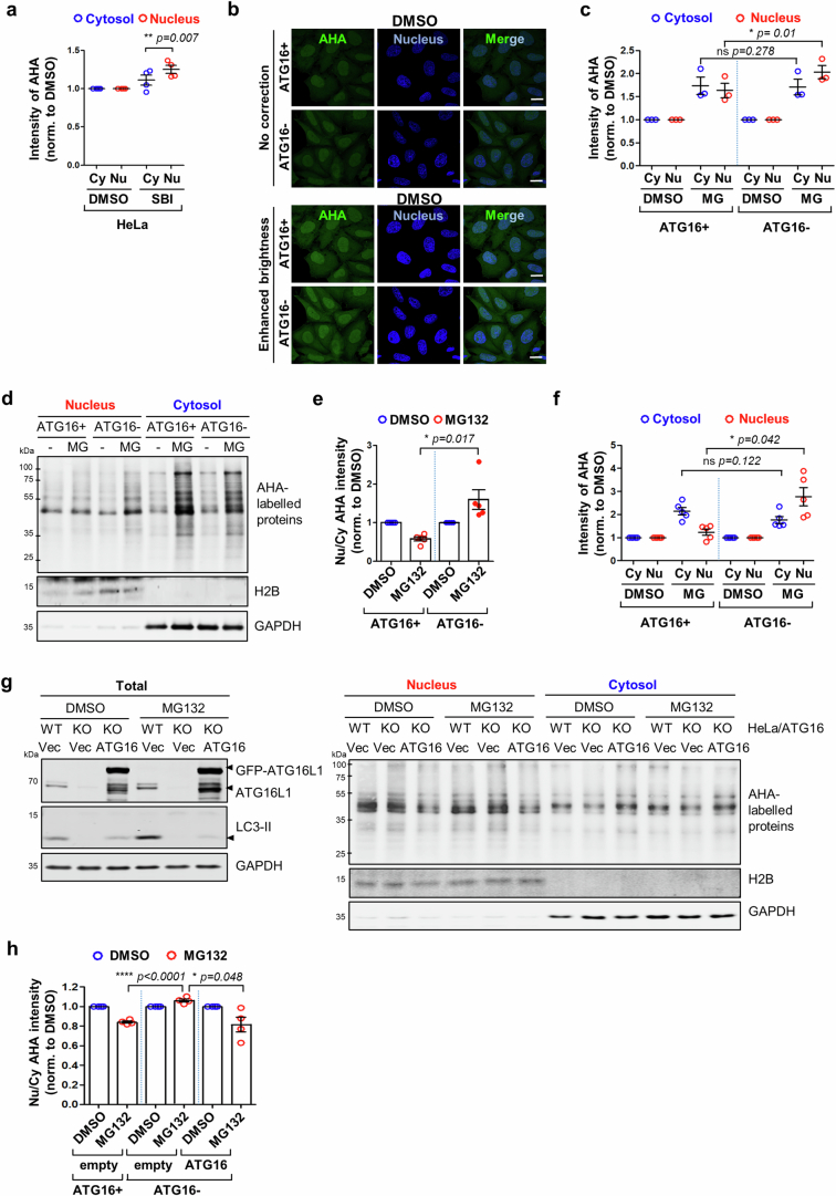
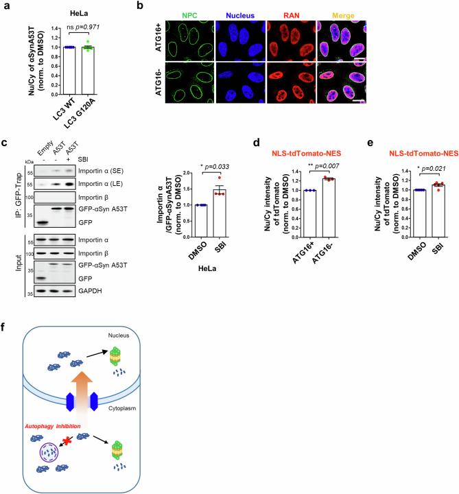
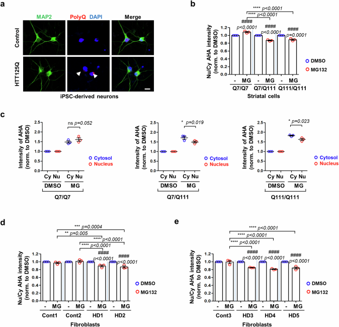
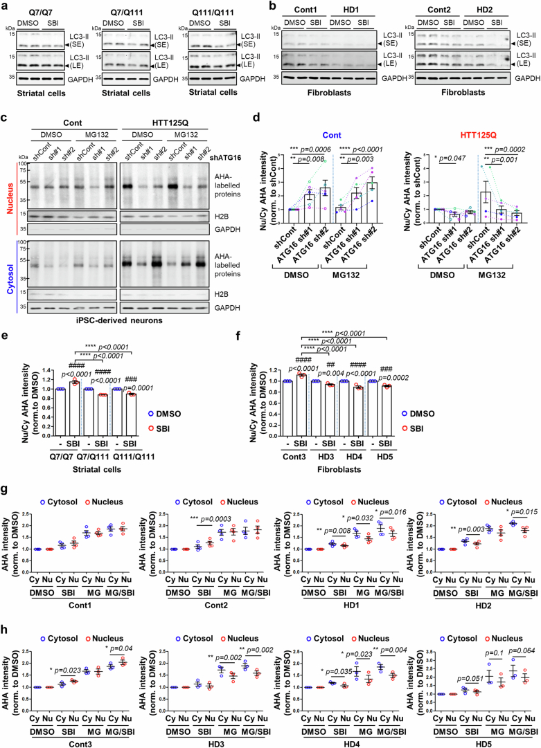
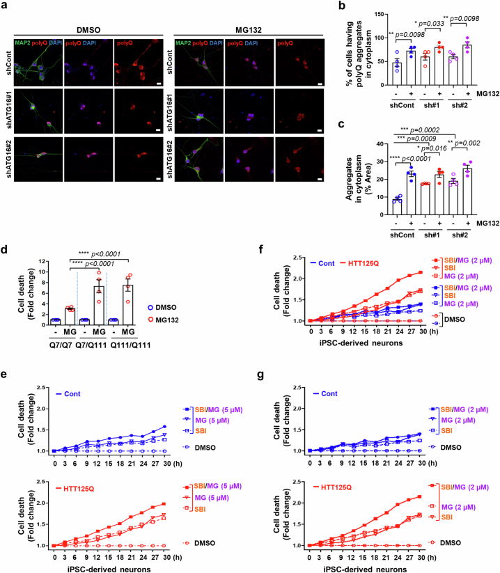
Autophagy is a conserved pathway where cytoplasmic contents are engulfed by autophagosomes, which then fuse with lysosomes enabling their degradation. Mutations in core autophagy genes cause neurological conditions, and autophagy defects are seen in neurodegenerative diseases such as Parkinson's disease and Huntington's disease. Thus, we have sought to understand the cellular pathway perturbations that autophagy-perturbed cells are vulnerable to by seeking negative genetic interactions such as synthetic lethality in autophagy-null human cells using available data from yeast screens. These revealed that loss of proteasome and nuclear pore complex components cause synergistic viability changes akin to synthetic fitness loss in autophagy-null cells. This can be attributed to the cytoplasm-to-nuclear transport of proteins during autophagy deficiency and subsequent degradation of these erstwhile cytoplasmic proteins by nuclear proteasomes. As both autophagy and cytoplasm-to-nuclear transport are defective in Huntington's disease, such cells are more vulnerable to perturbations of proteostasis due to these synthetic interactions.


Conflict of interest statement

D.C.R. is a consultant for Aladdin Healthcare Technologies, Mindrank AI, Nido Biosciences, Drishti Discoveries, Retro Biosciences, PAQ Therapeutics and Alexion Pharma Intl Ops Limited. G.B. is a founder and Director of Function Rx. The other authors declare no competing interests.

Figures

Fig. 1
Fig. 1. Identification of negative genetic interactors with autophagy compromise.
a, Schematic representation of FACS-based CRISPR/Cas9 synthetic lethality (SL) screen to identify SL regulators required for survival during autophagy compromise. b, Quantification of cell viability assessed the percentage of BFP+ (blue fluorescent protein-positive (BFP), gRNA-infected) cells normalized to the number of total cells after transduction with gRNAs that are present in BFP+ cells, as the lentivirus vector carrying the gRNAs also expresses BFP. c, Quantification of cell viability with gRNAs for the indicated SL candidate genes (gPSMD7, gNUP98 and gNUP133) in autophagy-incompetent cells (HeLa/ATG16L1 knockout (KO) (ATG16)/Cas9 (top), HeLa/ATG9 KO (ATG9)/Cas9 (bottom)) compared with their respective controls (autophagy-competent cells) (n = 3 technical triplicates after initial infected plates split into three plates of a 96-well plate for ATG16L1 WT versus KO cells and ATG9 WT versus KO cells. Percentage of BFP+ cells/total cells at 3 days set to 100% in all conditions to allow us to determine relative loss of cells over time in autophagy-competent versus autophagy-incompetent cells; two-tailed unpaired t-test). d, Immunoblots for SL candidate protein levels after each siRNA knockdown (representative blot from three biological repeats). e, Knockdown of PSMD7 enhances cell death in ATG16L1 KO measured by LDH assay (day 3) (n = 3 independent experiments; two-tailed paired t-test). f, Illustration of readouts of Incucyte cell death assay (measured by Incucyte live-cell imaging). Scale bar, 600 μm. g, Quantification of CellTox Green fluorescence intensity after siRNA-mediated knockdown of NUP98 or NUP133 (day 5) versus scramble control siRNA (SC) (n = 3 independent experiments; two-way analysis of variance (ANOVA) with post hoc Tukey test). h, Combined effect of autophagy inhibition (SBI-0206965 (SBI) 5 μM) with proteasome inhibition (bortezomib (Bz) 1 μM) and/or nuclear import inhibition (importazole (IPZ) 2 μM) on HeLa cell death (Incucyte cell death assay with CellTox Green fluorescence). When we compute areas under the curve for four biological replicates, then SBI versus SBI + Bz, P = 0.013; SBI versus SBI + IPZ, P = 0.047 (one-tailed paired t-test). Values are mean ± s.e.m. For comparisons between ATG16+ and ATG16 cell lines: * P < 0.05; ** P < 0.01; *** P = 0.001; for comparisons to relevant ATG16+ and ATG16 control within cell line: # P < 0.05, ## P < 0.01, ### P < 0.001, #### P < 0.0001. Source numerical data and unprocessed blots are available in source data. Source data
Fig. 2
Fig. 2. Autophagy depletion causes nuclear translocation of erstwhile cytoplasmic autophagic substrates.
a, Autophagy substrate A53T α-Syn localized more in the nucleus of ATG16L1 KO (ATG16) cells compared with ATG16L1 WT (ATG16+) cells treated with proteasome inhibitor (MG132 (MG, 10 μM, 6 h) or Bz (2 μM, 6 h)) by cell fractionation. b, Quantification of western blots (representative in a) showing relative changes of A53T α-Syn in nucleus and cytosol within cell lines caused by proteasome inhibitors (DMSO = 1) (n = 4 independent experiments; two-way ANOVA with post hoc Tukey test). ce, Nuclear FRAP in ATG16L1 WT and KO cells expressing either GFP-empty or GFP-A53T α-Syn. The initial recovery slope (0–32 s) rate of d (n = 3 independent experiments; two-tailed paired t-test) (c). Nuclear FRAP curves show faster recovery of GFP-A53T α-Syn in ATG16L1 KO cells compared with WT cells, while this effect was not seen with GFP-empty (d). Representative images of ATG16L1 WT and KO cells expressing either GFP-empty or GFP-A53T α-Syn before and after photobleaching and after recovery (up to 3 min or 5 min) (e). Arrowhead indicates photobleached cell. Scale bar, 10 µm. f, Increased nuclear aggresomes by Proteostat dye in ATG16L1 WT and KO cells expressing A53T α-Syn treated with MG (n = 3 independent experiments; two-tailed paired t-test). g, Increased nuclear aggresomes in ATG16L1 KO compared with WT with MG (2 μM, 15 h) (n = 4 independent experiments; two-tailed paired t-test). Scale bar, 20 µm. h, Knockdown of NUP98 or NUP133 inhibits A53T α-Syn shuttling into nucleus in ATG16L1 KO cells compared with WT cells, by immunostaining (n = 5 independent experiments; two-way ANOVA with post hoc Tukey test). i,j, Localization of AHA-labelled proteins by immunostaining upon either autophagy inhibition with SBI (5 µM, 15 h) in HeLa (i) (n = 4) or proteasome inhibition (MG, 2 µM, 15 h) in ATG16L1 WT and KO cells (j) (n = 3 independent experiments; two-tailed paired t-test). Scale bar, 20 µm. k, Overexpression of ATG16L1 rescues mislocalization of AHA-labelled proteins in ATG16L1 KO cells by western blotting (n = 5 independent experiments; one-way ANOVA with post hoc Tukey test). l, Schematic representation shows idealized protein levels/localization upon proteasome inhibition or NPC disruption in WT and autophagy-null cells. Values are mean ± s.e.m. Source numerical data and unprocessed blots are available in source data. DMSO, dimethylsulfoxide. Source data
Fig. 3
Fig. 3. Loss of AKIRIN2 decreases nuclear proteasome-mediated protein degradation in autophagy-deficient cells.
a,b, Knockdown of AKIRIN2 with distinct siRNAs inhibits nuclear localization of proteasome subunits PSMA5 (a) and PSMB4 (b). Cells were transfected with AKIRIN2 siRNAs (#1, #2 and #3) for 5 days in ATG16L1 WT and -null cells. Cells were fixed and labelled for endogenous AKIRIN (red), DAPI (nucleus, blue) and either PSMA5 (green, a) or PSMB4 (green, b). Quantification of nucleus/cytosol-localized proteasome subunit (PSMA5 (a), PSMB4 (b) (~n = 30 cells in each condition with three different siRNA oligonucleotides; ****P < 0.0001 versus DMSO; one-way ANOVA with post hoc Dunnett test) – single experiment to validate published results (right). Yellow dashes in a and b outline AKIRIN2-knockdown cells. Scale bar, 20 μm. c,d, Knockdown of AKIRIN2 with distinct siRNAs (si#1, si#2 and si#3) inhibits A53T α-Syn degradation in the nucleus assessed by cell fractionation (day 3). Nuclear A53T α-Syn accumulated in ATG16L1 KO (ATG16) cells after AKIRIN2 knockdown (c). Relative changes of GFP-A53T α-Syn levels in nucleus and cytosol within each cell line after AKIRIN2 knockdown (scramble (Sc) = 1) (n = 5 independent experiments; *P < 0.05, **P < 0.01, ****P < 0.0001 versus Sc; one-way ANOVA with post hoc Dunnett test) (d). e, Knockdown of AKIRIN2 enhances cytotoxicity (green fluorescence) in ATG16L1 KO cells measured by Incucyte live-cell imaging. Scale bar, 600 μm. f, Quantification of CellTox Green fluorescence intensity after knockdown of AKIRIN2 (day 5) (n = 4 independent experiments; ###P < 0.001, ####P < 0.0001 versus Sc; ***P < 0.001, ****P < 0.0001 for relative changes induced by specific siRNA in WT versus KO cells; two-way ANOVA with post hoc Tukey test). g, Schematic representation shows idealized protein levels/localization upon nuclear proteasome inhibition in WT and autophagy-null cells. Data used for two-way ANOVA derived control values as the sum of all data from that experiment, where controls were originally normalized to 1 (as shown in the graphs), divided by the number of conditions analysed (see Methods). Data analysed by one-way ANOVA, where ATG16+ and ATG16 data were not compared, were analysed separately. Values are mean ± s.e.m. Source numerical data and unprocessed blots are available in source data. DAPI, 4,6-diamidino-2-phenylindole. Source data
Fig. 4
Fig. 4. Cytoplasm-to-nucleus shuttling of bulk proteins is inhibited in Huntington’s disease cells.
a, Inhibition of autophagy using distinct shRNA targeting ATG16L1 (#1 and #2) decreases LC3-II and ATG16L1 levels in control (Cont) and iPS cell-derived neurons from a juvenile patient with HD originally carrying 125 CAGs (HTT125Q) with or without MG132 (MG, 1 µM for 15 h). Representative blot of three biological repeats. b,c, NPC disruption in HTT125Q-derived neurons was detected by staining of either NPC or RAN, compared with Cont iPS cell-derived neurons (b). Arrows indicate the signals of NPC or RAN in the cytoplasm. Scale bar, 10 μm. Quantified data represents mislocalization of NPC (left) or RAN (right) in HTT125Q-derived neurons (c). Values are mean ± s.e.m. (n = 3 independent experiments; *P < 0.05 versus Cont; two-tailed paired t-test). White dashes in b indicate nuclear outline. d, Decreased amount of nuclear-localized newly synthesized proteins (AHA-labelled proteins) in HTT125Q-derived neurons upon MG132 (2 µM for 15 h) treatment, compared with Cont iPS cell-derived neurons. Values are mean ± s.e.m. (n = 4 independent experiments; NS, not significant; *P < 0.05 versus DMSO; two-tailed paired t-test). e, Autophagy compromise exacerbated mislocalization of newly synthesized proteins in cytosol upon MG132 (1 µM for 15 h) treatment in HTT125Q-derived neurons (right) compared to Cont iPS cell-derived neurons (left). Values are mean ± s.e.m. (n = 5 independent experiments; *P < 0.05, **P < 0.01, ***P < 0.001, ****P < 0.0001 versus shCont; one-way ANOVA with post hoc Dunnett test). Data used for one-way ANOVA, where DMSO and MG132 data were not compared, were analysed separately. f, Schematic representation shows idealized protein localization/abundance upon proteasome inhibition, autophagy inhibition or combined proteasome/autophagy inhibition in the HD context. Source numerical data and unprocessed blots are available in source data. Source data
Fig. 5
Fig. 5. Defects in cytoplasm-to-nucleus shuttling of bulk proteins exacerbate cell death in Huntington’s disease cells.
a, Real-time CellTox Green fluorescence as a result of cell death is monitored by Incucyte in a time-dependent manner. Exacerbated cell death by combined inhibition of autophagy and proteasome (SBI and MG, respectively) in HTT125Q-derived neurons. These graphs do not show error bars, which make the graphs less clear. The raw data are shown in the data files with P values. When we compute areas under the curve for three biological replicates, then SBI versus SBI + MG in Cont iPS cell-derived neurons P = 0.029; SBI versus SBI + MG in HTT125Q-derived neurons P = 0.049; SBI in Cont versus HTT125Q-derived neurons P = 0.078; MG in Cont versus HTT125Q-derived neurons P = 0.036; SBI + MG in Cont versus HTT125Q-derived neurons P = 0.016 (one-tailed paired t-test). b, Left graph: enhanced cell death caused by combined inhibition of autophagy and proteasome in mouse striatal cells expressing Q7/Q111 compared with each inhibitor alone. Right graph: increased cell death caused by autophagy inhibition (SBI) in Q7/Q111 compared with Q7/Q7 striatal cells (expanded scale from the same data in the left-hand graph to clarify effects of SBI to enable ease of comparison). When we compute areas under the curve for four biological replicates normalized to DMSO values showing Q7/Q7 and Q7/Q111 striatal cells treated with proteasome inhibitor MG132 (MG, 2 µM), autophagy inhibitor SBI (5 µM) and/or both (SBI + MG) in a time-dependent manner, then SBI versus SBI + MG (in Q7/Q7 cells): P = 0.036, SBI versus SBI + MG (in Q7/Q111 cells): P = 0.015, SBI in Q7/Q7 versus Q7/Q111: P = 0.274, MG in Q7/Q7 versus Q7/Q111: P = 0.024, SBI + MG in Q7/Q7 versus Q7/Q111: P = 0.029 (one-tailed paired t-test). c, Schematic summary for our study. Cytoplasmic substrates accumulating during autophagy depletion move into the nucleus via nuclear pores to be degraded by nuclear proteasomes. In the HD context, defective cytoplasm-to-nuclear transport enhances susceptibility to autophagy inhibition leading to cell dysfunction. Source numerical data are available in source data. Source data
Extended Data Fig. 1
Extended Data Fig. 1. Synthetic lethality CRISPR/Cas9 screen.
a, Numbers of human orthologues of yeast genes with negative genetic (NG) or synthetic lethal (SL) interactions with core autophagy genes, identified using databases (genetic interactions from YeastMine and human orthologues from HGCN) in silico. Number of core autophagy genes showing negative genetic/synthetic lethal interactions with specific genes is shown on x axis. b, Establishment of Cas9 stable lines in both HeLa/ATG16L1 WT and ATG16L1 KO (upper panel) and HeLa/ATG9 WT and ATG9 KO (lower panel) cells. Cells stably expressing Cas9 plasmid were selected. Cas9 enzyme cutting efficiency after gRNA plasmid infection was measured by FACS (flow cytometer) in ATG16L1 WT and KO (upper panel) and in ATG9 WT and KO (lower panel). Cas9 stable cell line (the pool) was assessed for Cas9 cutting efficiency with a lentiviral vector encoding BFP, GFP and a sgRNA against GFP. The percentage of BFP + /GFP- (edited) to BFP + /GFP+ (total transduced) cells was analysed in Cas9-negative and Cas9-positive cells by flow cytometer. (x axis shows BFP fluorescence, y-axis shows GFP fluorescence; Q1 (BFP-/ GFP+), Q2 (BFP + /GFP+), Q3 (BFP-/GFP-), and Q4 (BFP + /GFP-)). Source numerical data are available in source data. Source data
Extended Data Fig. 2
Extended Data Fig. 2. Results of synthetic lethality CRISPR/Cas9 screen in either autophagy-competent or -incompetent cells.
a, b, Potential candidates from synthetic lethality (SL) CRISPR/Cas9 screen in ATG16L1 wild-type (WT) (ATG16+) and knockout (KO) (ATG16-) (a) and in ATG9 WT (ATG9+) and KO (ATG9-) cells (b) assessed at different time points after gRNA transduction. Two non-targeting gRNA sequences (gNt-43 and gNt-55) were employed as negative controls in the experiment. Values are mean ± S.E.M. (n = 3 technical triplicates after initial infected plates split into 3 plates of 96-well plate for ATG16L1 WT vs. KO cells and ATG9 WT vs. KO cells. % of BFP+ cells/total cells at 3 days (Day 3) set to 100 % in all conditions to allow us to determine relative loss of cells over time in autophagy-competent versus autophagy-incompetent cells; ns = not significant, * p < 0.05, ** p < 0.01; two-tailed unpaired t-test). Significantly decreased % of BFP+ cells indicating gRNA-infected cells (increased toxicity) in autophagy-null vs. WT cells indicates negative genetic/synthetic lethal interactions. Source numerical data are available in source data. LSM8 was only analysed in ATG16+ and ATG16- cells and was not pursued further in this study. Source data
Extended Data Fig. 3
Extended Data Fig. 3. Autophagy inhibition enhances cell death upon proteasome inhibition and/or nuclear import inhibition.
a, Representative images showing the cell death measured CellTox Green by real-time Incucyte in HeLa/ATG16L1 wild-type (ATG16+) and null cells (ATG16-). Cells were double-transfected with siRNA against either Sc (Scrambled), NUP98 or NUP133, respectively, and stained with CellTox Green dye, then monitored by real-time Incucyte. Data in Day 5 (72 h after siRNA double transfection) is shown in Fig. 1f,g. Red boxes indicate times where there is synthetic lethality for the different siRNAs in ATG16-. Scale bar, 200 µm. b-d, Proteasome inhibitors (MG132 (MG) (b, d) and Bz (c, d)) inhibit proteasome activity measured by Ub-G76V-GFP, a ubiquitin fusion degradation (UFD) reporter, at different time points (b-c; 24 h and d; 6 h) and different concentrations in HeLa cells. (Value are mean ± S.E.M; n = 3 biological independent experiments; **** p < 0.0001 vs. DMSO; one-way ANOVA with post hoc Dunnett test) e, Effect of proteasome inhibitor MG132 (MG, for 24 h) on cell death measured by LDH assay in HeLa/ATG16L1 WT (ATG16+) and KO (ATG16-) cells. Values are mean ± S.E.M. (n = 4 independent experiments; # p < 0.05, ## p < 0.01 vs. DMSO; * p < 0.05, ** p < 0.01 for relative changes induced by MG in WT vs. KO cells; two-tailed paired t-test). f, Effect of proteasome inhibitor Bortezomib (Bz, 1 µM for 24 h) on cell death in ATG16L1 WT and KO. Cell death was detected by LDH assay. Values are mean ± S.E.M. (n = 4 independent experiments; ## p < 0.01 vs. DMSO; * p < 0.05 for relative changes induced by Bz in WT vs. KO cells; two-tailed paired t-test). g-i, HeLa cells were transfected with NLS–tdTomato-NES, a shuttling construct containing both an NLS and an NES fused to tdTomato (red). Cells were treated with nuclear import inhibitors (ivermectin (IVM) and importazole (IPZ)). (g and h) Quantification of ratio of nucleus/cytosol-localized tdTomato intensity in HeLa/ATG16L1 wild-type and null cells at different concentrations and the different time points (IVM 25 µM and IPZ 25 µM for 6 h (g); IVM 10 µM, IPZ 2, 5 µM for 15 h (h) – data from one control experiment to confirm that the nuclear import inhibitors indeed affect this process) (Values are mean ± S.E.M; n = 30 cells in each condition with different time points and different concentrations; **** p < 0.0001 vs. DMSO; one-way ANOVA with post hoc Dunnett test). i, Representative pictures showing that nuclear import inhibitors (IVM 25 µM and IPZ 25 µM for 6 h) cause mislocalization of the NLS–tdTomato-NES to the cytoplasm in HeLa cells. Scale bar, 20 μm. These nuclear import inhibitors clearly increased the cytoplasmic pool of the NLS–tdTomato-NES reporter in steady-state – the failure to induce complete nuclear exclusion is consistent with other studies using these compounds with other NLS-containing substrates,, but may also reflect incomplete activities of the inhibitors at these concentrations, and/or the possibility of a weakly functional NES in this reporter construct. j, The effect of autophagy inhibition (SBI, 5 µM for 24 h) combined with nuclear import inhibition by ivermectin (IVM, 10 µM for 24 h) on cell death measured by CellTox Green in HeLa cells. Values are mean ± S.E.M. (n = 4 independent experiments; *** p < 0.001 for relative changes induced by SBI (autophagy inhibition) or IVM (nuclear import inhibition) vs. combined inhibition; one-way ANOVA with post hoc Dunnett test). k, Effect of autophagy/ULK1 inhibitor SBI-0206965 (SBI) on LC3-II levels which correlated with autophagosome numbers in HeLa cells. Quantified data represents autophagy inhibition by SBI on LC3-II level. Blots are representative of three biologically independent experiments. Values are mean ± S.E.M. (* p < 0.05 vs. DMSO; two-tailed paired t-test). l-o, Combined effect of autophagy inhibition with proteasome and/or nuclear import inhibition on cell death were measured by real-time cell death assay (CellTox Green) at 3 h intervals. HeLa cells were treated with indicated distinct inhibitors in growth media with CellTox Green dye for 24 h. Cell death were measured with CellTox Green fluorescence/total area by Incucyte live-cell imaging. (l) Autophagy inhibition (SBI, 5 µM), proteasome inhibition (MG132 (MG), 2 µM), nuclear import inhibition (Ivermectin (IVM), 10 µM). (m) Autophagy inhibition (SBI, 5 µM), proteasome inhibition (MG, 5 µM), nuclear import inhibition (IVM, 10 µM). (n) Autophagy inhibition (SBI, 5 µM), proteasome inhibition (MG, 5 µM), nuclear import inhibition (Importazole (IPZ), 5 µM). (o) Autophagy inhibition (SBI, 5 µM), proteasome inhibition (Bortezomib (Bz), 1 µM), nuclear import inhibition (IVM, 10 µM). Autophagy inhibition plus proteasome inhibitor and/or nuclear import inhibitor stimulates more cell death than each inhibition alone. Each graph of cell death represents same order of toxicity. Source numerical data and unprocessed blots are available in source data. Source data
Extended Data Fig. 4
Extended Data Fig. 4. Autophagy inhibition causes autophagic substrate shuttling into the nucleus.
a, b, Autophagy substrate αSyn A53T localizes in the nucleus more in ATG9 KO (ATG9-) compared to WT (ATG9+) cells. Immunoblots for GFP-αSyn A53T localization in nucleus and cytosol fraction upon proteasome inhibition with MG132 (MG,10 µM) or bortezomib (Bz, 2 µM) for 6 h in ATG9 WT and KO (a). (b) Quantification of relative changes of A53T α-Syn in nucleus and cytosol within ATG9 WT and KO cells (DMSO = 1) induced by proteasome inhibitors. Values are mean ± S.E.M. (n = 3 independent experiments; ** p < 0.01, *** p < 0.001, **** p < 0.0001 for relative changes induced by proteasome inhibitors in WT vs. KO cells; two-way ANOVA with post hoc Tukey test). c, d, Nuclear fluorescence recovery after photobleaching (FRAP) in ATG16L1 WT and KO cells expressing either GFP-empty or GFP-A53T α-Syn. FRAP was measured by bleaching the entire nucleus and monitoring fluorescence recovery up to 3 min for GFP or 5 min for GFP-A53T α-Syn, respectively, in line with the nuclear fluorescence recovery kinetics of these different proteins shown in Fig. 2c–e. c and d, Nuclear FRAP was determined as the change in ratio of Nucleus/Cytosol fluorescence, respectively, in cells where we examined a region of interest of the nucleus that had been previously photobleached and a region of the cytoplasm that had not been photobleached. c, The rate showing the initial recovery slope (0–32 s) of d. Values are mean ± S.E.M. (n = 3 independent experiments; * p < 0.01; two-tailed paired t-test). d, Mean FRAP curves showing that the change in ratio of Nucleus/Cytosol fluorescence of GFP-A53T αSyn in ATG16L1 KO cells recovered faster than in WT cells, while this effect was not seen with GFP-empty. Values are mean ± S.E.M. (n = 3 independent experiments) e, Representative images show the effect of GFP-αSyn A53T on the number of nuclear-localized aggresomes upon proteasome inhibition described in Fig. 2f. Arrowhead indicates nuclear-localized aggresomes. Images are representative of three biologically independent experiments. Scale bar, 20 µm. f, Control data for Fig. 2g showing baseline aggresome staining in DMSO. Figure 2g shows MG132 effect. Images are representative of four biologically independent experiments in the condition of DMSO. Scale bar, 20 µm. g-i, Inhibition of nuclear pore complex (NPC) blocks autophagic substrate shuttling into nucleus more in ATG16L1 KO cells compared to WT cells. g, Representative immunoblots for i showing GFP-αSyn A53T mutant localization in nucleus or cytosol fraction upon either NUP98 (si98) or NUP133 (si133) knockdown in ATG16L1 WT and KO. Blots are representative of four biologically independent experiments. h, Knockdown of NUP98 and NUP133 on total protein levels in nucleus or cytosol fraction by Coomassie staining. i, Values are mean ± S.E.M. (n = 4 independent experiments; #### p < 0.0001 vs. Sc; ** p < 0.01, **** p < 0.0001 for relative changes induced by specific siRNA in ATG16L1 WT (blue dots) vs KO (red dots) cells; two-way ANOVA with post hoc Tuckey test). j, Representative immunoblots showing that for nuclear import inhibition by importin α/β inhibitor ivermectin (IVM) blocked αSyn A53T shuttling into the nucleus in ATG16L1 KO cells assessed by cell fractionation. (right panel) Quantified data represents the ratio of nuclear/cytosol-localized αSyn A53T. Values are mean ± S.E.M. (n = 3 independent experiments; ns = not significant, * p < 0.05, ** p < 0.01 vs. DMSO; two-tailed paired t-test). k, Representative blots showing the binding of αSyn A53T with importin α in ATG16L1 KO cells compared to WT. Cells were transfected with either GFP-empty or GFP-αSyn A53T and then immunoprecipitates obtained using GFP-trap were processed for immunoblotting to detect importin α and importin β. (right panel) Quantification shows the amount of importin α -bound GFP-αSyn A53T (Values are mean ± S.E.M; n = 3 biological independent experiments; ns = not significant ATG16+ vs. ATG16- cells overexpressing GFP-αSyn A53T (AT); two-tailed paired t-test). l, Representative immunoblots showing that nuclear import inhibition by importin β inhibitor importazole (IPZ) did not affect αSyn A53T shuttling into the nucleus in ATG16L1 KO cells assessed by cell fractionation. (right panel) Quantified data represents the ratio of nuclear/cytosol-localized αSyn A53T. Values are mean ± S.E.M. (n = 4 independent experiments; ns = not significant vs. DMSO; two-tailed paired t-test). Source numerical data and unprocessed blots are available in source data. Source data
Extended Data Fig. 5
Extended Data Fig. 5. Puromycin-induced misfolded protein inclusions are found in the nucleus in ATG16L1 KO cells.
a, ATG16L1 WT (ATG16+) and KO (ATG16-) HeLa cells were treated with O-propargyl-puromycin (OP-Puro, DRIPs) with or without cycloheximide (CHX, 50 µg/ml) as a control for 2 h. Cells were fixed and OP-Puro and ubiquitin-positive structures were visualized using confocal microscopy. Arrow indicates DRIPs in the nucleus. Scale bar, 10 µm. (Representative images from 3 biological repeats) b, ATG16L1 WT and KO HeLa cells were treated with puromycin for 4 h. Cells were fixed and ubiquitin-positive structures were visualized using confocal microscopy. Total area of ubiquitin-positive structures (Ub+) in nucleus and entire cell was quantified. Scale bar, 20 µm. Values are mean ± S.E.M. (n = 3 independent experiments; * p < 0.05 vs. WT cells; two-tailed paired t-test). Source numerical data are available in source data. Source data
Extended Data Fig. 6
Extended Data Fig. 6. Bulk proteins overflow into nucleus under the autophagy depletion.
a, Localization of newly synthesized proteins (AHA-labelled proteins) in ATG16L1 WT (ATG16+) and KO (ATG16-) cells treated with autophagy kinase inhibitor SBI-0206965 (SBI, 5 µM) assessed by Click-iT assay for immunostaining. Data showing a nucleus/cytosol intensity ratio of AHA-labelled proteins in Fig. 2i is divided between AHA-labelled protein intensity in nucleus (Nu, red) and cytosol (Cy, blue) upon SBI treatment. Values are mean ± S.E.M. (n = 4 biologically independent experiments; ** p < 0.01 vs. DMSO; two-tailed paired t-test) b, c, Localization of newly synthesized proteins (AHA-labelled proteins) in ATG16L1 WT (ATG16+) and KO (ATG16-) cells treated with MG132 (MG) using immunofluorescence. b, Control data for AHA labelling in ATG16L1 WT and KO cells in DMSO by immunofluorescence. MG132 data are in Fig. 2j. Upper panel (No correction) shows the raw image micrographs at exposures that match Fig. 2j, for which this is the control. Lower panel (Brightness enhancement) shows increased brightness so that AHA signal can be more easily seen. Scale bar, 20 µm. c, Data from the nucleus/cytosol intensity ratio of AHA-labelled proteins in Fig. 2j is divided between AHA-labelled protein intensity in nucleus (Nu, red) and cytosol (Cy, blue) upon MG132 treatment. Values are mean ± S.E.M. (n = 3 biologically independent experiments; ns = not significant; * p < 0.05 for relative changes induced by MG in WT vs. KO cells; two-tailed paired t-test). d-f, Localization of newly synthesized proteins (AHA-labelled proteins) in ATG16L1 WT (ATG16+) and KO (ATG16-) cells treated with MG132 (MG) using immunoblotting after nucleus and cytosol fractionation. e, Quantified data from the intensity ratio of nucleus/cytosol-localized AHA-labelled proteins upon MG132 treatment. Values are mean ± S.E.M. Blots (d) are representative of 5 biologically independent experiments. (* p < 0.05 for relative changes induced by MG in WT vs. KO cells; two-tailed paired t-test). f, Data from the nucleus/cytosol intensity ratio of AHA-labelled proteins in Extended Data Fig. 6e is divided between AHA-labelled protein intensity in nucleus (Nu, red) and cytosol (Cy, blue) upon MG132 treatment. Values are mean ± S.E.M. (n = 5 biologically independent experiments; ns = not significant; * p < 0.05 for relative changes induced by MG in WT vs. KO cells; two-tailed paired t-test). g, h, Effect of autophagy rescue by ATG16L1 on newly synthesized protein localization in ATG16L1 KO cells treated with MG132. g, Representative blots show ATG16L1 rescue on autophagy in ATG16L1 KO (left) and localization of newly synthesized proteins in nucleus or cytosol under the ATG16L1 rescued condition (right) described in Fig. 2k. h, Intensity of Nucleus/Cytosol-localized AHA-labelled proteins by immunofluorescence. Values are mean ± S.E.M. (n = 4 independent experiments; **** p < 0.0001 for relative changes induced by MG132 in WT/empty vs. KO/empty cells, * p < 0.05 for relative changes induced by MG132 in KO/empty vs. KO/GFP-ATG16 overexpression (ATG16); two-tailed paired t-test). Source numerical data and unprocessed blots are available in source data. Source data
Extended Data Fig. 7
Extended Data Fig. 7. Autophagy compromise causes bulk proteins ‘overflow’.
a, Quantification of the ratio of nucleus/cytosol-localized αSyn A53T upon either LC3 wild-type (LC3 WT) or non-lipidated form (LC3 G120A) overexpression, by immunostaining. HeLa cells were transfected pHM6-αSyn A53T (HA tag) with either GFP-LC3 wild-type (WT) or GFP-LC3 G120A mutant and then fixed for immunostaining using HA antibody to confirm the localization of αSyn A53T (Value are mean ± S.E.M, n = 6 biological independent experiments; ns = not significant vs. LC3 WT; two-tailed paired t-test). b, HeLa/ATG16L1 wild-type and null cells were fixed and labelled for NPC (nuclear pore complex, green), RAN (Ras-related nuclear protein, red), and DAPI (nucleus, blue). This experiment was performed once simply to confirm an intact NPC in ATG16L1 WT and KO cells. Scale bar, 20 μm. c, Representative blots showing the binding of αSyn A53T with importin α upon autophagy inhibition (SBI, 5 uM) in HeLa cells. Cells were transfected with either GFP-empty or GFP-αSyn A53T and then cells were treated with SBI. Then, immunoprecipitates obtained using GFP-trap were processed for immunoblotting to detect importin α and importin β. (Right graph) Quantification shows the amount of importin α bound GFP-αSyn A53T (Value are mean ± S.E.M; n = 4 biological independent experiments; * p < 0.05 vs. DMSO; two-tailed paired t-test). d and e, Cells were transfected with NLS–tdTomato-NES which is a shuttling reporter containing both an NLS and an NES fused to tdTomato. d, Quantification of a ratio of nucleus/cytosol-localized tdTomato intensity in HeLa/ATG16L1 wild-type (ATG16+) and null cells (ATG16-) (Value are mean ± S.E.M, n = 3 independent experiments; ** p < 0.01 vs. ATG16 + ; two-tailed paired t-test). e, Quantification of a ratio of nucleus/cytosol-localized tdTomato intensity upon SBI treatment for 15 h in HeLa cells. (Value are mean ± S.E.M, n = 5 biological independent experiments; * p < 0.05 vs. DMSO; two-tailed paired t-test) f, Schematic representation shows autophagy compromise leads to an “overflow” of bulk proteins (likely autophagy substrates) into the nucleus for degradation by nuclear proteasomes. Source numerical data and unprocessed blots are available in source data. Source data
Extended Data Fig. 8
Extended Data Fig. 8. Relocation of newly synthesized proteins to the nucleus is inhibited in HD upon proteasome inhibition.
a, Validation of HTT125Q-derived neurons by immunofluorescence compared to control iPSC-derived neurons using polyQ antibody and MAP2 antibody (neuronal marker). Arrowhead indicates huntingtin aggregates. Scale bar, 10 μm. b and c, Reduced nuclear localization of newly synthesized proteins (AHA-labelled proteins) in mouse striatal cells expressing homozygous mutant (Q111/Q111) or heterozygous mutant (Q7/Q111) huntingtin upon MG132 treatment (MG, 1 µM for 15 h) compared with Q7/Q7 (wild-type). b, Quantified data from the intensity ratio of nucleus/cytosol-localized AHA-labelled proteins upon MG132 treatment. Values are mean ± S.E.M. (n = 3 independent experiments; #### p < 0.0001 vs. DMSO; **** p < 0.0001 for relative changes induced by MG132 in wild-type vs. HD striatal cells; two-way ANOVA with post hoc Tukey test). c, Data from the nucleus/cytosol intensity ratio of AHA-labelled proteins in (b) is divided between AHA-labelled protein intensity in nucleus (Nu, red) and cytosol (Cy, blue) upon MG132 treatment. Values are mean ± S.E.M. (n = 3 independent experiments; ns = not significant; * p < 0.05 for relative changes induced by MG132 in cytosol vs. nucleus. d, Localization of newly synthesized proteins in HD fibroblasts expressing mHTT-polyQ65 (HD1) or mHTT-polyQ80 (HD2) compared with control (Cont1, Cont2). Decreased nuclear localization of newly synthesized proteins in HD fibroblasts (HD1, HD2) compared with Controls (Cont1, Cont2) treated with MG132 (1 µM for 15 h). Values are mean ± S.E.M. (n = 3 independent experiments; #### p < 0.0001 vs. DMSO; ** p < 0.01,*** p < 0.001, **** p < 0.0001 for relative changes induced by MG132 in Cont vs. HD fibroblasts; two-way ANOVA with post hoc Tukey test). e, Localization of newly synthesized proteins in HD fibroblast (HD3, HD4, HD5) compared with control (Cont3). Decreased nuclear localization of newly synthesized proteins in HD fibroblasts compared with control (Cont3) upon MG132 treatment (1 µM for 15 h). Values are mean ± S.E.M. (n = 3 independent experiments; #### p < 0.0001 vs. DMSO; **** p < 0.0001 for relative changes induced by MG132 in Cont vs. HD fibroblasts; two-way ANOVA with post hoc Tukey test). Source numerical data are available in source data. Source data
Extended Data Fig. 9
Extended Data Fig. 9. Cytoplasm to nucleus trafficking of AHA-labelled proteins is inhibited in HD in response to both autophagy and proteasome inhibition, leading to detrimental consequences.
a, Confirmation of autophagy inhibition (LC3-II) by SBI (5 µM for 15 h) in mouse striatal cells expressing wild-type (Q7/Q7), mutant (Q111/Q111) or heterozygous (Q7/Q111). b, Confirmation of autophagy inhibition (LC3-II) by SBI treatment (10 µM for 15 h) in HD fibroblasts (HD1, HD2) compared to control (Cont1, Cont2). a and b are experiments performed once to validate behaviour of SBI compound that has been used extensively in the literatures and in the lab in other contexts. c and d, Autophagy compromise exacerbates mislocalization of newly synthesized proteins (AHA-labelled proteins) in cytosol upon MG132. Control iPSC-derived neurons and HTT125Q-derived neurons were infected with shCont or shATG16 (sh#1, sh#2). After 4 days, neurons were treated with MG132 (1 µM) for 15 h. Effect of autophagy compromise (shATG16 (sh#1, sh#2)) on mislocalised newly synthesized proteins (AHA-labelled proteins) in HTT125Q-derived neuron with or without MG132 by immunoblotting described in Fig. 4e. Raw data from Fig. 4e quantified data is shown in d. In Fig. 4e, the graphs show the ratio of nucleus-localized AHA-labelled proteins/cytosol-localized AHA-labelled proteins (Nu/Cy AHA intensity) normalized to shCont in each condition (either DMSO (1st lane of the graph) or MG132 treatment (4th lane of the graph), respectively). (d) The graphs show the ratio of Nu/Cy AHA intensity was normalized to shCont with DMSO treatment (1st Lane) in Control iPSC-derived neurons (left) and HTT125Q-derived neurons (right). Each dot with same colour indicates the same biological repeat experiment. (Value are mean ± S.E.M, n = 5 biologically independent experiments; * p < 0.05, ** p < 0.01, *** p < 0.001, **** p < 0.0001 vs. shCont; one-way ANOVA with post hoc Dunnett test) Data used for one-way ANOVA where DMSO and MG132 were not compared and we analysed separately shown in Fig. 4e. e, Autophagy inhibition (SBI 5 µM for 15 h) decreases newly synthesized proteins in nucleus compared to the cytosol in mouse striatal cells expressing Q111/Q111 and Q7/Q111 versus Q7/Q7 (wild-type). We did these experiments at the same time as Extended Data Fig. 8b,c and these shared the same untreated/DMSO treated condition. Values are mean ± S.E.M. (n = 3 independent experiments; ### p < 0.001, #### p < 0.0001 vs. DMSO; **** p < 0.0001 for relative changes induced by SBI in wild-type vs. HD striatal cells; two-way ANOVA with post hoc Tukey test). f, Autophagy inhibition (SBI 10 µM for 15 h) inhibits nuclear localization of newly synthesized proteins in HD fibroblasts (HD3, HD4, HD5) compared to control (Cont3). Values are mean ± S.E.M. (n = 3 independent experiments; ## p < 0.01, ### p < 0.001, #### p < 0.0001 vs. DMSO; **** p < 0.0001 for relative changes induced by SBI in Cont vs. HD fibroblasts; two-way ANOVA with post hoc Tukey test). g, h, Nucleus (red) and cytosol (blue) localized newly synthesized proteins in response to autophagy inhibitor (SBI, 10 µM for 15 h) and/or proteasome inhibitor (MG132 1 µM for 15 h) in Cont (Cont1, Cont2, Cont3) and HD fibroblasts (HD1, HD2, HD3, HD4, HD5). Decreased nuclear-localized newly synthesized proteins in HD fibroblasts versus control upon autophagy inhibition and/or proteasome inhibition. Values are mean ± S.E.M. (Cont1 (n = 4 independent experiments), Cont2 (n = 4 independent experiments), Cont3 (n = 3 independent experiments), HD1 (n = 4 independent experiments), HD2 (n = 4 independent experiments), HD3 (n = 3 independent experiments), HD4 (n = 3 independent experiments), HD5 (n = 3 independent experiments); * p < 0.05,** p < 0.01,*** p < 0.001 for nucleus vs. cytosol intensity; two-tailed paired t-test). Data in (Extended Data Figs. 8e and 9f) are divided between the AHA-labelled protein intensity in nucleus and cytosol in the condition of proteasome inhibition (Extended Data Fig. 8e), autophagy inhibition (Extended Data Fig. 9f) and both in (h) for ease of viewing. Source numerical data and unprocessed blots are available in source data. Source data
Extended Data Fig. 10
Extended Data Fig. 10. Cytoplasm to nucleus trafficking of AHA-labelled proteins is inhibited in HD in response to both autophagy and proteasome inhibition, leading to detrimental consequences.
a-c, HTT125Q-derived neurons were infected with either shControl (shCont) or shATG16 (sh#1, sh#2) for 4 days and then neurons were treated with either DMSO or MG132 for 15 h. a, Neurons were fixed and labelled with PolyQ (red), neuronal marker MAP2 (green) and DAPI (blue). Scale bar, 10 μm. b, Quantification of the percentage of cells having polyQ aggregates in cytoplasm of the cell body (Value are mean ± S.E.M, n = 4 biological independent experiments; * p < 0.05, ** p < 0.01 vs. DMSO; two-way ANOVA with post hoc Tukey test). c, Quantification of the percentage of cytoplasmic area in the cell body occupied by aggregates upon autophagy compromise and/or proteasome inhibition (Value are mean ± S.E.M, n = 4 biological independent experiments; * p < 0.05, ** p < 0.01, *** p < 0.001, **** p < 0.0001 vs. DMSO; two-way ANOVA with post hoc Tukey test). d, The effect of prolonged proteasome inhibition on cell death in HD mouse striatal cells. Cell death was detected by CellTox Green after MG132 treatment (1 µM) for 24 h. (Value are mean ± S.E.M, n = 4 biological independent experiments; **** p < 0.0001 for relative changes induced by MG132 in Cont vs. HD striatal cells; two-way ANOVA with post hoc Tukey test) e-g, Effect of autophagy inhibition (SBI) and/or proteasome inhibition (MG) on cell death in control iPSC-derived neurons and HTT125Q-derived neurons. Cell death measured CellTox Green fluorescence by Incucyte live-cell imaging in a time-dependent manner. The cell death experiments in Fig. 5a are shown with a high concentration of MG132 (Fig. 5a, Extended Data Fig. 10e (Cont and HTT125Q, respectively)) and the same experiment with a low concentration of MG132 is shown in (f and g) for ease of viewing. e, Representative graph with three biologically independent experiments described in Fig. 5a. Cell death by autophagy inhibition (SBI, 10 µM) and/or proteasome inhibition (MG, 5 µM) in control iPSC-derived neurons (top) and HTT125Q-derived neurons (bottom). Data in Fig. 5a are divided between control and HTT125Q-derived neurons in (e) for ease of viewing. f, g, Graphs showing cell death in Control and HTT125Q iPSC-derived neurons treated with proteasome inhibitor MG132 (MG, 2 µM), autophagy inhibitor SBI (10 µM) and/or both (SBI + MG). Data in (g) show the data extracted from (f) restricted to DMSO, SBI, MG and SBI + MG in control iPSC-derived neurons (top) and HTT125Q-derived neurons (bottom) to enable ease of comparisons. These graphs do not show error bars which make the graphs too messy. The raw data is shown in the data files with P values (n = 3 biologically independent experiments; one-tailed paired t-tests). When we compute areas under the curve for 3 biological replicates, then SBI vs. SBI + MG in Cont iPSC-derived neurons p = 0.032; SBI vs SBI + MG in HTT125Q-derived neurons p = 0.073; SBI in Cont vs. HTT125Q-derived neurons p = 0.078; MG in Cont vs. HTT125Q-derived neurons p = 0.025; SBI + MG in Cont vs. HTT125Q-derived neurons p = 0.022 (one-tailed paired t-tests). Autophagy inhibition plus proteasome inhibition causes more cell death than each inhibition alone in HTT125Q-derived neurons compared to control iPSC-derived neurons. Source numerical data are available in source data. Source data

References

    1. Menzies, F. M., Fleming, A. & Rubinsztein, D. C. Compromised autophagy and neurodegenerative diseases. Nat. Rev. Neurosci.16, 345–357 (2015). - PubMed
    1. Fleming, A. et al. The different autophagy degradation pathways and neurodegeneration. Neuron110, 935–966 (2022). - PMC - PubMed
    1. Ravikumar, B. et al. Inhibition of mTOR induces autophagy and reduces toxicity of polyglutamine expansions in fly and mouse models of Huntington disease. Nat. Genet.36, 585–595 (2004). - PubMed
    1. Rubinsztein, D. C., Codogno, P. & Levine, B. Autophagy modulation as a potential therapeutic target for diverse diseases. Nat. Rev. Drug Discov.11, 709–730 (2012). - PMC - PubMed
    1. Costanzo, M. et al. Global genetic networks and the genotype-to-phenotype relationship. Cell177, 85–100 (2019). - PMC - PubMed

Publication types

MeSH terms

Substances