. 2024 Aug 29;25(1):674.
doi: 10.1186/s12891-024-07737-y.

A CCL2/MCP-1 antagonist attenuates fibrosis of the infrapatellar fat pad in a rat model of arthritis

Affiliations

A CCL2/MCP-1 antagonist attenuates fibrosis of the infrapatellar fat pad in a rat model of arthritis

Hideya Yoshimura et al. BMC Musculoskelet Disord. .

Abstract

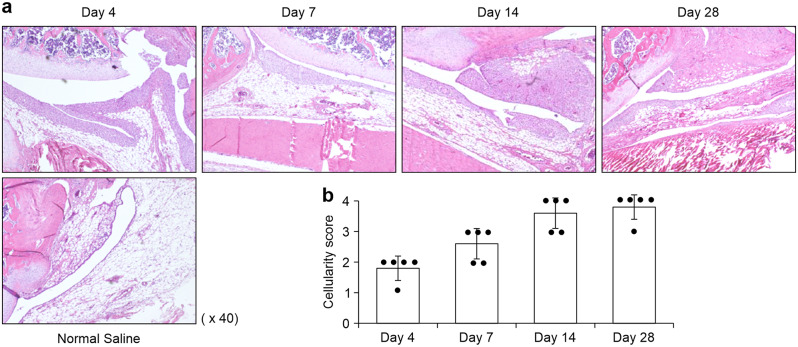
Background: Fibrosis of the infrapatellar fat pad (IFP) is a feature of osteoarthritis and contributes substantially to the pain and dysfunction in patients' joints. However, the underlying mechanisms remain unclear. C-C motif chemokine ligand-2 (CCL2) plays a central role in tissue fibrosis. Thus, we aimed to investigate the role of CCL2 in the development of IFP fibrosis in a rat model of arthritis, hypothesizing that a CCL2 antagonist could mitigate fibrotic progression.

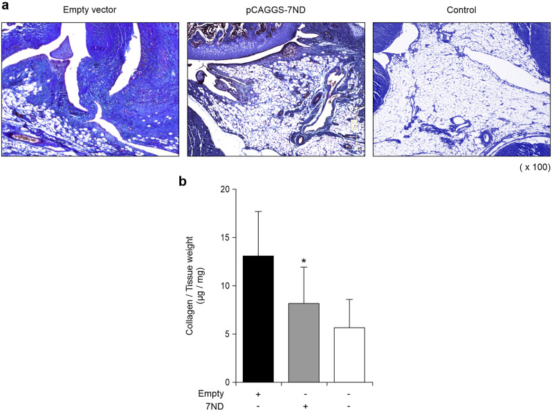
Methods: We induced arthritis in male Wistar rats using intra-articular injections of carrageenan. Furthermore, to evaluate the effects of a CCL2 antagonist on protein expression and collagen deposition in the IFP of the rats, we transferred an N-terminal-truncated CCL2 gene into a rat model via electroporation-mediated intramuscular injection. Macrophage infiltration and collagen deposition in the IFP were analyzed in vivo. Groups were compared using the Mann-Whitney U test and Student's t-test.

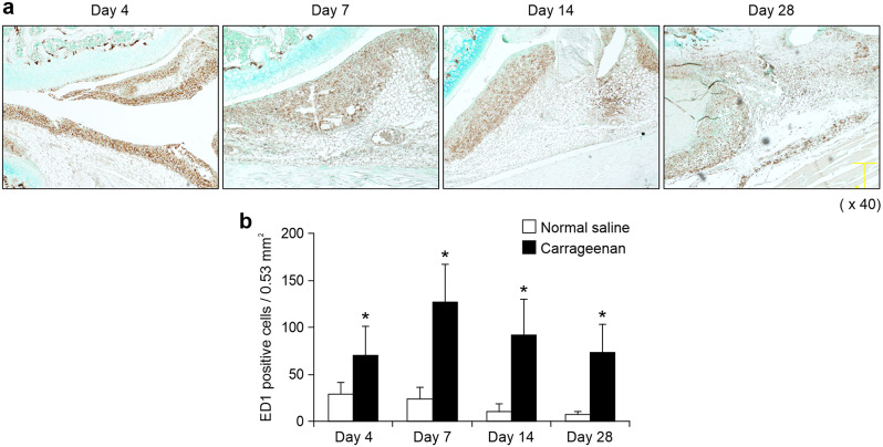
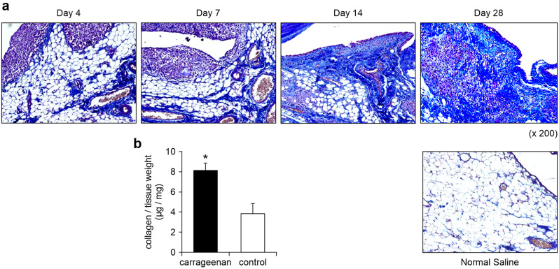
Results: We identified infiltrating macrophages as well as increases in CCL2 and TGF-β levels as collagen deposition progressed. Gene transfer of the CCL2-antagonist before arthritis induction attenuated collagen deposition remarkably.

Conclusions: We provide initial evidence that anti-CCL2 gene therapy can effectively suppress the development of IFP fibrosis in a rat model. Thus, targeting CCL2 holds promise as a therapeutic strategy for managing tissue fibrosis in osteoarthritis patients.

Keywords: CCL2; Fibrosis; Gene therapy; Infrapatellar fat pad; Macrophages; Osteoarthritis.


Conflict of interest statement

The authors declare no competing interests.

Figures

Fig. 1
Fig. 1
Time course of carrageenan-induced arthritis. A) Representative images of sagittal sections of IFP stained with HE. B) Semiquantitative evaluation of IFP inflammation. Each dot indicated the mean value at each time point. Data were also indicated as mean and SD value
Fig. 2
Fig. 2
Time course of macrophage infiltration in IFP. A) Immunohistochemical analysis of ED1-positive macrophages. Representative images of sagittal sections of IFP are shown. a) Analysis of ED1-positive cells in the IFP. The density of macrophages is highest 7 days after the onset of arthritis and remains elevated for 4 weeks. * indicates that the value was significantly different (P < 0.05) from the control
Fig. 3
Fig. 3
Enzyme-linked immunosorbent assay (ELISA) of knee joints in carrageenan-treated rats. a) ELISA of CCL2 protein levels from synovial tissue homogenates. CCL2 production peaks on day 14 and remains high on day 28. b) TGF-β protein levels in synovial tissues measured by ELISA are significantly higher in carrageenan-induced arthritis at each time point compared with levels in non-arthritic tissues. There were 10 rats per group at each time point in all experiments. Values are presented as the mean and standard deviation (n = 10). *P < 0.05 versus control
Fig. 4
Fig. 4
Collagen distribution in the synovial tissue area of the IFP fibrosis rat model. (a) As determined by Masson’s trichrome staining, the carrageenan-induced arthritis shows a wide deposition of collagen fibers along the IFP and synovial membrane on day 28. Representative joint sections from five rats are shown (Original magnification ×200). (b) Quantitative analysis of collagen deposition in carrageenan-induced arthritis. Collagen in IFP was measured using a collagen detection kit. A significant increase in collagen deposition in carrageenan-induced arthritis is seen on day 28. Values are presented as the mean and standard deviation of experiments involving 10 rats per group. *P < 0.05 versus control
Fig. 5
Fig. 5
Serum concentrations of 7ND after injection of the plasmid vector. Rats were treated with pCAGGS-7ND or an empty vector (n = 7 in each group). Equal amounts of serum were obtained from pCAGGS-7ND-treated animals at the indicated times, followed by measurement of the 7ND concentration using an ELISA. A significant amount of 7ND is detected in the serum from pCAGGS-7ND-treated animals within the second week after injection. Human CCL2 protein is not detected in empty vector-treated rats. The mean values from seven rats in each experiment are shown. ND = not detected
Fig. 6
Fig. 6
Amelioration of morphologic lesions and quantitative analysis of CCL2 in 7ND-treated animals. a) Rats were treated with 7ND or an empty vector 7 days prior to the intraarticular injection of carrageenan and sacrificed on day 28. Representative images on day28 from 7ND- or empty vector-treated rats are shown. b) The graph shows the density of ED-1-positive macrophages in the IFP and synovial tissues. c) Cellularity score of IFP from 7ND- or empty vector-treated rats on day28. d) ELISA of CCL2 protein levels from IFP in 7ND- or empty vector-treated rats. There were 15 rats per group in each experiment, and the values are presented as the mean and standard deviation. *P < 0.05 vs. control
Fig. 7
Fig. 7
Amelioration of collagen deposition in 7ND-treated animals. a) Masson’s trichrome-stained sections demonstrating the distribution of collagen proteins. Representative photographs are shown, and there were 5 rats per group in each experiment. b) Rats were treated with 7ND 7 days before the administration of carrageenan. Collagen production was determined by the Sircol collagen assay, and there were 15 rats per group in each experiment. Values are presented as the mean and standard deviation (n = 15). *P < 0.05 versus empty vector-treated rats

References

    1. Nakagawa Y, Tsuji K, Nakamura T, Katagiri H, Ozeki N, Shioda M, An JS, Yoshida R, Sekiya I, Koga H. Association of Infrapatellar Fat Pad Fibrosis at 3 months after ACL Reconstruction with short-term clinical outcomes and inflammatory cytokine levels in the synovial fluid. Orthop J Sports Med. 2023;11(4). - PMC - PubMed
    1. Belluzzi E, Stocco E, Pozzuoli A, Granzotto M, Porzionato A, Vettor R, De Caro R, Ruggieri P, Ramonda R, Rossato M et al. Contribution of infrapatellar fat pad and synovial membrane to knee osteoarthritis pain. Biomed Res Int 2019: 6390182, 2019. - PMC - PubMed
    1. Clockaerts S, Bastiaansen-Jenniskens YM, Runhaar J, Van Osch GJVM, Verhaar JAN, De Clerck LS, et al. The infrapatellar fat pad should be considered as an active osteoarthritic joint tissue: a narrative review. Osteoarthritis Cartilage. 2010;18:876–82. 10.1016/j.joca.2010.03.014 - DOI - PubMed
    1. Sellam J, Berenbaum F. The role of synovitis in pathophysiology and clinical symptoms of osteoarthritis. Nat Rev Rheumatol. 2010;6:625–35. 10.1038/nrrheum.2010.159 - DOI - PubMed
    1. Eymard F, Pigenet A, Citadelle D, Flouzat-Lachaniette CH, Poignard A, Benelli C, et al. Induction of an inflammatory and prodegradative phenotype in autologous fibroblast-like synoviocytes by the infrapatellar fat pad from patients with knee osteoarthritis. Arthritis Rheumatol. 2014;66:2165–74. 10.1002/art.38657 - DOI - PubMed

LinkOut - more resources