. 2024 Oct 1;123(19):3463-3477.
doi: 10.1016/j.bpj.2024.08.022. Epub 2024 Aug 28.

Advanced sampling simulations of coupled folding and binding of phage P22 N-peptide to boxB RNA

Affiliations

Advanced sampling simulations of coupled folding and binding of phage P22 N-peptide to boxB RNA

Luis Vollmers et al. Biophys J. .

Abstract

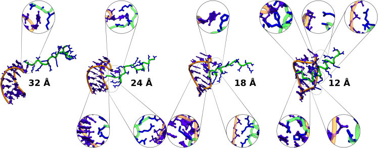
Protein-RNA interactions are crucially important for numerous cellular processes and often involve coupled folding and binding of peptide segments upon association. The Nut-utilization site (N)-protein of bacteriophages contains an N-terminal arginine-rich motif that undergoes such a folding transition upon binding to the boxB RNA hairpin loop target structure. Molecular dynamics free energy simulations were used to calculate the absolute binding free energy of the N-peptide of bacteriophage P22 in complex with the boxB RNA hairpin motif at different salt concentrations and using two different water force field models. We obtained good agreement with experiment also at different salt concentrations for the TIP4P-D water model that has a stabilizing effect on unfolded protein structures. It allowed us to estimate the free energy contribution resulting from restricting the molecules' spatial and conformational freedom upon binding, which makes a large opposing contribution to binding. In a second set of umbrella sampling simulations to dissociate/associate the complex along a separation coordinate, we analyzed the onset of preorientation of the N-peptide and onset of structure formation relative to the RNA and its dependence on the salt concentration. Peptide orientation and conformational transitions are significantly coupled to the first contact formation between peptide and RNA. The initial contacts are mostly formed between peptide residues and the boxB hairpin loop nucleotides. A complete transition to an α-helical bound peptide conformation occurs only at a late stage of the binding process a few angstroms before the complexed state has been reached. However, the N-peptide orients also at distances beyond the contact distance such that the sizable positive charge points toward the RNA's center-of-mass. Our result may have important implications for understanding protein- and peptide-RNA complex formation frequently involving coupled folding and association processes.


Conflict of interest statement

Declaration of interests The authors declare that they have no competing financial interests or personal relationships that could have appeared to influence the work reported in this paper.

Figures

Figure 1
Figure 1
Structure of the 19 residue bacteriophage P22 N-peptide (green cartoon, sequence: NAKTRRHERRRKLAIERDT) in complex with the 15-mer boxB RNA hairpin motif (orange and purple, sequence: 3′-GCGCUGACAAAGCGC) (15). The three main contact areas are illustrated in three separate panels, with (a) indicating the K3 contacting the guanine (G6) and adenine (A7) nucleobases, (b) showing loop base C8 contacting one arginine residue and an isoleucine residue, and (c) capturing the two most prominent arginine contacts to the phosphate backbone. For clarity hydrogen atoms are omitted. Peptide atoms are in licorice and RNA atoms in CPK representation (PDB: 1A4T (15)).
Figure 2
Figure 2
Illustration of three center-of-mass anchors and virtual bonds of different atom groups in boxB RNA (orange/purple, molecule; black, anchor beads) and the N-peptide (green/blue, molecule; white, anchor beads). The central anchor of each molecule, P0 and Q0, are defined as the centers-of-mass of all nonhydrogen atoms. P1 and P2 are defined as the centers-of-mass (nonhydrogen atoms) of residue G6 and A11, respectively. Q1 and Q2 are defined as the centers-of-mass (nonhydrogen atoms) of residue LYS3 and GLU8, respectively. The spherical angles of θ and ϕ are defined by the virtual bonds P1-P0-Q0 and P2-P1-P0-Q0. The orientational angles Ψ,Θ, and Φ are defined by the virtual bonds P1-P0-Q0-Q1, P0-Q0-Q1, and P0-Q0-Q1-Q2. The equilibrium angles for the axial and orientational restraining potentials are (in radians), Θ0 = 2.53, Φ0 = 2.7, Ψ0 = −3.13, θ0 = 1.49, ϕ0 = −2.88, the force constant is in all cases, k= 100.0 kcal mol−1 rad−2.
Figure 3
Figure 3
Calculated RMSD of only nonhydrogen atoms of the RNA (thin line) and peptide (bold line) with respect to the crystal structure during unrestrained MD simulations. Different line colors indicate results at three different salt concentrations (resulting in three RMSD curves for peptide and RNA, respectively). Note, a Gaussian filter with a window size of 20 was used to improve clarity. The average peptide-RNA center-of-mass distances at each ion concentration are also listed with their standard deviations in parentheses.
Figure 4
Figure 4
Comparison of the potential of mean force curves with standard errors obtained from the WR method. Standard errors of the mean are depicted as bars.
Figure 5
Figure 5
Calculated free energy change along the N-peptide boxB separation center-of-mass distance coordinate using the TIP3P or TIP4P-D water models and three salt concentrations. No additional restraints on conformation or partner orientation are included. Standard errors of the mean are depicted as bars.
Figure 6
Figure 6
Spherical distribution (in terms of the polar and azimuthal angles θ and ϕ) of the N-peptide relative to the boxB RNA at different center-of-mass distances. The panels above each plot illustrate the distribution of peptide (cyan) and RNA (blue) partners by best superposition of representative snapshots. The distribution and relative orientation becomes increasingly nonrandom with decreasing center-of-mass distances (see also Figs. S4–S7).
Figure 7
Figure 7
Rolling time-average of the dot products between the peptide’s dipole moment (calculated relative to the center-of-mass of the peptide) and the normalized center-of-mass vector (upper panels) and between the normalized dipole moment vector and the normalized center-of-mass vector. A stepsize of 5000 frames and a window size of 20000 frames was used. In the latter case only the direction and not the magnitude of the dipole vector is considered. Standard errors of the mean are depicted as bars.
Figure 8
Figure 8
Total variation distance DTV between bulk orientations and orientations adopted during the US simulations. Low values for DTV correspond to bulk-like behavior and indicate the absence of ordered configurational ensembles. Boundaries, ΞDTV, are extracted as the middle of three columns with consistently increasing DTV for decreasing ri. For example, for TIP3P water with low salt concentration, this would be 34 Å. A generalized logistic curve is fitted to each data set as a line (70).
Figure 9
Figure 9
Upper panels: average closest contact distance rcc with error bars corresponding to δrcc versus RNA-peptide center-of-mass distance. The lines depict a fitted function of the form f(x)=a+blog(cexp(xd)+e). Lower panels indicate the frequency of a contact closer than 4 Å, fcc. Here the line depicts a fitted, generalized logistic curve (70).
Figure 10
Figure 10
Average deviation of the N-peptide and boxB RNA conformation from the native bound structures along the HREUS center-of-mass distance coordinate. The lines represent a generalized logistic function fit (70). The average RMSD was calculated for all nonhydrogen atoms with respect to the individual peptide and RNA conformation in the experimental complex structure. Standard errors of the mean are depicted as bars.
Figure 11
Figure 11
Representative binding process rendered for the TIP4P-D water model with 81 mM salt concentration. The peptide approaches from the top of the hairpin loop and folds into the major groove over time. The RNA and peptide are shown in their usual color schemes. The RNA atoms are represented via balls and sticks, while the peptide side chains are shown as licorice in the detail plots.

Similar articles

References

    1. Bahadur R.P., Zacharias M., Janin J. Dissecting protein–RNA recognition sites. Nucleic Acids Res. 2008;36:2705–2716. - PMC - PubMed
    1. Lund M., Vrbka L., Jungwirth P. Specific Ion Binding to Nonpolar Surface Patches of Proteins. J. Am. Chem. Soc. 2008;130:11582–11583. - PubMed
    1. Patel D.J. Adaptive recognition in RNA complexes with peptides and protein modules. Curr. Opin. Struct. Biol. 1999;9:74–87. - PubMed
    1. Chen Y., Varani G. Engineering RNA-binding proteins for biology. FEBS J. 2013;280:3734–3754. - PMC - PubMed
    1. Allers J., Shamoo Y. Structure-based analysis of protein-RNA interactions using the program ENTANGLE. J. Mol. Biol. 2001;311:75–86. - PubMed

Publication types

LinkOut - more resources