. 2024 Dec 3;23(12):1743-1760.
doi: 10.1158/1535-7163.MCT-23-0931.

Immunoproteasome Activation Expands the MHC Class I Immunopeptidome, Unmasks Neoantigens, and Enhances T-cell Anti-Myeloma Activity

Affiliations

Immunoproteasome Activation Expands the MHC Class I Immunopeptidome, Unmasks Neoantigens, and Enhances T-cell Anti-Myeloma Activity

Priyanka S Rana et al. Mol Cancer Ther. .

Abstract

Proteasomes generate antigenic peptides that are presented on the tumor surface to cytotoxic T-lymphocytes. Immunoproteasomes are highly specialized proteasome variants that are expressed at higher levels in antigen-presenting cells and contain replacements of the three constitutive proteasome catalytic subunits to generate peptides with a hydrophobic C-terminus that fit within the groove of MHC class I (MHC-I) molecules. A hallmark of cancer is the ability to evade immunosurveillance by disrupting the antigen presentation machinery and downregulating MHC-I antigen presentation. High-throughput screening was performed to identify compound A, a novel molecule that selectively increased immunoproteasome activity and expanded the number and diversity of MHC-I-bound peptides presented on multiple myeloma cells. Compound A increased the presentation of individual MHC-I-bound peptides by >100-fold and unmasked tumor-specific neoantigens on myeloma cells. Global proteomic integral stability assays determined that compound A binds to the proteasome structural subunit PSMA1 and promotes association of the proteasome activator PA28α/β (PSME1/PSME2) with immunoproteasomes. CRISPR/Cas9 silencing of PSMA1, PSME1, or PSME2 as well as treatment with immunoproteasome-specific suicide inhibitors abolished the effects of compound A on antigen presentation. Treatment of multiple myeloma cell lines and patient bone marrow-derived CD138+ cells with compound A increased the anti-myeloma activity of allogenic and autologous T cells. Compound A was well-tolerated in vivo and co-treatment with allogeneic T cells reduced the growth of myeloma xenotransplants in NOD/SCID gamma mice. Taken together, our results demonstrate the paradigm shifting impact of immunoproteasome activators to diversify the antigenic landscape, expand the immunopeptidome, potentiate T-cell-directed therapy, and reveal actionable neoantigens for personalized T-cell immunotherapy.


Conflict of interest statement

E. Malek reports being on the advisory board for Bristol Myers Squibb, Janssen, and Amgen; research funding from MedPacto Inc.; and being a speaker for Bristol Myers Squibb and Janssen. D.J. Adams reports personal fees and other support from Convelo Therapeutics outside the submitted work. J.J. Driscoll reports a patent for UH Ventures pending. No disclosures were reported by the other authors.

Figures
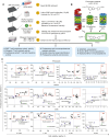
Figure 1.
Figure 1.
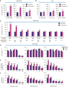
Identification of novel molecules that increased proteasomal ChT-like activity. A, Schematic representation of the cell-based HTS. RPMI-8226 cells were treated with a library of 9,600 novel molecules and the effect on proteasomal ChT-like activity measured. Top hits were identified that increased proteasome ChT-like activity by ≥50%, and the selected primary hits were further evaluated for their effects on cell viability, antigen presentation, and T-cell–mediated cytotoxicity in murine and human cell–based assays. B, Schematic of the HTS using the cell-permeable fluorogenic substrate LLVY-R110 that is specifically cleaved by proteasomes and immunoproteasomes. C, Effect of the hits on cell viability and proteasome activity in RPMI-8226, ARH-77, and U266 cells. Red boxes indicate compounds that increased proteasome activity ≥50% and maintained cell viability ≥90%. HTS, high-throughput screens; tuba-A, tubastatin A.
Figure 2.
Figure 2.
Effect of compound A on proteasomal catalytic activities. A, Effect of compound A on proteasomal peptide-hydrolyzing activity in RPMI-8226 cells. Cells were treated with the fluorogenic peptide substrates Suc-LLVY-MCA, LLVY-R110, and Ac-ANW-MCA for 1 to 3 hours. Fluorescent intensity was monitored as a direct measurement of substrate hydrolysis. B, Effect of compound A on proteasomal peptide-hydrolyzing activity in HeLa cells. C, Effect of compound A on a series of fluorogenic substrates specifically cleaved by proteasome and immunoproteasome catalytic sites. LLVY-R110 and Suc-LLVY-MCA are cleaved by both the β5c subunit and β5i subunit, whereas ANC-ANW-AMC is cleaved specifically only by the β5i subunit. Z-LLL-AMC is cleaved by β5c, Ac-WLA-AMC is cleaved by β5c, Boc-LRR-AMC is cleaved by the β2c and β2i subunits, Ac-RLR-AMC is cleaved by the β2c and β2i subunits, Z-ARR-AMC is cleaved by β2c and β2i subunits, Z-LLE-AMC is cleaved by the β1c subunit, and ANC-PAL-AMC is cleaved by the β1i subunit. Lysates from RPMI-8226 cells were incubated with the indicated proteasome substrates at concentrations of 15 μmol/L for up to 3 hours, and fluorescent intensity monitored was a direct measure of the activity of proteasomal subunit–specific catalytic activity. D, Effect of immunoproteasome-specific inhibitors on multiple myeloma viability as determined by the 2,3-bis[2-methoxy-4-nitro-S-sulfophenynl]-2H-tetrazolium-5 carboxanilide inner salt assay. E and F, Effect of treatment with immunoproteasome-specific inhibitors on the ability of compound A to modulate proteasomal catalytic activity in multiple myeloma cells. RPMI-8226 cells were treated with the immunoproteasome inhibitors ONX-0912, ONX-0914, and KZR-616 at the indicated concentrations with and without compound A. Proteasomal ChT-like activity was then measured by recording LLVY-R110 hydrolysis as mentioned earlier. Immunoproteasome-specific activity was measured by recording Ac-ANW-AMC hydrolysis. Bioassays were performed in triplicate.
Figure 3.
Figure 3.
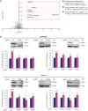
Global PISA assay to identify compound A target proteins. Results of 1D PISA identified four proteins that demonstrated statistically significant P values after treatment at both 6 and 24 hours compared with vehicle samples. Red box indicates the most significantly stabilized target proteins following treatment of RPMI-8226 cells with compound A after both 6 and 24 hours. The proteasome-specific protein PSMA1 is indicated by a red dot. Gray dots indicate proteins with −log2FC <1 or P value > 0.05 at either time point. B, Effect of PSME1, PSME2, and PSMA1 on the effect of compound A on proteasome activity. Effect of compound A on immunoproteasome activity was measured using Ac-ANW-AMC in cells with KO of PSME1, PSME2, and PSMA1. Immunoblots show CRISPR/CAS9-mediated PSME1, PSME2, and PSMA1 KO in RPMI-8226 and ARH-77 cells followed by the effect on proteasomal catalytic activities. IB, immunoblot; KO, knockout.
Figure 4.
Figure 4.
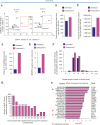
Compound A effect on SIINFEKL–MHC-I presentation and the murine immunopeptidome. A, Effect of top eight hits that increased proteasomal ChT-like activity with SIINFEKL–MHC-I complex presentation on lysis of E.G7-Ova cells by the B3Z T-cell hybridoma. E.G7-Ova cells were pretreated with compounds (1 μmol/L), after which the cells were washed and co-cultured with the H-2Kb–restricted B3Z T cells genetically engineered to express a TCR that specifically recognizes the SIINFEKL–H2Kb complex. E.G7-Ova cells were co-cultured with B3Z cells at an E:T ratio of 3:1 for 24 and 48 hours. B, Effect of compound A on the number of peptides eluted from MHC-I (M1/42) of E.G7-ova cells. C, Effect of compound A on total peptide peak intensity after elution with mouse MHC-I (M1/42). D, Effect of compound A on E.G7-Ova presentation of SIINFEKL–MHC-I. E, Parallel reaction monitoring analysis of lysates spiked with the SIINFEKL peptide show that pretreatment with compound A resulted in an increase in SIINFEKL peptide copy number bound/cell by >1.5-fold. F, The number of peptides eluted from MHC-I (M1/42) were 8 to 9 amino acids in length, with no significant difference between treated and untreated samples. G, Pretreatment of E.G7-Ova cells with compound A increased the presentation of MHC-I–specific peptides by up to 50-fold. More than 450 peptides were increased by 2-fold with compound A treatment. H, Rank order of genes of the top 20 MHC-I–restricted peptides upregulated in cells treated with compound A. Red stars indicate peptide that has undergone posttranslational methionine oxidation. FC, fold change.
Figure 5.
Figure 5.
Effect of compound A on the myeloma immunopeptidome. A, Effect of compound A on number of peptides eluted from MHC-I (W6/32) in RPMI-8226 cells. Cells were pretreated with compound A (1 μmol/L) for 48 hours, after which they were pelleted, lysed, and immunoprecipitated with human MHC-I (W6/32). Acid elution resulted in the identification of MHC-I peptide abundance in cells treated with compound A relative to untreated cells. B, Total peak intensity of peptides eluted from pan–MHC-I (W6/32) was increased by two-fold with compound A. C, Length of peptides eluted from MHC-I (W6/32). D, Pretreatment of RPMI-8226 cells with compound A increased the presentation of MHC-I–specific peptides up to 200-fold. More than 1,500 peptides were increased two-fold after treatment of cells with compound A. E, Waterfall plot representing peptides upregulated by compound A and downregulated by bortezomib treatment of RPMI-8226 cells. F, Effect of compound A on antigen peptide terminal residue. Greater than 80% of the top 50 peptides upregulated by compound A had a hydrophobic terminal aliphatic or aromatic residue. G, Genes that encode the respective MHC-I–restricted peptides upregulated and downregulated after compound A or bortezomib treatment. H, Rank order of genes that encode the 20 MHC-I–restricted peptides most upregulated after compound A treatment. BTZ, bortezomib; FC, fold change.
Figure 6.
Figure 6.
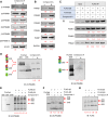
Effect of compound A on immunoproteasome subunit β5i /PSMB8 association with the proteasome activator and immunoproteasome. A, Effect of compound A on proteasome and immunoproteasome catalytic subunit levels. B, Effect of compound A on proteasome activator subunit levels. C, Effect of compound A on PSME1 and PSME2 association with FLAG-tagged β5i/PSMB8 and FLAG-tagged β5/PSMB5 in cell lysates. D, Effect of compound A treatment on the incorporation of β5i/PSMB8 and β5/PSMB5 into endogenous 20S/26S/30S proteasomal complexes. Complexes were detected by native-PAGE and immunoblotting. E, F, and G, Effect of compound A on FLAG-tagged β5/PSMB5 and FLAG-tagged β5i/PSMB8 high molecular weight proteasomal complexes in RPMI-8226 lysates. Purified 20S, 26S, and 20S immunoproteasomes were loaded as controls for migration distance and β5i/PSMB8 or β5/PSMB5 composition. Membranes were probed with antibodies specific to β5/PSMB5 (E), β5i/PSMB8 (F), and FLAG (G). IB, immunoblot.
Figure 7.
Figure 7.
Effect of compound A on allogenic and autologous T-cell anti-myeloma cytotoxicity. A, RPMI-8226 cells were treated with compound A and probed with pan–MHC-I or MHC-I subtype allele-specific antibodies, after which the samples were analyzed by flow cytometry. Pretreatment with compound A significantly increased pan–MHC-I HLA-ABC and HLA-B surface presentation in all MMCLs assayed. B, RPMI-8226 were pretreated with compound A for 48 hours, after which they were washed and co-cultured with healthy CD8+ cytotoxic T cells for 18 hours at E:T ratios of 1:1, 2:1, and 3:1. Compound A increased CTL-mediated cytotoxicity of tumor cells, which was measured by gating for CD138+, annexin V+, and PI+ cells at E:T 2:1 and 3:1. Pretreatment of MMCLs and T cells alone with compound A resulted in ∼5 to 10% cell death, which was enhanced further when multiple myeloma cells were co-cultured at E:T ratios of 2:1 and 3:1. T cells alone also did not show significant cell death when pretreated with compound A. There was <5% multiple myeloma cell death detected with or without compound A treatment. Similar results were observed with CD8+ T cells alone. C, Patient multiple myeloma CD138+ cells were treated with compound A followed by co-culturing with CD8+ patient-derived autologous T cells (E:T = 2:1). Cytotoxicity was measured by gating for CD138+, annexin V+, and PI+ cells. D and E, Effect of compound A on PDL1 or PDL2 expression in MMCLs. D and E, Surface markers were quantitated by flow cytometry. TGFβ and BTZ were included as positive controls as they are known to upregulate PDL1/L2 expression. F, Effect of compound A on the viability of RPMI-8226 cells alone or combined with ICIs and CD8+ T cells. Multiple myeloma and CD8+ T cells were separately treated with or without compound A and the indicated agents (BTZ, TGFβ, and ICIs). Following co-culture, the relative percent of viable multiple myeloma cells was quantitated by annexin V and PI staining and flow cytometry. Assays were performed in triplicate. Ann-V, annexin V; BTZ, bortezomib.

References

    1. Yang K, Halima A, Chan TA. Antigen presentation in cancer—mechanisms and clinical implications for immunotherapy. Nat Rev Clin Oncol 2023;20:604–23. - PubMed
    1. Waldman AD, Fritz JM, Lenardo MJ. A guide to cancer immunotherapy: from T cell basic science to clinical practice. Nat Rev Immunol 2020;20:651–68. - PMC - PubMed
    1. Sharma P, Hu-Lieskovan S, Wargo JA, Ribas A. Primary, adaptive, and acquired resistance to cancer immunotherapy. Cell 2017;168:707–23. - PMC - PubMed
    1. Sijts EJ, Kloetzel PM. The role of the proteasome in the generation of MHC class I ligands and immune responses. Cell Mol Life Sci 2011;68:1491–502. - PMC - PubMed
    1. Desjardins M. Antigen cross-presentation: proteasome location, location, location. EMBO J 2019;38:e102799. - PMC - PubMed

MeSH terms

Substances