. 2024 Aug 26:12:e17866.
doi: 10.7717/peerj.17866. eCollection 2024.

Variation in floral morphology, histochemistry, and floral visitors of three sympatric morning glory species

Affiliations

Variation in floral morphology, histochemistry, and floral visitors of three sympatric morning glory species

Awapa Jirabanjongjit et al. PeerJ. .

Abstract

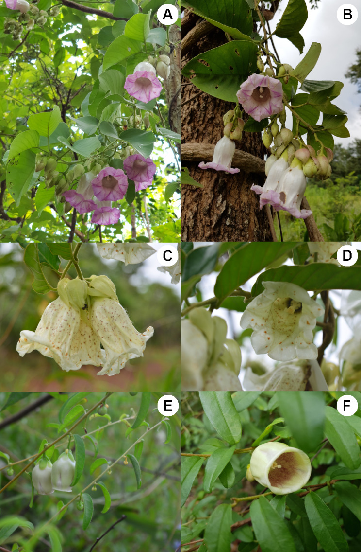
Three morning glory species in the genus Argyreia Lour., A. lycioides (Choisy) Traiperm & Rattanakrajang, A. mekongensis Gagnep & Courchet, and A. versicolor (Kerr) Staples & Traiperm, were found co-occurring and co-flowering. Argyreia mekongensis and A. versicolor are rare, while A. lycioides is near threatened and distributed throughout Myanmar and Thailand. We investigated key floral characters (floral morphology and phenology, as well as the micromorphology of the floral nectary disc and staminal trichomes) and screened for important chemical compounds hypothesized to contribute to pollinator attraction. Our findings demonstrate that some aspects of floral morphology (e.g., corolla size, limb presence, and floral color) of the three studied congeners exhibit significant differences. Moreover, pollinator composition appears to be influenced by floral shape and size; morning glory species with wider corolla tubes were pollinated by larger bees. The morphology of the floral nectary disc was similar in all species, while variation in staminal trichomes was observed across species. Glandular trichomes were found in all three species, while non-glandular trichomes were found only in A. versicolor. Histochemical results revealed different compounds in the floral nectary and staminal trichomes of each species, which may contribute to both floral attraction and defense. These findings demonstrate some segregation of floral visitors among sympatric co-flowering morning glory species, which appears to be influenced by the macro- and micromorphology of flowers and their chemical compounds. Moreover, understanding the floral morphology and chemical attractants of these sympatric co-flowering Argyreia species may help to maintain their common pollinators in order to conserve these rare and endangered species, especially A. versicolor.

Keywords: Argyreia; Biodiversity; Convolvulaceae; Histochemistry; Plant conservation; Pollinator; Trichome; Xylocopa.


Conflict of interest statement

The authors declare there are no competing interests.

Figures

Figure 1
Figure 1. Photos showing the floral characters of three sympatric Argyreia species.
(A, B) A. versicolor, (C, D) A. mekongensis, and (E, F) A. lycioides. Photos A, B and F were taken by Yotsawate Sirichamon, and C, D and E were taken by Tripatchara Atiratana.
Figure 2
Figure 2. A comparison of quantitative floral characters in three sympatric Argyreia species.
(A) Corolla length. (B) Corolla tube diameter. (C) Stamen length. (D) Pistil length. Circles and error bars denote means and standard errors. Species with different lowercase letters are significantly different (p < 0.05).
Figure 3
Figure 3. Animal visitors of three sympatric species of Argyreia.
(A) Xylocopa latipes visiting A. versicolor. (B) X. aestuans visiting A. versicolor. (C) X. latipes visiting A. mekongensis. (D) X. aestuans visiting A. mekongensis. (E) Amegilla sp. visiting A. mekongensis. (F) Mylabris phalerata beetle consuming the corolla of A. mekongensis. (G) Cinnyris jugularis sunbird robbing nectar from A. mekongensis. (H) Unknown bee species (Anthophila) visiting A. lycioides. (I) Unknown wasp species (Vespidae) visiting A. lycioides. (J) Cinnyris jugularis sunbird robbing nectar from A. lycioides. Photos credited to Awapa Jirabanjongjit.
Figure 4
Figure 4. Visitation rates of animal visitors observed at A. lycioides flowers.
(A) 2019 (n = 4 plants) and in (B) 2020 (n = 4 plants). The blue circles and error bar denote means and standard errors, while pastel-colored jittered points show the distributions of the raw data. Note: Only Anthophila and Vespidae contacted floral stigmas and anthers, and are the potential pollinators of A. lycioides.
Figure 5
Figure 5. Results of histochemical analysis conducted in A. versicolor.
(A) Transversal section of the floral nectary showing the presence of terpenes; positive staining shown at the blue arrow pointing to a nectary duct (ND) and the nectary epidermis (EP). (B) Staminal trichomes showing the presence of terpenes; positive staining shown by the blue arrow pointing to the apical gland cell. (C) Unstained long glandular trichomes at the base of staminal filaments; red arrows pointing to rounded cylindrical (RCY) and obovoid (OBO) apical gland cells. (D) Longitudinal section of the floral nectary showing the presence of flavonoids under a fluorescence microscope; positive staining shown by the white arrow pointing to the nectary disc. (E) Unstained short glandular trichomes at the middle of staminal filaments; red arrow pointing to an apical gland cell that is globose shaped (GLO). (F) Staminal trichomes stained with Naturstoff reagent and viewed under fluorescence microscope reveal the presence of flavonoids; strong staining shown at the blue arrow, white arrow pointing to an apical cell of a non-glandular trichome. Photos credited to Awapa Jirabanjongjit.
Figure 6
Figure 6. Results of histochemical analysis conducted in A. mekongensis.
(A) Transversal section of the floral nectary showing the presence of terpenes; positive staining shown at the red arrows pointing to the nectary duct (ND) and nectary epidermis (EP). (B) Staminal trichomes tested positive for terpenes; blue arrows point to apical gland cells showing terpenes inside of the glands. (C) Transversal section of the floral nectary stained with Sudan Black B showing the presence of lipids; positive staining shown at the red arrow pointing to the nectary duct (ND). (D) Staminal trichomes stained with Sudan Black B tested positive for lipids; red arrows point to apical gland cells containing lipids, and demonstrate the different types of gland cells: BE, bell-shaped; RCO, rounded conical. (E) Transversal section of the floral nectary stained with Sudan III showing the presence of lipids in black. (G) Transversal section of the floral nectary stained with Naturstoff reagent and viewed under a fluorescence microscope showing the presence of flavonoids. (F, H) Staminal trichomes stained with Sudan III tested positive for lipids; blue arrows point to lipids inside of the apical gland cells, while red arrows demonstrate the different types of gland cells: RCY, rounded cylindrical; CON, convex; GLO, globose. Other abbreviations: N, nectary; OV, ovary; EP, epidermis. Photos credited to Awapa Jirabanjongjit.
Figure 7
Figure 7. Results of histochemical analysis conducted in A. lycioides.
(A) Longitudinal section of the floral nectary showing the presence of terpenes. (B) Staminal trichomes tested positive for terpenes; positive staining shown at the red arrows, which also demonstrate some of the different types of apical gland cells: GLO, globose; RCY, rounded cylindrical. (C) Longitudinal section of the floral nectary stained with Naturstoff reagent and viewed under a fluorescence microscope showing the presence of flavonoids. (D) Unstained glandular trichomes; red arrows demonstrate some of the different types of apical gland cells: PY, pyriform; OBO, obovoid. Other abbreviations: N, nectary; OV, ovary; BS, basal cells; S, stalk. Photos credited to Awapa Jirabanjongjit.

References

    1. Agati G, Tattini M. Multiple functional roles of flavonoids in photoprotection. New Phytologist. 2010;186(4):786–793. doi: 10.1111/j.1469-8137.2010.03269.x. - DOI - PubMed
    1. Alonso WR, Rajaonarivony JI, Gershenzon J, Croteau R. Purification of 4S-limonene synthase, a monoterpene cyclase from the glandular trichomes of peppermint (Mentha x piperita) and spearmint (Mentha spicata) Journal of Biological Chemistry. 1992;267(11):7582–7587. - PubMed
    1. Antonious GF. Production and quantification of methyl ketones in wild tomato accessions. Journal of Environmental Science and Health, Part B. 2001;36(6):835–848. doi: 10.1081/PFC-100107416. - DOI - PubMed
    1. Bailey SF, Hargreaves AL, Hechtenthal SD, Laird RA, Latty TM, Reid TG, Teucher AC, Tindall JR. Empty flowers as a pollination-enhancement strategy. Evolutionary Ecology Research. 2007;9(8):1245–1262.
    1. Baker HG. Chemical constituents of nectar in relation to pollination mechanisms and phylogeny. In: Nitecki M, editor. Biochemical aspects of evolutionary biology. University of Chicago Press; Chicago: 1982. pp. 131–171.

LinkOut - more resources