

Case Reports
doi: 10.1002/ccr3.9343.
eCollection 2024 Sep.
Polypoid endometriosis-An exceptional subtype of endometriosis mimicking an aggressive pelvic cancer
Affiliations
- PMID: 39210927
- PMCID: PMC11358198
- DOI: 10.1002/ccr3.9343
Item in Clipboard
Case Reports
Polypoid endometriosis-An exceptional subtype of endometriosis mimicking an aggressive pelvic cancer
Clin Case Rep.
.
Abstract
Polypoid endometriosis is a rare manifestation of endometriosis, which may mimic pelvic cancer. This subtype commonly encountered in post-menopausal women may be wrongly mistaken for a neoplasm on clinical, radiological, perioperative or pathologic assessments leading to inadequate treatment.
Keywords: endometriosis; pelvic cancer; polypoid endometriosis; post‐menopausal.
© 2024 The Author(s). Clinical Case Reports published by John Wiley & Sons Ltd.
Conflict of interest statement
The authors declare no conflicts of interest.
Figures
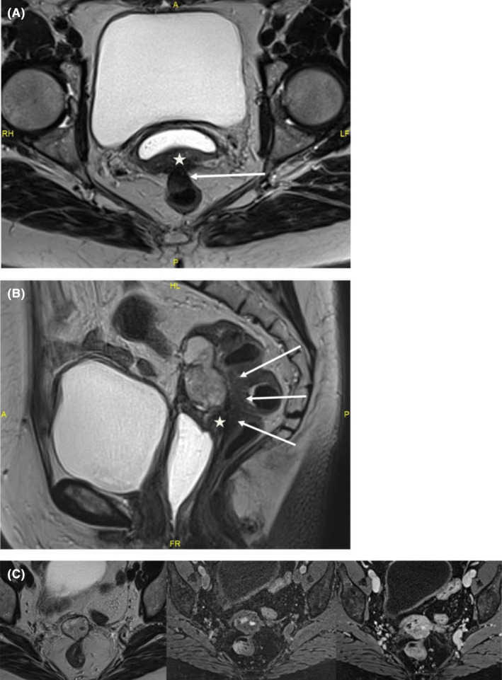
MRI: (A) T2W axial image: endometriotic nodule of posterior vaginal wall (star) with contiguous transmural digestive involvement (arrows). (B) T2W sagittal image: the lesion is close to a fibrous endometriotic nodule of the posterior vaginal fornix and of the rectovaginal septum (star). Transmural involvement of the anterior wall of the middle rectum with typical “mushroom cap” in the sagittal plane (arrows). (C) T2W‐T1W‐T1W post contrast axial images: mainly solid lesion of the vaginal stump in heterogeneous T2 hypersignal, with hemorrhagic remodeling in T1 hypersignal, homogeneous enhancement after contrast injection.
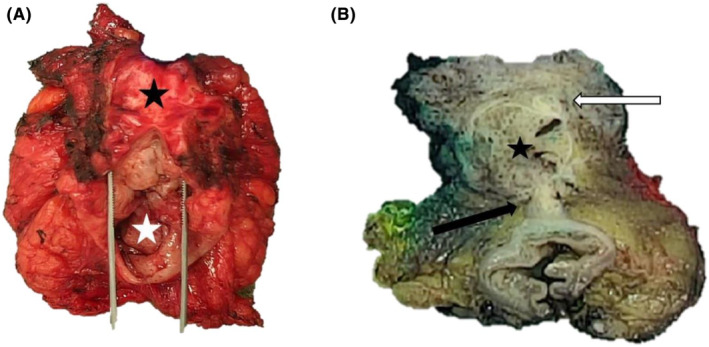
Macroscopic photographs of the posterior pelvectomy with endometriosis lesions before and after formalin fixation. Gross findings of the resected specimen at first examination before fixation, with the vaginal wall (black star) attached to the rectum (white star) (A). Post‐fixation transversal section revealed a firm and gray‐tan, round cystic mass (star), located between and protruding through the vaginal chorion (white arrow) and the anterior rectal wall (black arrow) (B).
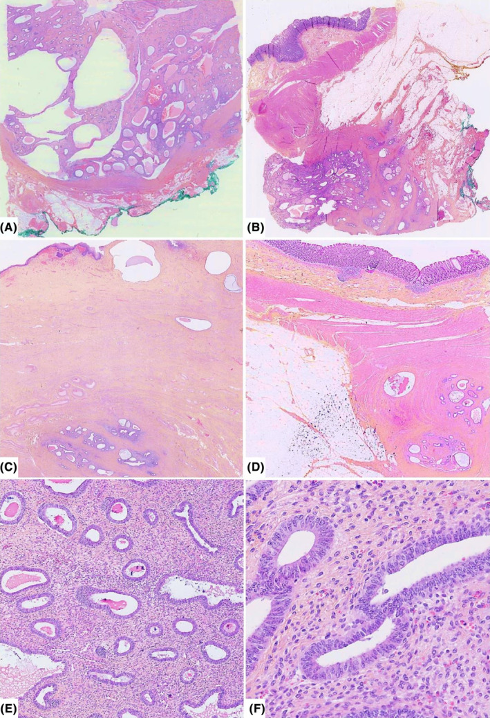
Representative H&E histologic sections of deep florid endometriosis involving the vagina and the rectum at different magnifications. Lesions forming polypoid and cystic masses reminding benign endometrial polyp (A and B), with mural infiltrative aspects of the vaginal chorion and the anterior rectal wall (C and D), respectively, at low magnification. Mild and high power views show endometrial‐type glands dispersed within an abundant fibrous stroma with focal hemorrhage (E) and lined by pseudostratified columnar cells without cytonuclear atypia and low mitotic activity (F).
References
-
- Dunselman GA, Vermeulen N, Becker C, et al. ESHRE guideline: management of women with endometriosis. Hum Reprod. 2014;29(3):400‐412. - PubMed
-
- Parker RL, Dadmanesh F, Young RH, Clement PB. Polypoid endometriosis: a clinicopathologic analysis of 24 cases and a review of the literature. Am J Surg Pathol. 2004;28(3):285‐297. - PubMed
-
- Jaiman S, Gundabattula SR, Pochiraju M, Sangireddy JR. Polypoid endometriosis of the cervix: a case report and review of the literature. Arch Gynecol Obstet. 2014;289(4):915‐920. - PubMed
Publication types
LinkOut - more resources
Full Text Sources
