Review
. 2024 Aug 15:15:1426418.
doi: 10.3389/fimmu.2024.1426418. eCollection 2024.

Stem-like CD8+ T cells in cancer

Affiliations
Review

Stem-like CD8+ T cells in cancer

Chelsea Steiner et al. Front Immunol. .

Abstract

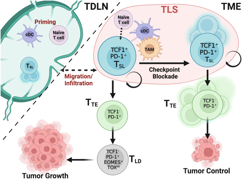
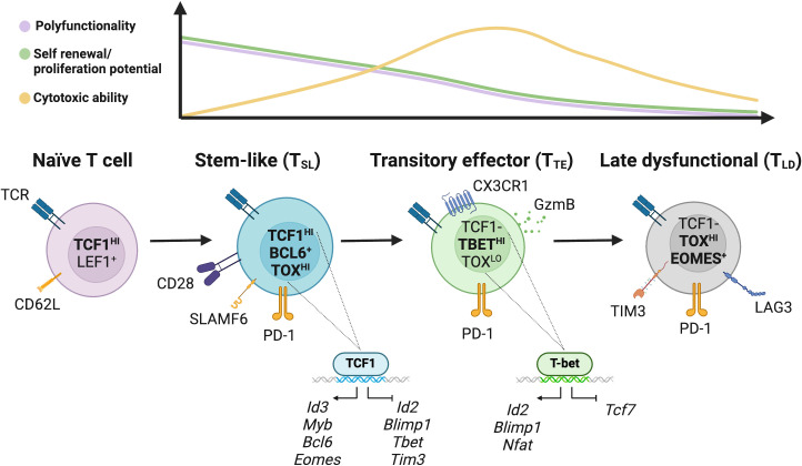
Stem-like CD8+ T cells (TSL) are a subset of immune cells with superior persistence and antitumor immunity. They are TCF1+ PD-1+ and important for the expansion of tumor specific CD8+ T cells in response to checkpoint blockade immunotherapy. In acute infections, naïve CD8+ T cells differentiate into effector and memory CD8+ T cells; in cancer and chronic infections, persistent antigen stimulation can lead to T cell exhaustion. Recent studies have highlighted the dichotomy between late dysfunctional (or exhausted) T cells (TLD) that are TCF1- PD-1+ and self-renewing TCF1+ PD-1+ TSL from which they derive. TCF1+ TSL cells are considered to have stem cell-like properties akin to memory T cell populations and can give rise to cytotoxic effector and transitory T cell phenotypes (TTE) which mediate tumor control. In this review, we will discuss recent advances made in research on the formation and expansion of TSL, as well as distinct niches required for their differentiation and maintenance in the setting of cancer. We will also discuss potential strategies to generate these cells, with clinical implications for stemness enhancement in vaccine design, immune checkpoint blockade (ICB), and adoptive T cell therapies.

Keywords: cancer models; chronic viral infection; immune; stem-like CD8 T cells (TSL); tertiary lymphoid structure (TLS); tumor microenvironment (TME).


Conflict of interest statement

The authors declare that the research was conducted in the absence of any commercial or financial relationships that could be construed as a potential conflict of interest.

Figures

Figure 1
Figure 1
TSL cells drive and maintain CD8+ T cell responses in cancer after ICB. Naïve and TSL CD8+ T cells are primed and activated in the tumor draining lymph node (TDLN) or tertiary lymphoid structures (TLS) within the tumor by conventional dendritic cells (cDCs) that present tumor derived antigen. A portion of these activated TSL cells reside in the TDLN and maintain a reservoir that migrate and infiltrate the tumor microenvironment (TME). Maintenance of TSL cells has yet to be fully determined within these immunological niches. Without ICB, following activation, TSL cells infiltrate tumors and rapidly undergo exhaustion in the presence of persistent antigen stimulation. While transitory effector CD8+ T cells (TTE) cells differentiate from TSL cells, TTE quickly adopt a late dysfunctional (TLD) phenotype but can carry a level of some tumor control through cytotoxic cytokines and tumor cell targeting. Upon ICB, the TSL population undergoes self-renewal and proliferation, giving rise to the TTE subset and this supports the majority of the CD8+ T cell antitumoral response, leading to tumor control.
Figure 2
Figure 2
Model of the characteristics and differentiation of CD8+ T cell states in cancer. The transcription factors TCF1 and LEF1, as well as the adhesion and lymphocyte homing molecules CD62L, are highly expressed in naïve T cells. Downstream, the population of stem-like CD8+ T (TSL) cells with strong polyfunctionality and self-renewal ability is defined by TCF1. These cells have a strong proliferation capacity, are primarily quiescent in vivo, and are able to support the CD8+ T cell response. By suppressing the expression of effector-associated genes like Id2, Blimp-1, Tbet and Tbx21 and stimulating memory-associated genes like Eomes, Myb, Bcl-6, TCF1 facilitates the generation, maintenance, and functionality of these cells. Phenotypically, TSL cells display CD28 and ICOS costimulatory markers, low or moderate amounts of PD-1, SLAMF6, and CXCR5. TSL cells expand upon ICB and both maintain the TSL reservoir and differentiate into further subpopulations. Differentiation of TSL give rise to downstream transitory effector CD8+ T cells (TTE) that express high PD-1 receptor, proliferate rapidly in steady state down regulating TCF1 expression, and upregulate T-BET. T-BET inhibits TOX-mediated development of late dysfunctional T cells (TLD) phenotype in TTE cells. Transitory cells exhibit the expression of CX3 chemokine receptor 1 (CX3CR1) and PD-1. TTE proliferate to help target and eliminate tumor cells. After chronic antigen stimulation, these TTE cells develop into TLD cells which are characterized by high expression of checkpoint receptors (PD-1, TIM3, LAG3, CTLA-4, TIGIT, and CD101), poor polyfunctionality, low proliferation capacity, but retain some cytotoxic potential.

References

    1. Sharma P, Allison JP. Dissecting the mechanisms of immune checkpoint therapy. Nat Rev Immunol. (2020) 20:75–6. doi: 10.1038/s41577-020-0275-8 - DOI - PubMed
    1. Brummelman J, Mazza EMC, Alvisi G, Colombo FS, Grilli A, Mikulak J, et al. . High-dimensional single cell analysis identifies stem-like cytotoxic CD8(+) T cells infiltrating human tumors. J Exp Med. (2018) 215:2520–35. doi: 10.1084/jem.20180684 - DOI - PMC - PubMed
    1. Siddiqui I, Schaeuble K, Chennupati V, Fuertes Marraco SA, Calderon-Copete S, Pais Ferreira D, et al. . Intratumoral Tcf1(+)PD-1(+)CD8(+) T cells with stem-like properties promote tumor control in response to vaccination and checkpoint blockade immunotherapy. Immunity. (2019) 50:195–211 e110. doi: 10.1016/j.immuni.2018.12.021 - DOI - PubMed
    1. Miller BC, Sen DR, Al Abosy R, Bi K, Virkud YV, LaFleur MW, et al. . Subsets of exhausted CD8(+) T cells differentially mediate tumor control and respond to checkpoint blockade. Nat Immunol. (2019) 20:326–36. doi: 10.1038/s41590-019-0312-6 - DOI - PMC - PubMed
    1. Im SJ, Hashimoto M, Gerner MY, Lee J, Kissick HT, Burger MC, et al. . Defining CD8+ T cells that provide the proliferative burst after PD-1 therapy. Nature. (2016) 537:417–21. doi: 10.1038/nature19330 - DOI - PMC - PubMed

Substances