. 2024 Aug 28;3(9):e70002.
doi: 10.1002/jex2.70002. eCollection 2024 Sep.

Profiling biomanufactured extracellular vesicles of human forebrain spheroids in a Vertical-Wheel Bioreactor

Affiliations

Profiling biomanufactured extracellular vesicles of human forebrain spheroids in a Vertical-Wheel Bioreactor

Chang Liu et al. J Extracell Biol. .

Abstract

Extracellular vesicles (EVs) secreted by human brain cells have great potential as cell-free therapies in various diseases, including stroke. However, because of the significant amount of EVs needed in preclinical and clinical trials, EV application is still challenging. Vertical-Wheel Bioreactors (VWBRs) have designed features that allow for scaling up the generation of human forebrain spheroid EVs under low shear stress. In this study, EV secretion by human forebrain spheroids derived from induced pluripotent stem cells as 3D aggregates and on Synthemax II microcarriers in VWBRs were investigated with static aggregate culture as a control. The spheroids were characterized by metabolite and transcriptome analysis. The isolated EVs were characterized by nanoparticle tracking analysis, electron microscopy, and Western blot. The EV cargo was analyzed using proteomics and miRNA sequencing. The in vitro functional assays of an oxygen and glucose-deprived stroke model were conducted. Proof of concept in vivo study was performed, too. Human forebrain spheroid differentiated on microcarriers showed a higher growth rate than 3D aggregates. Microcarrier culture had lower glucose consumption per million cells and lower glycolysis gene expression but higher EV biogenesis genes. EVs from the three culture conditions showed no differences in size, but the yields from high to low were microcarrier cultures, dynamic aggregates, and static aggregates. The cargo is enriched with proteins (proteomics) and miRNAs (miRNA-seq), promoting axon guidance, reducing apoptosis, scavenging reactive oxygen species, and regulating immune responses. Human forebrain spheroid EVs demonstrated the ability to improve recovery in an in vitro stroke model and in vivo. Human forebrain spheroid differentiation in VWBR significantly increased the EV yields (up to 240-750 fold) and EV biogenesis compared to static differentiation due to the dynamic microenvironment and metabolism change. The biomanufactured EVs from VWBRs have exosomal characteristics and more therapeutic cargo and are functional in in vitro assays, which paves the way for future in vivo stroke studies.

Keywords: Vertical‐Wheel Bioreactor; aggregates; extracellular vesicles; human forebrain spheroids; microcarriers; multi‐omics.


Conflict of interest statement

The authors declare no conflicts of interest.

Figures
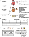
FIGURE 1
FIGURE 1
Schematics of (a) Human iPSC seeding plan, (b) differentiation timeline, and (c) experimental design. Agg, aggregates grown in the VWBR; MC, cells attached to Matrigel‐coated microcarriers in the VWBR; 6‐well, aggregates grown as static culture in 6‐well plates.
FIGURE 2
FIGURE 2
Forebrain spheroid differentiation from human iPSCs on microcarriers and as aggregates in VWBRs (Run 2 data). (a) Culture morphology over time. (b) Cell density, expansion fold, and pH kinetics. H, high seeding density; L, low seeding density. Scale bar of 4×: 500 µm. Scale bar of 10×: 200 µm.
FIGURE 3
FIGURE 3
Metabolite analysis of human iPSC‐derived forebrain spheroids in VWBRs. (a) Changes in glucose consumption, lactate generation, and the ratio of lactate generation to glucose consumption. (b) Changes in glutamine consumption, NH4 + generation, and the ratio of NH4 + generation to glutamine consumption. (c) Metabolite concentrations normalized to cell numbers. (d) Changes in K+ and Ca2+ over time.
FIGURE 4
FIGURE 4
mRNA sequencing of cells grown in the three culture conditions. (a) PCA plot to show the cluster of Agg, MC, and 6‐well. (b) Heatmap illustration of top DEGs among the three culture conditions. (c, e) Volcano plot to show the comparison of DEGs between two culture conditions. (d, f) Network plots for gene set enrichment analysis.
FIGURE 5
FIGURE 5
Neural differentiation, EV biogenesis, and metabolic pathway analysis by RT‐qPCR. (a) Autophagy genes, ESCRT‐independent and ‐dependent genes. (b) Glycolytic genes, pentose phosphate genes and differentiation genes. ESCRT, endosomal sorting complex required for transport. (c) Genes selected from mRNA‐Seq dataset. * indicates < 0.05.
FIGURE 6
FIGURE 6
EV isolation and characterizations (Run 2 data). (a) The representative particle size distribution measured by nanoparticle tracking analysis. (b) EV mean and mode sizes. (c) EV particle numbers normalized to cell numbers. (d) EV particle numbers normalized to protein amounts. (e) Western blot assay of exosomal markers (right) and Ponceau S staing of gel (left). (f) TEM images to show exosome morphology. The double layer was indicated by a pair of pointing arrows. Scale bar: 200 nm. * indicates < 0.05. N.S. not significant.
FIGURE 7
FIGURE 7
Proteomics analysis of forebrain spheroid EV protein cargo. (a) PCA plot to show the cluster of Agg, MC, and 6‐well. (b) Venn diagram of DEPs for the three EV groups. (c, e) Volcano plot to show the comparison of DEPs between two culture conditions. (d, f) Ridgeline diagram of DEP enriched pathways. (g) Proteins selected from proteomics data to match RT‐qPCR data as in Fig.5. (h) Correlation between cell mRNA sequencing data and EV proteomics.
FIGURE 8
FIGURE 8
miRNA sequencing of forebrain spheroid EV miRNA cargo. (a) PCA plot to show the cluster of Agg cell, MC cell, Agg EV and MC EV. (b) The count of miRNAs identified in each sample. (c) Heatmap illustration of top DEGs among the four conditions. (d) DEGs identified in cells and EVs (Agg vs. MC). (e, h) Heatmap of DEGs in MC and Agg (EV vs. cell). (f, i) Volcano plot of DEGs in MC and Agg (EV vs. cell). (g, j) KEGG pathway analysis for the top miRNAs. Enlarged images are provided for (e), (g), (h) and (j) in supplementary materials.
FIGURE 8
FIGURE 8
miRNA sequencing of forebrain spheroid EV miRNA cargo. (a) PCA plot to show the cluster of Agg cell, MC cell, Agg EV and MC EV. (b) The count of miRNAs identified in each sample. (c) Heatmap illustration of top DEGs among the four conditions. (d) DEGs identified in cells and EVs (Agg vs. MC). (e, h) Heatmap of DEGs in MC and Agg (EV vs. cell). (f, i) Volcano plot of DEGs in MC and Agg (EV vs. cell). (g, j) KEGG pathway analysis for the top miRNAs. Enlarged images are provided for (e), (g), (h) and (j) in supplementary materials.
FIGURE 9
FIGURE 9
In vitro functional assays in iFCo OGD stroke model for EV therapeutic evaluation. RT‐qPCR results of EV treatment after OGD. Genes were involved in carbon metabolism (a–c), proliferation and apoptosis (d, e), ROS scavenge (f) and immune response (g, h). OGD, oxygen and glucose deprivation; ROS, reactive oxygen species; TCA, tricarboxylic acid. * indicates < 0.05. N.S., not significant.

References

    1. Abdollahi, S. (2021). Extracellular vesicles from organoids and 3D culture systems. Biotechnology and Bioengineering, 118(3), 1029–1049. - PubMed
    1. Apodaca, L. A. , Baddour, A. A. D. , Garcia, C. , Alikhani, L. , Giedzinski, E. , Ru, N. , Agrawal, A. , Acharya, M. M. , & Baulch, J. E. (2021). Human neural stem cell‐derived extracellular vesicles mitigate hallmarks of Alzheimer's disease. Alzheimer's Research & Therapy, 13(1), 1–18. - PMC - PubMed
    1. Arthur, P. , Kandoi, K. , Sun, L. , Kalvala, A. , Kuthleria, S. , Bhattacharaya, S. , Kulkarni, T. , Nimma, R. , Li, Y. , Lamba, D. A. , & Sing, M. (2023). Biophysical, molecular and proteomic profiling of human retinal organoids derived exosomes. Pharmacological Research, 40(4), 801–816. - PMC - PubMed
    1. Benito‐Kwiecinski, S. , & Lancaster, M. A. (2020). Brain organoids: Human neurodevelopment in a dish. Cold Spring Harbor Perspectives in Biology, 12(8), a035709. - PMC - PubMed
    1. Borys, B. S. , Dang, T. , So, T. , Rohani, L. , Revay, T. , Walsh, T. , Thompson, M. , Argiropoulos, B. , Rancourt, D. E. , & Jung, S. (2021). Overcoming bioprocess bottlenecks in the large‐scale expansion of high‐quality hiPSC aggregates in vertical‐wheel stirred suspension bioreactors. Stem Cell Research & Therapy, 12(1), 1–19. - PMC - PubMed

LinkOut - more resources