. 2024 Jul 26;27(8):110591.
doi: 10.1016/j.isci.2024.110591. eCollection 2024 Aug 16.

Dual effect of N-terminal deletion of cardiac myosin essential light chain in mitigating cardiomyopathy

Affiliations

Dual effect of N-terminal deletion of cardiac myosin essential light chain in mitigating cardiomyopathy

Yoel H Sitbon et al. iScience. .

Abstract

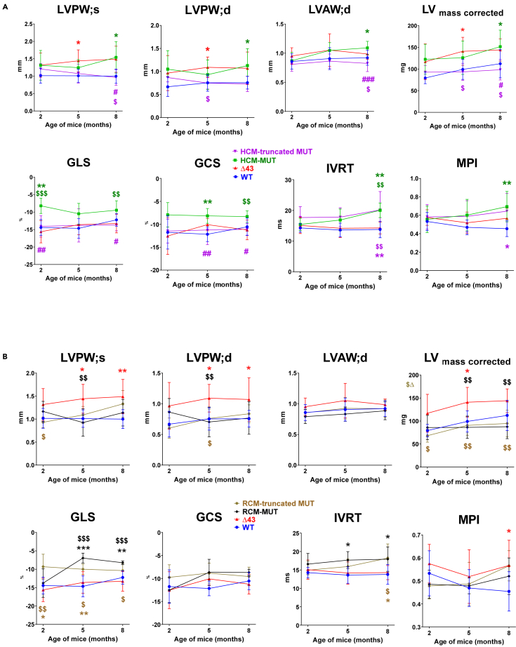
We investigated the role of the N-terminus (residues 1-43) of the myosin essential light chain (N-ELC) in regulating cardiac function in hypertrophic (HCM-A57G) and restrictive (RCM-E143K) cardiomyopathy mice. Both models were cross-genotyped with N-ELC-truncated Δ43 mice, and the offspring were studied using echocardiography and muscle contractile mechanics. In A57G×Δ43 mice, Δ43 expression improved heart function and reduced hypertrophy and fibrosis. No improvements were seen in E143K×Δ43 compared to RCM-E143K mice. HCM-mutant pathology involved an impaired N-ELC tension sensor, disrupted N-ELC-actin interactions, an altered force-pCa relationship, and a destabilized myosin's super-relaxed state. Removal of the malfunctioning N-ELC sensor led to functional rescue in HCM-truncated mutant hearts. However, the RCM mutation could not be rescued by N-ELC deletion, likely due to its proximity to the myosin motor domain, affecting lever-arm rigidity and myosin function. This study provides insights into the role of N-ELC in the development and potential rescue of ELC-mutant cardiomyopathy.

Keywords: Biochemical mechanism; Medical biochemistry; Molecular medicine; Pathophysiology.


Conflict of interest statement

The authors declare no competing interests.

Figures

None
Graphical abstract
Figure 1
Figure 1
Evaluation of heart function in ELC mouse models of cardiomyopathy Echocardiography measurements were conducted in 2, 5, and 8-month-old WT, Δ43, HCM-mutant, and HCM-truncated mutant mice (A) and WT, Δ43, RCM-mutant, and RCM-truncated mutant mice (B). Parameters measured included LVPW (left ventricular posterior wall thickness in systole and diastole), LVAW (LV anterior wall), and LV mass. Additionally, speckle-tracking-based strain analysis was performed, measuring GLS (global longitudinal strain) and GCS (global circumferential strain). Evaluation of isovolumetric relaxation time (IVRT) and myocardial performance index (MPI) was also conducted. The data represent the mean of n = N° mice (shown in Tables S1–S3) ±SD, with statistical analysis performed using one-way ANOVA with Tukey’s multiple comparisons test. Significance is indicated by ∗p versus WT, $p versus Δ43, and #p versus HCM-mutant.
Figure 2
Figure 2
Assessment of fibrosis and collagen content in the left ventricles (LV) of ∼8-month-old WT, HCM, RCM, Δ43, HCM-truncated, and RCM-truncated mutant mice (A) Representative images of picrosirius red-stained LV in mice. Scale bar, 100 μm. (B) SuperPlots showing the percentage of fibrotic area quantified from a total of 4 WT (2 F and 2 M), 3 HCM-mutant (2 F and 1 M), 4 RCM-mutant (2 F and 2 M), 3 Δ43 (2 F and 1 M), 3 HCM-truncated (3 F), and 3 RCM-truncated (2 F and 1 M) mutant mice. This analysis includes 4–7 LV images per animal and 16–22 per group. The data per animal are presented using large, color-coded symbols (circles for WT, squares for HCM-mutant, diamonds for RCM-mutant, triangles for Δ43, inverse triangles for HCM-truncated, and hexagons for RCM-truncated mutant hearts). The respective image measurements are indicated by small, color-coded symbols. Open symbols indicate female (F), and closed symbols indicate male (M) mice. Data are presented as the mean of n = Nº images ±SD (standard deviation), with significance calculated using one-way ANOVA with Tukey’s multiple comparisons test.
Figure 3
Figure 3
TEM images of LV samples from ∼8-month-old female ELC mutant mice (A) Representative images at 3000x and 5000× magnification of LV samples from WT, HCM-mutant, RCM-mutant, Δ43, HCM-truncated mutant, and RCM-truncated mutant mice. Scale bars, 1 μm (3000x) and 800 nm (5000x). (B) Number of mitochondria per μm2 in WT, HCM-mutant, RCM-mutant, Δ43, HCM-truncated mutant, and RCM-truncated mutant hearts analyzed using 3000x images (5 images per animal). Note the significantly increased number of mitochondria in RCM-mutant versus WT, HCM-mutant, and Δ43 mice. (C) Mitochondrial cross-section area (in μm2) for the group of mice depicted in B. Note the increased mitochondrial area in HCM-truncated mutant versus HCM-mutant hearts. Data are mean of n = Nº images ±SD and analyzed using one-way ANOVA with Tukey’s multiple comparisons test.
Figure 4
Figure 4
Partial deletion of N-ELC rescues the SRX↔DRX equilibrium in ∼8-month-old cross-genotype models (A and B) HCM-truncated mutant and B. RCM-truncated mutant mice. Values are means of n = N° mice ±SD with significance (p) determined using one-way ANOVA with Tukey’s multiple comparisons test. Open symbols represent female mice, and closed symbols represent male mice.
Figure 5
Figure 5
Assessment of contractile function in ELC mouse models of cardiomyopathy Isometric steady-state force and force-pCa relationship were conducted in LVPM fibers from ∼8-month-old WT, HCM-mutant, Δ43, and HCM-truncated mutant (A) and WT, RCM-mutant, Δ43, and RCM-truncated mutant (B) groups. The left panels illustrate maximal tension at pCa4, while the right panels depict the Ca2+ sensitivity of force development. The data are presented as means ± SD of n = Nº animals (5–10 fibers per heart). Statistical significance was determined using one-way ANOVA with Tukey’s multiple comparisons test. Open symbols represent female mice, and closed symbols represent male mice.
Figure 6
Figure 6
Effect of N-ELC deletion on myosin RLC phosphorylation in ∼8-month-old double ELC mutant mice (A) Representative western blot of 8 M urea extracts from left ventricle (LV) samples separated by urea-PAGE. Phosphorylated (+P-RLC) and non-phosphorylated forms of RLC were separated based on differences in their isoelectric points (pI) and detected using a myosin RLC-specific antibody (CT-1) developed in our laboratory. (B) Quantification of myosin RLC phosphorylation in the hearts of WT, HCM-mutant, RCM-mutant, Δ43, HCM-truncated mutant, and RCM-truncated mutant mice, represented as SuperPlots. Data for each animal (three animals per group) are depicted using large, color-coded symbols, with triangles representing male mice and circles depicting female mice. Individual mouse measurements are indicated by small, color-coded symbols, with error bars denoting standard deviations. Data are presented as the mean of n = Nº animals ±SD with significance calculated using one-way ANOVA with Tukey’s multiple comparisons test.
Figure 7
Figure 7
I-TASSER modeled structures of the human ventricular WT, Δ43, A57G, and E143K ELC mutants Notably, the A57G mutation leads to changes in the spatial arrangement of the N-terminus when compared to the WT and the E143K mutant. These changes are likely attributed to the presence of a helical structure(s) between amino acids 29–43 in A57G-ELC, which is not observed in the WT or E143K.

References

    1. Alcalai R., Seidman J.G., Seidman C.E. Genetic Basis of Hypertrophic Cardiomyopathy: From Bench to the Clinics. J. Cardiovasc. Electrophysiol. 2008;19:104–110. doi: 10.1111/j.1540-8167.2007.00965.x. - DOI - PubMed
    1. Seidman C.E., Seidman J.G. Identifying sarcomere gene mutations in hypertrophic cardiomyopathy: a personal history. Circ. Res. 2011;108:743–750. doi: 10.1161/CIRCRESAHA.110.223834. - DOI - PMC - PubMed
    1. Semsarian C., Ingles J., Maron M.S., Maron B.J. New perspectives on the prevalence of hypertrophic cardiomyopathy. J. Am. Coll. Cardiol. 2015;65:1249–1254. doi: 10.1016/j.jacc.2015.01.019. - DOI - PubMed
    1. Semsarian C., Ingles J., Wilde A.A.M. Sudden cardiac death in the young: the molecular autopsy and a practical approach to surviving relatives. Eur. Heart J. 2015;36:1290–1296. doi: 10.1093/eurheartj/ehv063. - DOI - PubMed
    1. Yadav S., Sitbon Y.H., Kazmierczak K., Szczesna-Cordary D. Hereditary heart disease: pathophysiology, clinical presentation, and animal models of HCM, RCM, and DCM associated with mutations in cardiac myosin light chains. Pflügers Archiv. 2019;471:683–699. doi: 10.1007/s00424-019-02257-4. - DOI - PMC - PubMed

LinkOut - more resources