Review
. 2024 Jul 30;16(7):e65742.
doi: 10.7759/cureus.65742. eCollection 2024 Jul.

Oral Granulomatous Disorders: A Diagnostic Insight

Affiliations
Review

Oral Granulomatous Disorders: A Diagnostic Insight

Durba Roychowdhury et al. Cureus. .

Abstract

Granulomatous inflammation represents a unique pattern of chronic inflammation observed in a restricted form of infectious and certain non-infectious diseases. The formation of granulomas typically involves immune responses. Granulomatous disorders encompass a broad spectrum of conditions that share the common histological feature of granuloma formation. Their involvement in the oral soft and hard tissues is quite infrequent; however, their manifestation can pose a diagnostic challenge due to the diverse range of potential causes and the relatively non-specific appearance of the individual lesions. The ultimate outcome of a complex entails the formation of a granuloma, resulting from the interplay among an invading pathogen or antigen, chemical substance, medication, or other irritant, persistent presence of antigens in the bloodstream, activation of macrophages, initiation of Th1 cell response, B-cell overactivity, presence of circulating immune complexes, and a wide range of biological signaling molecules, ultimately leading to the development of fibrosis attributed to the actions of transforming and platelet-derived growth factor. This article emphasizes the clinicopathological diagnostic criteria of oral granulomatous disorders as a guide for treatment and management.

Keywords: caseation necrosis; granuloma; inflammation; management; non-caseating.


Conflict of interest statement

Conflicts of interest: In compliance with the ICMJE uniform disclosure form, all authors declare the following: Payment/services info: All authors have declared that no financial support was received from any organization for the submitted work. Financial relationships: All authors have declared that they have no financial relationships at present or within the previous three years with any organizations that might have an interest in the submitted work. Other relationships: All authors have declared that there are no other relationships or activities that could appear to have influenced the submitted work.

Figures

Figure 1
Figure 1. Pathogenesis of a granuloma.
IL: interleukin, IFN-γ: interferon-γ, TNF: tumor necrosis factor; MHC: major histocompatibility complex Permission has been obtained from the original publishers to reproduce this figure from the source [3].
Figure 2
Figure 2. Structure and cellular composition of granulomas.
Permission has been obtained from the original publishers to reproduce this figure from the source [13].
Figure 3
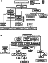
Figure 3. Diagnostic algorithm for a) pulmonary tuberculosis (TB) and b) extra-pulmonary tuberculosis (EPTB) as per the Revised National TB Control Program (RNTCP) guidelines.
TB: tuberculosis, AFB: acid-fast bacilli, RIF: rifampicin Permission has been obtained from the original publishers to reproduce this figure from the source [22].
Figure 4
Figure 4. Positive acid-fast bacilli (AFB) in sputum smear.
Permission has been obtained from the original publishers to reproduce this figure from the source [23].
Figure 5
Figure 5. Oral tuberculosis: photomicrograph revealing tuberculous granulomas along with Langhans giant cells (black arrow), epithelioid cells (red arrow), and lymphocytes (blue arrow) (x40).
Courtesy: College of Medicine & Sagore Dutta Hospital
Figure 6
Figure 6. Schematic illustration of the approach to the patient with suspected leprosy according to the WHO criteria.
Permission has been obtained from the original publishers to reproduce this figure from the source [27].
Figure 7
Figure 7. Paucibacillary (tuberculoid) leprosy. Well-formed granulomatou inflammation illustrating clusters of lymphocytes and histiocytes.
Permission has been obtained from the original publishers to reproduce this figure from the source [29].
Figure 8
Figure 8. Multibacillary (lepromatous) leprosy. Sheets of lymphocytes and histiocytes displaying dispersed vacuolated lepra cells (black arrow).
Permission has been obtained from the original publishers to reproduce this figure from the source [20].
Figure 9
Figure 9. Multibacillary (lepromatous) leprosy. Acid-fast stain demonstrating numerous small mycobacterial organisms(black arrow) observed individually or in clusters.
Permission has been obtained from the original publishers to reproduce this figure from the source [20].
Figure 10
Figure 10. Diagnostic algorithm for actinomycosis.
MALDI-TOF: matrix-assisted laser desorption ionization time-of-flight mass spectrometry Permission has been obtained from the original publishers to reproduce this figure from the source [31].
Figure 11
Figure 11. (a) Colony of actinomycotic organisms (black arrow) surrounded by polymorphonuclear leukocytes (x10). (b) Actinomycotic colony showing club-shaped filaments arranged in a radiating rosette pattern (red arrow) (x40).
Permission has been obtained from the original publishers to reproduce this figure from the source [20].
Figure 12
Figure 12. A step-by-step process for the diagnosis of invasive candidiasis (IC) and invasive aspergillosis (IA).
CVC: central venous catheter, TPN: total parenteral nutrition, BDG: β-d-glucan, GM: galactomannan, rt-PCR: real-time PCR, Mn: Mannan, A-Mn: anti-Mannan antibody, NPV: negative predictive value Permission has been obtained from the original publishers to reproduce this figure from the source [33].
Figure 13
Figure 13. Photomicrograph revealing scattered epithelioid macrophages intermixed with lymphocytes and plasma cells. Some macrophages comprise organisms of histoplasma capsulatum (arrows) (x40). Inset: high-power photomicrograph of a tissue section readily illustrates the small yeasts of histoplasma capsulatum (Grocott-Gomori methenamine silver stain).
Permission has been obtained from the original publishers to reproduce this figure from the source [23].
Figure 14
Figure 14. Photomicrograph shows fungal hyphae with a fruiting body (black arrow) of an Aspergillus spp. Inset: the high-power photomicrograph reveals the characteristic septate hyphae of Aspergillus spp. (Grocott-Gomori methenamine silver stain).
Permission has been obtained from the original publishers to reproduce this figure from the source [20].
Figure 15
Figure 15. Zygomycosis: the photomicrograph demonstrates the large, nonseptate fungal hyphae distinctive of the zygomycotic organisms.
Permission has been obtained from the original publishers to reproduce this figure from the source [20].
Figure 16
Figure 16. Proposed diagnostic methods for the evaluation of patients with suspicion of pulmonary Langerhan’s cell histiocytosis (PLCH).
Permission has been obtained from the original publishers to reproduce this figure from the source [36].
Figure 17
Figure 17. Langerhans cell histiocytosis: a) severe bone resorption involving the mandibular molar regions exhibiting similarities to advanced periodontitis; b) periapical radiograph shows marked bone resorption in the mandibular teeth in a young girl, resulting in a “floating-in-the-air” appearance; c) the lesion has extended beyond the bone producing this proliferative soft tissue mass.
Permission has been obtained from the original publishers to reproduce this figure from the source [20].
Figure 18
Figure 18. Diffuse infiltration of pale-staining Langerhans cells admixed with numerous red granular eosinophils. Inset: electron micrograph illustrating rod-shaped birbeck bodies (black arrows) in the cytoplasm of a Langerhans cell.
Permission has been obtained from the original publishers to reproduce this figure from the source [20].
Figure 19
Figure 19. Diagnostic methods for sarcoidosis.
Permission has been obtained from the original publishers to reproduce this figure from the source [38].
Figure 20
Figure 20. Sarcoidosis: photomicrograph of a labial minor salivary gland showing granulomatous inflammation distinguised by encircled aggregates of histiocytes, lymphocytes, and multinucleated giant cells. Inset: photomicrograph revealing a multinucleated giant cell with an intracytoplasmic asteroid body (black arrow).
Permission has been obtained from the original publishers to reproduce this figure from the source [20].
Figure 21
Figure 21. Algorithm for diagnosing Crohn's disease.
* esophagogastroduodenoscopy may be deemed, especially for children and/or in the presence of upper gastrointestinal symptoms; † choice of cross-sectional imaging technique depends on several factors. Permission has been obtained from the original publishers to reproduce this figure from the source [41].
Figure 22
Figure 22. Crohn’s disease: medium-power photomicrograph of an oral lesion showing a non-necrotizing granuloma in the submucosal connective tissue.
Permission has been obtained from the original publishers to reproduce this figure from the source [20].

References

    1. Mohan H, Mohan S. JP Medical Ltd. JP Medical Ltd; 2012. Essential pathology for dental students.
    1. Kumar V, Abbas A. K, Fausto N, Aster J.C. Robbins and Cotran Pathologic Basis of Disease. Vol. 43. Elsevier Health Sciences; 2012. Robbins and Cotran pathologic basis of disease; p. 77.
    1. A clinicopathological classification of granulomatous disorders. James DG. Postgrad Med J. 2000;76:457–465. - PMC - PubMed
    1. Granulomatous diseases of the oral tissues- an etiology & histopathology. Gotmare S, Tamgadge A, Bhalerao S, et al. 2007
    1. Macrophage structure | Britannica. 2024. https://www.britannica.com/science/macrophage https://www.britannica.com/science/macrophage

LinkOut - more resources