. 2024 Aug 15:11:1365783.
doi: 10.3389/fmolb.2024.1365783. eCollection 2024.

Enhanced enrichment of extracellular vesicles for laboratory and clinical research from drop-sized blood samples

Affiliations

Enhanced enrichment of extracellular vesicles for laboratory and clinical research from drop-sized blood samples

Alexa Guerrero-Alba et al. Front Mol Biosci. .

Abstract

In the realm of biomedical advancement, extracellular vesicles (EVs) are revolutionizing our capacity to diagnose, monitor, and predict disease progression. However, the comprehensive exploration and clinical application of EVs face significant limitations due to the current isolation techniques. The size exclusion chromatography, commercial precipitation reagents, and ultracentrifugation are frequently employed, necessitating skilled operators and entailing challenges related to consistency, reproducibility, quality, and yields. Notably, the formidable challenge of extracellular vesicle isolation persists when dealing with clinical samples of limited availability. This study addresses these challenges by aiming to devise a rapid, user-friendly, and high-recovery EVs isolation technique tailored for blood samples. The NTI-EXO precipitation method demonstrated a 5-fold increase in the recovery of serum EVs compared to current methodologies. Importantly, we illustrate that a mere two drops of blood (∼100 µL) suffice for the recovery of enriched EVs. The integrity and quality of these isolated EVs were rigorously assessed for the size, purity, and contaminants. This method was validated through the successful isolation of EVs from organ transplant recipients to detect disease-specific exosomal markers, including LKB1, SARS-CoV-2 spike protein, and PD-L1. In conclusion, NTI-EXO method can be used for small clinical samples, thereby advancing discoveries in the EV-centric domain and propelling the frontiers of biomedical research and clinical applications.

Keywords: EVS; blood; diagnosis; exosome; marker; transplant.


Conflict of interest statement

The authors declare that the research was conducted in the absence of any commercial or financial relationships that could be construed as a potential conflict of interest.

Figures

FIGURE 1
FIGURE 1
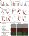
A comparison of NTI-EXO experimental conditions for the efficient recovery of EVs from human and porcine serum (A) A depiction of the NTI-EXO protocol yielding EVs from human and porcine serum. (B–C) EVs yields isolated from normal human serum from various combinations of NTI-EXO, PBS, and centrifugation speeds. (D) Characterization of human- and porcine-derived EVs using western blot. EVs recovered by NTI-EXO, TF (Thermo Fisher reagent), and UC (ultracentrifugation) were compared. CD9, CD63, CD86, ALIX, 20 s, Flot-1, NFkB1, and TSG101 were analyzed. Some of these markers were better enriched with the NTI-EXO method than commercial or standard (UC) methods. Twenty micrograms of the total protein were used for immunoblotting experiments. Experimental conditions A1-A3, B1-B3 and C1-C3 are individual isolation conditions. Values are given as mean ± S.D. of three independent experiments. Statistical significance was defined at **p < 0.01, and ***p < 0.001 compared to the corresponding control.
FIGURE 2
FIGURE 2
Comparative characterization of EVs isolated from human and porcine serum using NTI-EXO or ultracentrifugation. (A) Yields, (B) size, and (C) concentration of EVs isolated by different experimental conditions (see Figure 1B). Total protein yields were assayed by BCA; Size and concentration were analyzed using NanoSight data (NTA). (D) NanoSight plots displaying EV size and calculated mean, mode, and size characterization. (E) Cartoon showing overall EVs uptake assay. EVs were added to NIH-3T3 cells; UC-exosomes (reporter exosomes isolated using ultracentrifugation), TF-Exosomes (commercial kit isolated exosomes) and NTI-EXZO isolated exosomes. Statistical significance was defined at **p < 0.01, and ***p < 0.001 compared to the corresponding control.
FIGURE 3
FIGURE 3
Exosomal protein recovery and characterization from serum samples. (A, B) Total initial, recovered, and not recovered (waste) protein yields from NTI-EXO of porcine and normal human serum samples and 2 serum samples from lung transplant patients. Total isolated EV-enriched protein was analyzed using BCA and SDS-PAGE-Coomassie. (C, D) NanoSight data demonstrating distribution of EVs. Results show that based on initial plasma proteins, 20%–30% of EV-enriched proteins were recovered. Based on concentration, over 95% of EVs were isolated from 4 different samples. (E, F) Control serum and EVs isolated using NTI-EXO, TF or UC were analyzed for total protein and albumin (BCG assay). (G) Fold enrichment of total protein is compared in three methods for human and porcine serum. (H) EVs purity was further assayed for initial and carried over albumin and ApOA1 using immunoblotting. (I) Whole mount TEM images of EVs enriched using NTI-EXO precipitation of human and porcine serum and SEC. Scale bars, 100 nm. Statistical significance was defined at **p < 0.01, and ***p < 0.001 compared to the corresponding control.
FIGURE 4
FIGURE 4
Optimal NTI-EXO concentration to enrich EV isolation. The NTI-EXO method was used to isolate EVs under 100 nM in diameter. The NTI-EXO reagent at a ratio to final volume of 1X, 0.5X, 0.25X and 0.16X was used. (A) NanoSight detection of EV intensity versus size of the population. (B) Protein concentration of adding the reagent in a 1X ratio (blue) versus 2X ratio (black) was measured by BCA (C). Lowering pH with a low concentration of NTI-EXO improved EV recovery with EVs size at 120 nm (D). Sodium acetate pH 4.5 was added with NTI-EXO 1X, EV size measured ny NTA. (E, F). Western blots data demonstrates that NTI-EXO dilution reduced EV recovery, which can be compensated for by lowering the pH. This recovery was confirmed by immunoblotting CD63 and Flot-1 together with a loading control, LC-IgG (kappa light chain IgG). Band intensity was analyzed and plotted using ImageJ software. Statistical significance was defined at **p < 0.01, and ***p < 0.001 compared to the corresponding control.
FIGURE 5
FIGURE 5
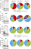
Comparison of particle size and marker composition of EVs isolated by SEC from human serum or by NTI-EXO from human, porcine, or mouse sera. The ExoView R200 platform was used for characterization. (A) Particle size and marker composition from SEC-isolated human EVs. NTI-EXO EV isolation results from (B) human serum, (C) porcine serum, and (D) mouse serum. The NTI- or SEC-isolated EVs were characterized for detection of CD81, CD63, and CD9 and the isotype control MIgG. Co-localization of serum-derived EVs was shown using 3 fluorescent channels and an overlay of fluorescent images. Data represents the CD81, CD63, and CD9 co-localization (%). Data are the mean ± SEM of 3 independent biological experiments with 3 technical replicates.
FIGURE 6
FIGURE 6
Validation of NTI-EXO by comparison with our previous disease marker results. Previous work with UC and commercial reagents was reproduced using the NTI-EXO EV isolation method. Frozen patient plasma samples were treated with NTI-EXO to isolate EVs. EVs isolated from a lung transplant patient sample: (A) total recovery, and (B) characterization using immunoblotting for LKB1 (Santa Cruz Biotechnology and Cell Signaling), CD9, and a loading control LC-IgG. (C, D) EVs isolated from plasma from a lung transplant patient with COVID-19 using NTI-EXO. Extracted EV samples were immunoblotted for SARS-CoV-2 spike protein, CD63, with loading control LCIgG. Disease markers as well as EV markers were detected in EV-enriched proteins isolated by TF or NTI-EXO.
FIGURE 7
FIGURE 7
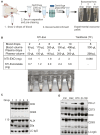
Breakthrough single drop-method to isolate EVs from mouse serum. (A) Depiction of method to isolate EVs from drop-sized samples to obtain a visible pellet of EVs. (B) Data from 2 independent experiments of porcine or mouse sera obtained from animals undergoing organ transplant studies. EVs were isolated from fresh serum or frozen plasma samples by NTI-EXO or TF. The NTI-EXO-Acetate method increased recovery. Visible pellets can be seen in the samples processed by the NTI-EXO method. (C) At 12 h of EVLP, porcine plasma was assayed for EV markers. From 1 to 5 drops of plasma were acquired and treated with NTI-EXO for EV isolation followed by protein measurement and immunoblotting for EV markers including CD9, CD63, ALIX, 20 s, and the loading control LC-IgG. Twenty micrograms of total protein were resolved for immunoblotting. (D) Two drops of plasma from kidney transplanted mice were used for EV isolation to monitor PD-L1, CD73, EV markers CD9 and CD63, and the loading control LC-IgG. Data suggest small-sized serum samples can be used successfully to characterize disease-related markers.

References

    1. Abd Elmageed Z. Y., Yang Y., Thomas R., Ranjan M., Mondal D., Moroz K., et al. (2014). Neoplastic reprogramming of patient-derived adipose stem cells by prostate cancer cell-associated exosomes. Stem Cells 32 (4), 983–997. 10.1002/stem.1619 - DOI - PMC - PubMed
    1. Agarwal A., Fanelli G., Letizia M., Tung S. L., Boardman D., Lechler R., et al. (2014). Regulatory T cell-derived exosomes: possible therapeutic and diagnostic tools in transplantation. Front. Immunol. 5, 555. 10.3389/fimmu.2014.00555 - DOI - PMC - PubMed
    1. Bachurski D., Schuldner M., Nguyen P. H., Malz A., Reiners K. S., Grenzi P. C., et al. (2019). Extracellular vesicle measurements with nanoparticle tracking analysis - an accuracy and repeatability comparison between NanoSight NS300 and ZetaView. J. Extracell. Vesicles 8 (1), 1596016. 10.1080/20013078.2019.1596016 - DOI - PMC - PubMed
    1. Bansal S., Limaye A. P., Lee J., Fleming T., Poulson C., Omar A., et al. (2021a). Circulating exosomes induced by respiratory viral infections in lung transplant recipients activate cellular stress, innate immune pathways and epithelial to mesenchymal transition. Transpl. Immunol. 69, 101480. 10.1016/j.trim.2021.101480 - DOI - PubMed
    1. Bansal S., Perincheri S., Fleming T., Poulson C., Tiffany B., Bremner R. M., et al. (2021b). Cutting edge: circulating exosomes with covid spike protein are induced by BNT162b2 (Pfizer-BioNTech) vaccination prior to development of antibodies: a novel mechanism for immune activation by mRNA vaccines. J. Immunol. 207 (10), 2405–2410. 10.4049/jimmunol.2100637 - DOI - PMC - PubMed

LinkOut - more resources