Review
. 2024 Aug 15:14:1421571.
doi: 10.3389/fcimb.2024.1421571. eCollection 2024.

Unraveling the complex interplay: immunopathology and immune evasion strategies of alphaviruses with emphasis on neurological implications

Affiliations
Review

Unraveling the complex interplay: immunopathology and immune evasion strategies of alphaviruses with emphasis on neurological implications

Raquel de Oliveira Souza et al. Front Cell Infect Microbiol. .

Abstract

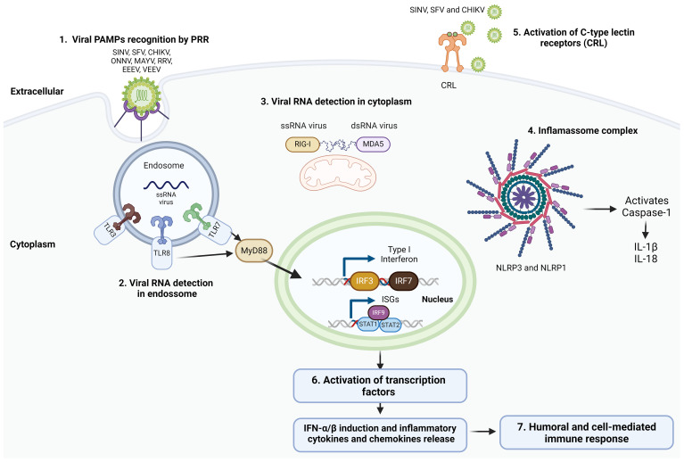
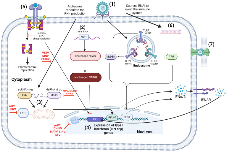
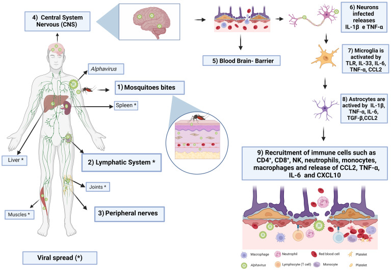
Arthritogenic alphaviruses pose a significant public health concern due to their ability to cause joint inflammation, with emerging evidence of potential neurological consequences. In this review, we examine the immunopathology and immune evasion strategies employed by these viruses, highlighting their complex mechanisms of pathogenesis and neurological implications. We delve into how these viruses manipulate host immune responses, modulate inflammatory pathways, and potentially establish persistent infections. Further, we explore their ability to breach the blood-brain barrier, triggering neurological complications, and how co-infections exacerbate neurological outcomes. This review synthesizes current research to provide a comprehensive overview of the immunopathological mechanisms driving arthritogenic alphavirus infections and their impact on neurological health. By highlighting knowledge gaps, it underscores the need for research to unravel the complexities of virus-host interactions. This deeper understanding is crucial for developing targeted therapies to address both joint and neurological manifestations of these infections.

Keywords: alphavirus; chikungunya; coinfection; immune evasion; immunopathology; mayaro; mouse; nervous system.


Conflict of interest statement

The authors declare that the research was conducted in the absence of any commercial or financial relationships that could be construed as a potential conflict of interest.

Figures

Figure 1
Figure 1
Alphavirus induced innate immune response. The innate immune response induced by alphaviruses involves a series of key mechanisms. (1) Alphavirus recognition is mediated by pattern recognition receptors (PRRs) that detect pathogen-associated molecular patterns (PAMPs) associated with the invading viruses. This recognition is facilitated by various receptors, including (2) TLR3, TLR7, and TLR8 located in endosomes, as well as (3) RIG-I-like receptors (RLRs) such as RIG-I and MDA5 found in the cytoplasm. These receptors detect viral RNA and initiate antiviral responses by activating signaling cascades that ultimately lead to the activation of transcription factors. (4) NOD-like receptors (NLRs), including NLRP3 and NLRP1, form inflammasomes that activate caspase-1, resulting in the production of pro-inflammatory cytokines IL-1β and IL-18. (5) C-type lectin receptors (CLRs) are also activated during alphavirus infection, playing a role in modulating the immune response. (6) The activation of PRRs triggers signaling cascades that culminate in the activation of transcription factors. This activation leads to the expression of type I interferons (IFNs), IFN-stimulated genes (ISGs), as well as the production of pro-inflammatory cytokines and chemokines. (7) These responses are crucial for mounting an effective antiviral humoral and cell-mediated immune response against alphaviruses. [Created with BioRender.com with license no. OL270LDGL6.].
Figure 2
Figure 2
Alphavirus immune evassion with emphasis on type I IFN responses. (1) Alphavirus attachment and entry are mediated by host pattern recognition receptors (PRRs), such as RIG, NOD, and TLRs, triggering the production of IFN-I. Once inside the host’s cells, alphaviruses interfere with the IFN-I system at various levels: (2) Inhibition of the cGAS-STING pathway. Following CHIKV infection, there is a significant decrease in cGAS expression, while STING expression remains relatively stable. The non-structural protein nsPl interacts with STING, affecting the activation of IFNs and the activation of the IFN-β promoter, typically controlled by cGAS-STING pathway; (3) CHIKV nsP2 and E1/E2, and SINV nsPI, inhibit the activation of the IFNβ-promoter triggered by the MDA5/RIG-I receptor signaling pathway; (4) The nsP2 from CHIKV, MAYV, SINV, SFV influence ISGs through modifications such as palmitoylation and phosphorylation to hinder interferon α/β and ISG production; (5) Inhibition of JAK-STAT signaling: CHIKV disrupts the phosphorylation of STAT1, essential for IFN-induced JAK-STAT signalling, aiding in immune evasion and enhancing viral replication. (6) Alphaviruses develop viral RNAi suppressors to evade RNAi-based defense mechanisms in eukaryote, as exemplified by the Semliki virus. (7) Alphaviruses like CHIKV employ strategies to avoid the adaptive immune response, including the formation of stable cellular extensions that shield the virus from neutralizing antibodies and facilitate efficient intercellular transmission. [Created with BioRender.com with license no. BG270SYVXK.].
Figure 3
Figure 3
Proposed mechanism of arthritogenic viruses’ pathogensis: from skin entry to neuroinflammation. (1) When a virus-infected mosquito bites a human, it injects saliva containing the virus through its proboscis, breaking the skin barrier. The virus initially infects dendritic cells and fibroblasts, triggering the activation of the innate- immune system through pattern recognition receptors (PRRs) like Toll-like receptors (TLRs) and RIG-I type receptors (RLRs). This initiates a series of immune responses, including the recruitment of macrophages and other immune cells, as well as the production of pro-inflammatory cytokines (such as IL-6 and TNF-α), chemokines (CCL2, CXCL10), and interferons (IFN-α. and IFNβ); (2) despite the body’s efforts to contain the virus, it reaches the nearest lymph node with the help of migratory immune cells like dendritic cells. In the lymph nodes, the virus replicates intensely before .entering the bloodstream, causing viremia. From there, the virus spreads to various organs like the liver, muscles, joints, spleen, and, in severe cases, the brain; (3) the virus reaches the brain through peripheral nerves and the bloodstream; (4) once in the Central Nervous System (CNS), (5) the virus compromises the blood-brain barriers integrity, using mechanisms like “Trojan horse,” transcytosis, or direct disruption to enter the sterile environment; (5) infected neurons release signals like TNF-α and IL-1β, activating glial cells; (6) microglia, specialized macrophages acting as sentinel cells, are the first to respond to neuronal infection by recognizing danger signals from neurons through receptors like Toll-like receptors; (8) this triggers the activation of microglia, followed by astrocyte activation. Activated astrocytes release various molecules like IL-1β, TNF-α, IL-6, IFN- γ, TGF-β, and CCL2, which promote neuroroinflammation and modulate blood-brain barrier permeability; (9) astrocytes further stimulate an inflammatory response by recruiting immune cells like CD4+T, CD8+T, NK cells, neutrophils, monocytes, and macrophages to the site of infection, releasing chemokines and cytokines like CXCL10, CCL2, TNF-α, and IL-6. [Created with Biorender.com with license no. DJ272BIEJ1.].

Similar articles

Cited by

References

    1. Adamu A., Li S., Gao F., Xue G. (2024). The role of neuroinflammation in neurodegenerative diseases: current understanding and future therapeutic targets. Front. Aging Neurosci. 16. doi: 10.3389/fnagi.2024.1347987 - DOI - PMC - PubMed
    1. Aguilar M. J. (1970). Pathological changes in brain and other target organs of infant and weanling mice after infection with non-neuroadapted Western equine encephalitis virus. Infect. Immun. 2, 533–542. doi: 10.1128/iai.2.5.533-542.1970 - DOI - PMC - PubMed
    1. Aguilar P. V., Estrada-Franco J. G., Navarro-Lopez R., Ferro C., Haddow A. D., Weaver S. C. (2011). Endemic Venezuelan equine encephalitis in the Americas: hidden under the dengue umbrella. Future Virol. 6, 721–740. doi: 10.2217/fvl.11.5 - DOI - PMC - PubMed
    1. Akhrymuk I., Frolov I., Frolova E. I. (2018). Sindbis Virus Infection Causes Cell Death by nsP2-Induced Transcriptional Shutoff or by nsP3-Dependent Translational Shutoff. J. Virol. 92 (23), e01388-18. doi: 10.1128/JVI.01388-18 - DOI - PMC - PubMed
    1. Albeituni S., Stiban J. (2019). Roles of ceramides and other sphingolipids in immune cell function and inflammation. Adv. Exp. Med. Biol. 1161, 169–191. doi: 10.1007/978-3-030-21735-8_15 - DOI - PubMed

MeSH terms

LinkOut - more resources