. 2024 Oct 7;221(10):e20231562.
doi: 10.1084/jem.20231562. Epub 2024 Aug 30.

RAC2 gain-of-function variants causing inborn error of immunity drive NLRP3 inflammasome activation

Affiliations

RAC2 gain-of-function variants causing inborn error of immunity drive NLRP3 inflammasome activation

Anne Doye et al. J Exp Med. .

Abstract

A growing number of patients presenting severe combined immunodeficiencies attributed to monoallelic RAC2 variants have been identified. The expression of the RHO GTPase RAC2 is restricted to the hematopoietic lineage. RAC2 variants have been described to cause immunodeficiencies associated with high frequency of infection, leukopenia, and autoinflammatory features. Here, we show that specific RAC2 activating mutations induce the NLRP3 inflammasome activation leading to the secretion of IL-1β and IL-18 from macrophages. This activation depends on the activation state of the RAC2 variant and is mediated by the downstream kinase PAK1. Inhibiting the RAC2-PAK1-NLRP3 inflammasome pathway might be considered as a potential treatment for these patients.


Conflict of interest statement

Disclosures: A. Doye, P. Chaintreuil, O. Visvikis, E. Verhoeyen, and L. Boyer reported a patent to EP23305994.8 pending and a patent to EP24306144.7 pending. S. Kracker reported a patent to EP23305994.8 pending. No other disclosures were reported.

Figures

Figure 1.
Figure 1.
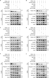
RAC2 Q61R, RAC2 D63V, RAC2 E62K, and RAC2 G12R mutants bind PAK1 RBD. Hemagglutinin-tagged RAC2 (WT) and mutants were expressed for 16 h in HEK293T cells. HA-RAC2 activation level measurements were assessed by GST–PAK–RBD pull-down assay followed by immunoblotting (IB). (A) RAC2 Q61R mutation was compared with WT and the constitutively active mutant RAC2 Q61E. (B) D57N, D63V, and E62K mutations were compared with WT and the constitutively active mutant RAC2 Q61E. (C) RAC2 G12R mutation was compared with WT and the constitutively active mutants RAC2 G12V and RAC2 Q61E. Data are representative for n = 3. Molecular weight (kD) is indicated in the figure. Source data are available for this figure: SourceData F1.
Figure 2.
Figure 2.
NLRP3 inflammasome activation is correlated to RAC2 activation level. HEK293T cells were transfected for 16 h with plasmids encoding the NLRP3 inflammasome components: myc-NLRP3, GFP-tagged ASC, mouse Caspase-1 (mCaspase1), and pro-IL-1β-Flag together with the WT form or the different mutants of RAC2 (HA-RAC2). Cleaved-IL-1β secretion in the culture supernatant (Sup) and protein expression in the cell lysates were assessed by immunoblotting. (A) RAC2 Q61R mutation was compared with WT and the constitutively active mutant RAC2 Q61E. (B) RAC2 D57N, D63V, and E62K mutations were compared with WT and the constitutively active mutant RAC2 Q61E. (C) G12R mutation was compared to WT and the constitutively active mutant RAC2 G12V. Data are representative for n = 3. Molecular weight (kD) is indicated in the figure. Source data are available for this figure: SourceData F2.
Figure 3.
Figure 3.
NLRP3 inflammasome activation induced by RAC2 active mutants is inhibited by inhibitors of the PAK–NLRP3 inflammasome signaling pathway. (A–F) HEK293T cells were transfected for 16 h with plasmids encoding the NLRP3 inflammasome components (Inflam. mix) together with the HA-RAC2 Q61E (A), HA-RAC2 Q61R (B), HA-RAC2 E62K (C), HA-RAC2 D63V (D), HA-RAC2 G12R (E), or HA-RAC2 G12V (F). Inhibitors of NLRP3 (MCC950), PAK (IPA3 and AZ13711265), or pan-Caspases (Emricasan) were added 6 h before transfection. Cleaved-IL-1β secretion in the culture supernatant (Sup) and protein expression in the cell lysates were assessed by immunoblotting. Data are representative for n = 3. Molecular weight (kD) is indicated in the figure. Source data are available for this figure: SourceData F3.
Figure 4.
Figure 4.
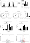
Caspase-1 activation triggered by RAC2 active variants in circulating myeloid cells and primary human monocyte–derived macrophages. (A–E) Whole blood circulating leukocytes from healthy donors (Control) or patient RAC2 E62K variant were incubated with the FAM–YVAD–FLICA probe for 1 h before fluorescent antibodies staining and analyzed by flow cytometry. CD14+ monocytes and CD66b+ granulocytes were gated as well as cells with low expression of CD14, CD66b, and CD16, corresponding to lymphocytes. Cells were analyzed for (A and B) FAM–YVAD–FLICA probe MFI or (C and D) for the presence of active Caspase-1 specks using Caspase-1-A and Caspase-1-H ratio analysis. Cells with Caspase-1 specks were defined by a high Caspase-1-A/Caspase-1-H ratio. Control conditions were repeated at least three times, and representative data are shown. (A) Overlay of the FAM–YVAD–FLICA fluorescence intensity measured in monocytes, granulocytes, and lymphocytes of healthy donors (Control) and patient variant RAC2 E62K. (B) Quantification of the FAM–YVAD–FLICA MFI measured in monocytes (Mo), granulocytes (Gr), and lymphocytes (Ly) of healthy donors (Control) and patient variant RAC2 E62K. Control conditions were repeated at least three times, and representative data are shown. (C and D) Gating of the monocytes and granulocytes with speck in healthy donors (Control) and patient variant RAC2 E62K using Caspase-1-A and Caspase-1-H ratio. (E) Graphic representation of the percentage of monocytes and granulocytes with Caspase-1 specks defined by a high Caspase-1-A/Caspase-1-H ratio in healthy donors (Control) and patient variant RAC2 E62K. (F and G) Human monocyte–derived macrophages were cotransduced for 72 h with Vpx containing virus-like particles and HA-tagged WT and mutated RAC2 encoding lentiviral vectors. Empty HIVSFFV-HARAC2-IRES-GFP vector (EV) was used as control. LPS was added for 8 h to induce the expression of pro-IL-1β. (F and G) Supernatants were analyzed using ELISA for (F) IL-1β and (G) IL-18. Data are mean ± SEM. Statistical analyses were performed using ordinary one-way ANOVA. *P < 0.05; **P < 0.01; ***P < 0.001; ****P < 0.0001. Data are representative for n = 3. (H) Volcano plot depicting differentially expressed genes in MOLM-13 transduced with RAC2 WT or RAC2 E62K compared to MOLM-13 transduced with empty vector. Red dots represent genes expressed at higher levels while blue dots represent genes with higher expression levels. Y axis denotes −log10 P values, and x axis shows log2 fold change values. Volcano plots were generated using VolcaNoseR. Data are representative for n = 5.
Figure S1.
Figure S1.
Gating strategies and validation of protocol. (A) PBMCs isolated from healthy donors were treated for 1 h with FAM–YVAD–FLICA before CD14 labeling and then analyzed by flow cytometry. Live cells were gated and doublets were excluded using SSC-A and SSC-H plot. CD14 high monocytes were analyzed for the presence of Caspase-1 speck using FITC-Caspase-1-A and FITC-Caspase-1-H. Monocytes with Caspase-1 specks were defined by a high Caspase-1-A/Caspase-1-H ratio. (B) PBMCs were treated 8 h with LPS (100 ng/ml) and 1 h with Nigericin (5 µM) (LPS + Nigericin) or with vehicle (Control). After 1 h incubation with the FAM–YVAD–FLICA probe cells were CD14 labeled and analyzed by flow cytometry. Experiments were repeated at least four times and representative data are shown. (A and B) The data were reused from Fig. 6 A. (C) Graphic representation showing the percentage of cells with Caspase-1 speck. (D) Overlay of the FAM–YVAD–FLICA fluorescence intensity measured in monocytes healthy donors (Control) and cells treated with LPS and Nigericin (LPS + Nigericin). (E) Graphic representation of data showing the MFI measured. Data are expressed as the mean ± SEM. Statistical analyses were performed using paired t test. *P < 0.05. Data are representative for n = 3.
Figure 5.
Figure 5.
The RAC2 A59S human patient mutation activates the NLRP3 inflammasome. (A) HA-RAC2 (WT), HA-RAC2 A59S, and HA-RAC2 E62K mutants were expressed for 16 h in HEK293T cells. HA-RAC2 activation level measurements were assessed by GST–PAK–RBD pull-down assays and immunoblotting (IB). (B–E) HEK293T cells were transfected for 16 h with plasmids encoding the NLRP3 inflammasome components (Inflam. mix) together with the WT or RAC2 E62K or RAC2 A59S forms. Cleaved IL-1β secretion in the culture supernatant (Sup) and protein expression in the cell lysates were assessed by immunoblotting. (C) Inhibitors of NLRP3 (MCC950), PAK (IPA3 or AZ 13711265), or pan-Caspases (Emricasan) were added 6 h before transfection. Data are representative for n = 3. (D and E) Inflammasome was reconstituted in HEK293T to evaluate the effect of the NLRP3 T659A mutation on IL-1β secretion compared to NLRP3 in cells expressing (D) RAC2 A59S or (E) RAC2 E62K. Data are representative for n = 3. Molecular weight (kD) is indicated in the figure. (F) U937 Cas9 (Cas9), NLRP3 KO, or GSDMD KO cells were cotransduced for 72 h with Vpx containing virus-like particles and HA-tagged RAC2 A59S or E62K encoding lentiviral vectors. Empty HIVSFFV-HARAC2-IRES-GFP vector was used as control. LPS was added for 8 h to induce the expression of pro-IL-1β. Supernatants were analyzed using ELISA for IL-1β. Data are mean ± SEM. Statistical analyses were performed using two-way ANOVA. ****P < 0.0001. Data are representative for n = 3. (G) LDH release was measured in the culture medium of U937 Cas9 (Cas9), NLRP3 KO, or NINJ1 KO cells expressing empty vector (Control), HA-tagged RAC2 A59S, or RAC2 E62K. The graph represents the percentage of cytotoxicity. Data are representative for n = 3. (H) Single-cell analysis for a blood sample of a patient harboring a RAC2 A59S variant and a representative control healthy donor blood sample drawn at the same time and analyzed in parallel are shown. Uniform Manifold Approximation and Projection plots of whole blood cells from control and RAC2 A59S patient donors. After alignment, joint clustering allows to detect 13 cell populations. pDC, plasmacytoid dendritic cell; mDC, myeloid dendritic cell; ISG, IFN-stimulated genes. (I) Violin plots showing the distribution of gene expression of IL-1β and NLRP3 genes in the cell clusters for both control (red) and RAC2 A59S patient (blue). NKT, natural killer T. Source data are available for this figure: SourceData F5.
Figure S2.
Figure S2.
Rac2 A59S– and E62K–triggered IL-1β secretion is GSDMD dependent. (A) U937 NINJ1 KO cells expressing empty vector (Control), HA-RAC2 A59S, or HA-RAC2 E62K were treated with LPS for 8 h to induce the expression of pro-IL-1β. Supernatants were analyzed using ELISA for IL-1β. Data are mean ± SEM. Statistical analyses were performed using ordinary one-way ANOVA. ***P < 0.001; ****P < 0.0001. Data are representative for n = 3. (B) LDH release was measured in the culture medium of U937 GSDMD KO cells expressing empty vector (Control) or HA-tagged RAC2 A59S or RAC2 E62K. The graph represents the percentage of cytotoxicity. Data are representative for n = 3.
Figure S3.
Figure S3.
RAC2 A59S variant patients have increased circulating monocytes and dendritic cells. Point-range plot of the analysis of the difference of cell proportions in clusters between control and RAC2 A59S patient samples identified in single-cell RNAseq. NKT, natural killer T; ISG, IFN-stimulated gene; FDR, false discovery rate.
Figure 6.
Figure 6.
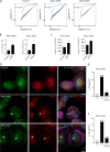
Monocytes and macrophages isolated from patients harboring the RAC2 E62K or RAC2 A59S mutations have an abnormal NLRP3 inflammasome activation. (A) PBMCs isolated from healthy donors (Control) or patients with RAC2 mutation (RAC2 A59S and RAC2 E62K) were treated for 1 h with FAM–YVAD–FLICA before CD14 labeling and then analyzed by flow cytometry. Cells with high expression of CD14, corresponding to monocytes, were gated and then analyzed for the presence of Caspase-1 speck using FITC-Caspase-1-A and FITC-Caspase-1-H. Monocytes with Caspase-1 speck were defined by a high Caspase-1-A/Caspase-1-H ratio. Control conditions were repeated at least three times, and representative data are shown. (B and C) Graphic representation of percentage of cells with Caspase-1 specks. Control conditions are expressed as the mean ± SEM of at least three independent experiments. (D) Monocyte-derived macrophages isolated from control healthy donors or patients presenting RAC2 A59S or RAC2 E62K variants were stained with anti-ASC (green), anti-NLRP3 (red), Hoechst 33342 for nuclei (blue), and phalloidin-Alexa 647 for actin filaments (magenta). The arrow indicates ASC speck colocalization with NLRP3 staining. Scale bar represents 10 µm. (E and F) Monocyte-derived macrophages isolated from patient RAC2 A59S (E) or a RAC2 E62K (F) variants were treated with LPS alone or LPS and MCC950 for 8 h to induce the expression of IL-1β in the absence or presence of NLRP3 inhibition, respectively. Non-treated cells (NT) were used as control. IL-1β secretion was measured using ELISA. The data were reused in Fig. S1, A and B, to illustrate the gating strategy. Data are mean ± SEM. Data are representative for n = 2.

References

    1. Accetta, D., Syverson G., Bonacci B., Reddy S., Bengtson C., Surfus J., Harbeck R., Huttenlocher A., Grossman W., Routes J., and Verbsky J.. 2011. Human phagocyte defect caused by a Rac2 mutation detected by means of neonatal screening for T-cell lymphopenia. J. Allergy Clin. Immunol. 127:535–538.e1: 2. 10.1016/j.jaci.2010.10.013 - DOI - PubMed
    1. Alkhairy, O.K., Rezaei N., Graham R.R., Abolhassani H., Borte S., Hultenby K., Wu C., Aghamohammadi A., Williams D.A., Behrens T.W., et al. . 2015. RAC2 loss-of-function mutation in 2 siblings with characteristics of common variable immunodeficiency. J. Allergy Clin. Immunol. 135:1380–1384.e1: 5. 10.1016/j.jaci.2014.10.039 - DOI - PMC - PubMed
    1. Ambruso, D.R., Knall C., Abell A.N., Panepinto J., Kurkchubasche A., Thurman G., Gonzalez-Aller C., Hiester A., deBoer M., Harbeck R.J., et al. . 2000. Human neutrophil immunodeficiency syndrome is associated with an inhibitory Rac2 mutation. Proc. Natl. Acad. Sci. USA. 97:4654–4659. 10.1073/pnas.080074897 - DOI - PMC - PubMed
    1. Arrington, M.E., Temple B., Schaefer A., and Campbell S.L.. 2020. The molecular basis for immune dysregulation by the hyperactivated E62K mutant of the GTPase RAC2. J. Biol. Chem. 295:12130–12142. 10.1074/jbc.RA120.012915 - DOI - PMC - PubMed
    1. Batistic, L. and Boyer L.. 2024. RAC2 gain-of-function variants causing inborn error of immunity drive NLRP3 inflammasome activation. Dryad. 10.5061/dryad.p8cz8w9zx - DOI

MeSH terms