. 2024 Nov 14;45(43):4601-4615.
doi: 10.1093/eurheartj/ehae546.

Colchicine prevents accelerated atherosclerosis in TET2-mutant clonal haematopoiesis

Affiliations

Colchicine prevents accelerated atherosclerosis in TET2-mutant clonal haematopoiesis

María A Zuriaga et al. Eur Heart J. .

Abstract

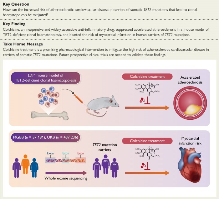
Background and aims: Somatic mutations in the TET2 gene that lead to clonal haematopoiesis (CH) are associated with accelerated atherosclerosis development in mice and a higher risk of atherosclerotic disease in humans. Mechanistically, these observations have been linked to exacerbated vascular inflammation. This study aimed to evaluate whether colchicine, a widely available and inexpensive anti-inflammatory drug, prevents the accelerated atherosclerosis associated with TET2-mutant CH.

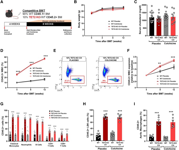
Methods: In mice, TET2-mutant CH was modelled using bone marrow transplantations in atherosclerosis-prone Ldlr-/- mice. Haematopoietic chimeras carrying initially 10% Tet2-/- haematopoietic cells were fed a high-cholesterol diet and treated with colchicine or placebo. In humans, whole-exome sequencing data and clinical data from 37 181 participants in the Mass General Brigham Biobank and 437 236 participants in the UK Biobank were analysed to examine the potential modifying effect of colchicine prescription on the relationship between CH and myocardial infarction.

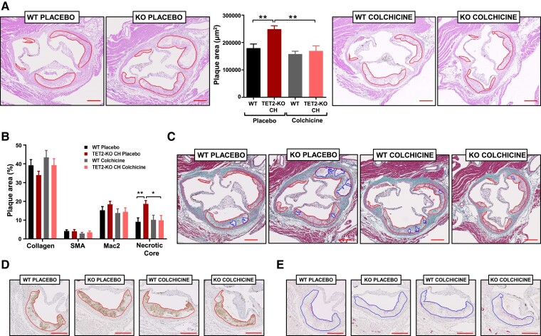
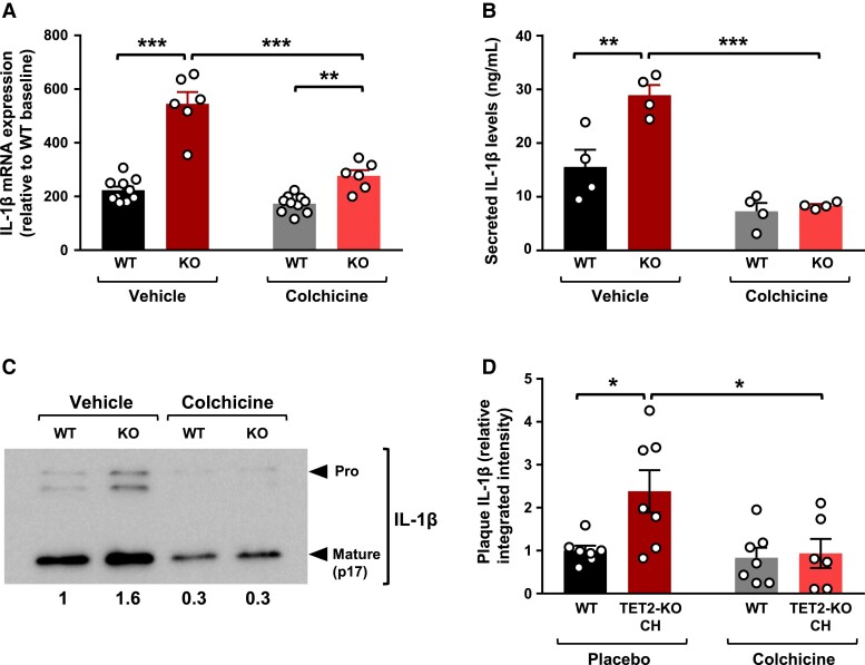
Results: Colchicine prevented accelerated atherosclerosis development in the mouse model of TET2-mutant CH, in parallel with suppression of interleukin-1β overproduction in conditions of TET2 loss of function. In humans, patients who were prescribed colchicine had attenuated associations between TET2 mutations and myocardial infarction. This interaction was not observed for other mutated genes.

Conclusions: These results highlight the potential value of colchicine to mitigate the higher cardiovascular risk of carriers of somatic TET2 mutations in blood cells. These observations set the basis for the development of clinical trials that evaluate the efficacy of precision medicine approaches tailored to the effects of specific mutations linked to CH.

Keywords: Atherosclerosis; CHIP; Colchicine; Inflammation; Precision medicine; TET2.


Figures

Structured Graphical Abstract
Clonal haematopoiesis (CH) driven by somatic mutations in the TET2 gene has been associated with heightened inflammation and an increased risk of atherosclerotic cardiovascular disease. In mice, we found that colchicine treatment prevented the development of accelerated atherosclerosis in a mouse model of TET2-deficient CH. In humans, analyses of sequencing data from two large biobanks showed that patients who were prescribed colchicine had attenuated associations between TET2-mutant CH and myocardial infarction. MGBB, Mass General Brigham Biobank; UKB, UK Biobank.
Figure 1
Figure 1
A mouse model of TET2-mutant clonal haematopoiesis and chronic colchicine treatment. (A) Summary of experimental design. (B) Mouse body weight throughout the duration of the experiment. (C) Serum cholesterol levels at endpoint. (D) Percentage of CD45.2+ cells in the white blood cell population at multiple timepoints, evaluated by flow cytometry. (E) Representative dot plots of the endpoint flow cytometry analysis of CD45.2+ white blood cells in TET2-KO clonal haematopoiesis mice treated with colchicine or placebo. (F) Expansion of CD45.2+ white blood cells relative to baseline chimerism. (G) Percentage of CD45.2+ cells in the main white blood cell populations in peripheral blood 12 weeks after the bone marrow transplantations, determined by flow cytometry. (H) Percentage of CD45.2+ cells in the bone marrow lineage-Sca1 + cKit + cell population at endpoint, determined by flow cytometry. (I) Percentage of CD45.2+ cells in aortic macrophages at endpoint, determined by flow cytometry (n = 5–6 pools of two aortae per genotype, as shown in the figure). All results are expressed as mean ± SEM (n = 17 placebo-treated WT mice; n = 16 placebo-treated TET2-KO clonal haematopoiesis mice; n = 13 colchicine-treated WT mice; n = 10 colchicine-treated TET2-KO clonal haematopoiesis mice unless otherwise indicated). Statistical significance was evaluated by repeated measures two-way analysis of variance with Tukey’s multiple comparisons tests in (D) and (F) and two-way analysis of variance with Sidak’s multiple comparison tests in (G), (H), and (I) (**P < .01, ***P < .001 for the comparison to each corresponding WT control). The error bars indicate SEM
Figure 2
Figure 2
Colchicine prevents the effects of TET2-mutant clonal haematopoiesis on experimental atherosclerosis in mice. (A) Aortic root plaque size in the different experimental groups. Representative images of haematoxylin and eosin-stained sections are shown; atherosclerotic plaques are delineated. (B) Histological analysis of plaque composition, quantified as percentage macrophage content (Mac2 antigen immunostaining), vascular smooth muscle cell content (smooth muscle α-actin immunostaining), collagen content (Masson’s trichrome staining), and necrotic core (collagen-free acellular regions in Masson’s trichrome-stained sections). (C) Representative images of Masson’s trichrome-stained sections; atherosclerotic plaques and necrotic areas are delineated. (D) Representative images of Mac2 antigen immunostaining. (E) Representative images of smooth muscle α-actin immunostaining. All results are expressed as mean ± SEM (n = 17 placebo-treated WT mice; n = 16 placebo-treated TET2-KO clonal haematopoiesis mice; n = 13 colchicine-treated WT mice; n = 10 colchicine-treated TET2-KO clonal haematopoiesis mice). Statistical significance was evaluated by two-way analysis of variance with Sidak’s multiple comparisons tests (*P < .05, **P < .01). The error bars indicate SEM
Figure 3
Figure 3
Colchicine inhibits elevated interleukin-1β production in conditions of TET2-mutant clonal haematopoiesis. (A) Quantitative real-time PCR analysis of interleukin-1β transcript expression in peritoneal macrophages isolated from Tet2−/− or +/+ mice (n = 10 WT, 6 KO, from two independent experiments) and stimulated for 4 h with 10 ng/mL lipopolysaccharide and 2 ng/mL interferon-γ in the presence of 1 μM colchicine or phosphate-buffered saline vehicle. mRNA levels are shown relative to the WT untreated group. (B) Enzyme-linked immunosorbent assay analysis of interleukin-1β protein levels in the supernatant of Tet2−/− and Tet2+/+ peritoneal macrophages (n = 4 mice) stimulated for 4 h with 10 ng/mL lipopolysaccharide and 2 ng/mL interferon-γ in the presence of 1 μM colchicine or phosphate-buffered saline vehicle, combined with a final 25 min incubation with 5 mM adenosine 5′-triphosphate. (C) Representative western blot analysis of pro-interleukin-1β and mature interleukin-1β protein levels in the culture supernatant of Tet2−/− and Tet2+/+ peritoneal macrophages. Numbers indicate mature interleukin-1β levels relative to vehicle-treated WT macrophages. (D) Immunofluorescence analysis of interleukin-1β protein levels in aortic root plaques of the different in vivo experimental groups (n = 7 mice per group), quantified as relative integrated fluorescence intensity normalized to plaque area. Statistical significance was evaluated by two-way analysis of variance with Sidak’s multiple comparisons tests (*P < .05, **P < .01, ***P < .001). The error bars indicate SEM

Comment in

Similar articles

Cited by

References

    1. Tall AR, Fuster JJ. Clonal hematopoiesis in cardiovascular disease and therapeutic implications. Nat Cardiovasc Res 2022;1:116–24. 10.1038/s44161-021-00015-3 - DOI - PMC - PubMed
    1. Natarajan P. Genomic aging, clonal hematopoiesis, and cardiovascular disease. Arterioscler Thromb Vasc Biol 2023;43:3–14. 10.1161/ATVBAHA.122.318181 - DOI - PMC - PubMed
    1. Kessler MD, Damask A, O'Keeffe S, Banerjee N, Li D, Watanabe K, et al. . Common and rare variant associations with clonal haematopoiesis phenotypes. Nature 2022;612:301–9. 10.1038/s41586-022-05448-9 - DOI - PMC - PubMed
    1. Bick AG, Weinstock JS, Nandakumar SK, Fulco CP, Bao EL, Zekavat SM, et al. . Inherited causes of clonal haematopoiesis in 97,691 whole genomes. Nature 2020;586:763–8. 10.1038/s41586-020-2819-2 - DOI - PMC - PubMed
    1. Jaiswal S, Fontanillas P, Flannick J, Manning A, Grauman PV, Mar BG, et al. . Age-related clonal hematopoiesis associated with adverse outcomes. N Engl J Med 2014;371:2488–98. 10.1056/NEJMoa1408617 - DOI - PMC - PubMed