. 2024 Aug 30;9(98):eadk9550.
doi: 10.1126/sciimmunol.adk9550. Epub 2024 Aug 30.

Germline-targeting HIV vaccination induces neutralizing antibodies to the CD4 binding site

Affiliations

Germline-targeting HIV vaccination induces neutralizing antibodies to the CD4 binding site

Tom G Caniels et al. Sci Immunol. .

Abstract

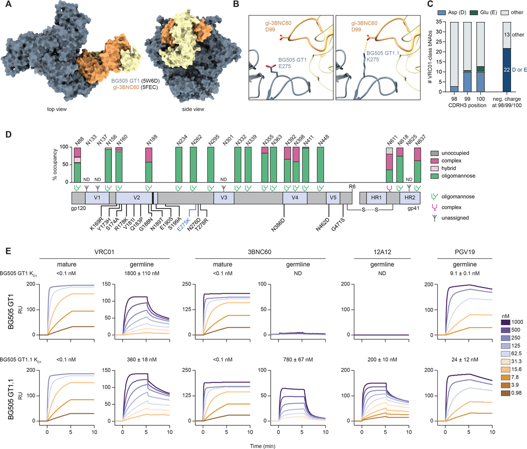
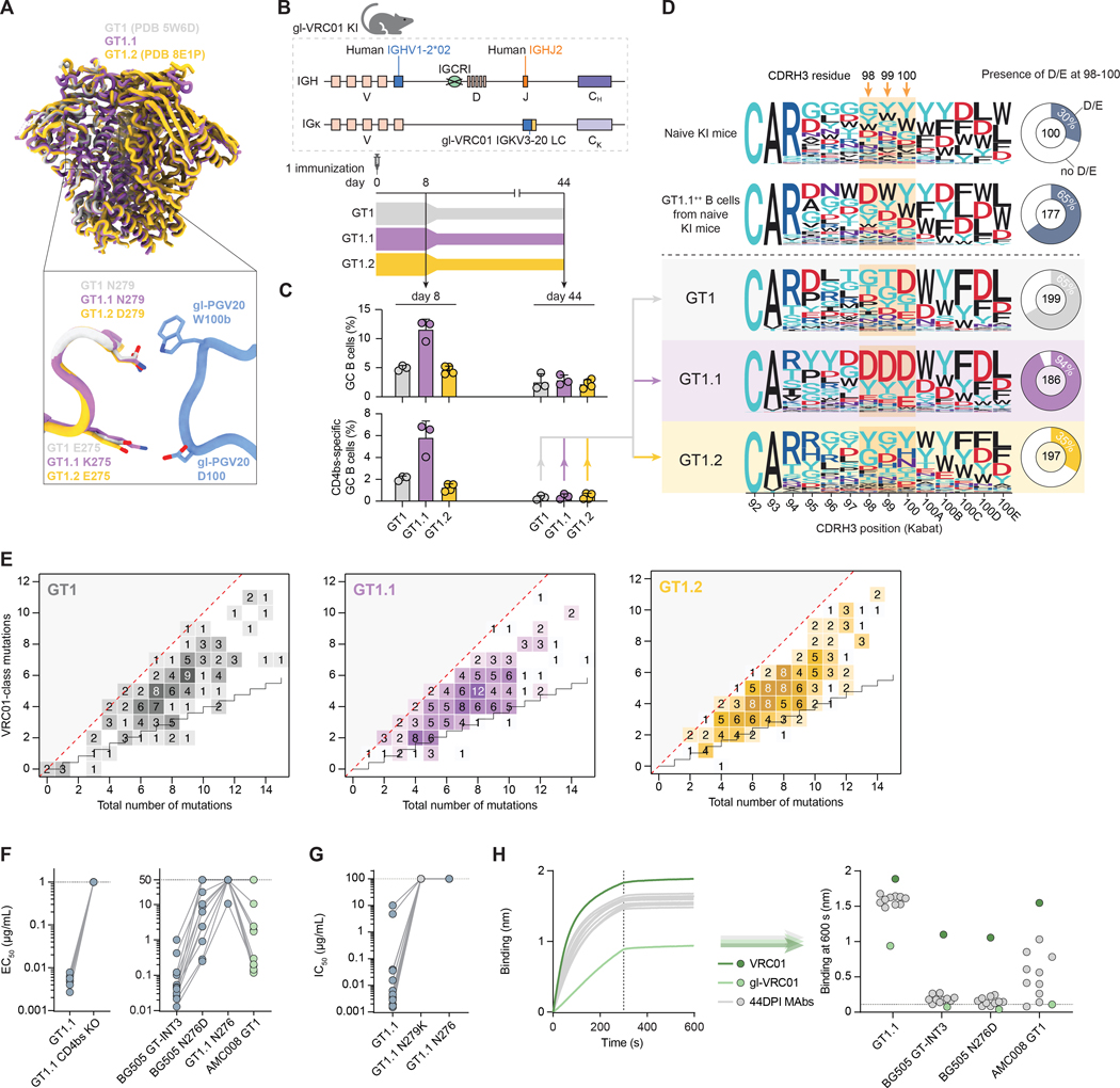
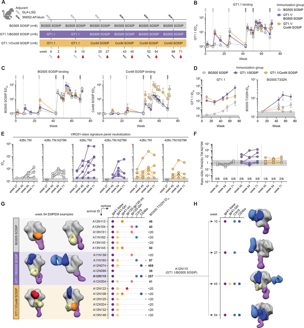
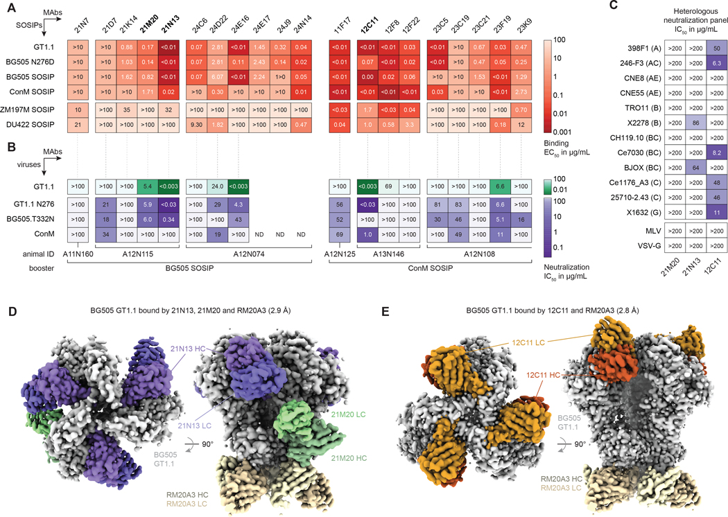
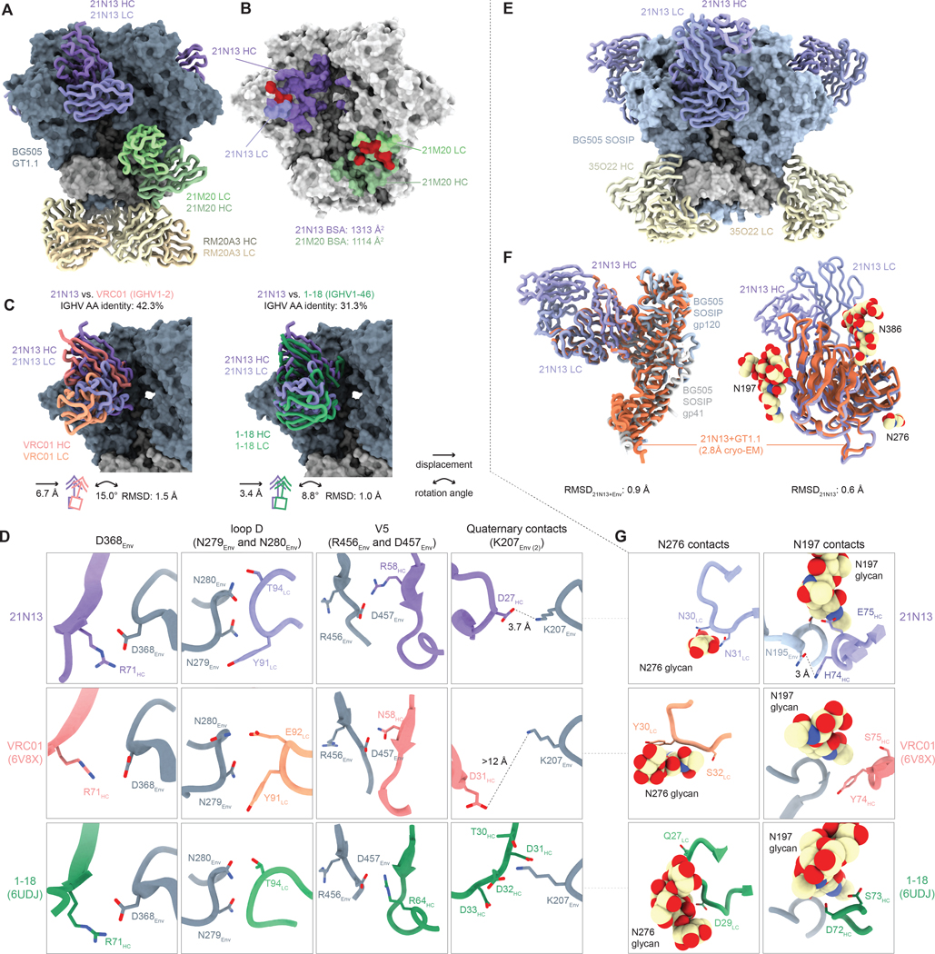
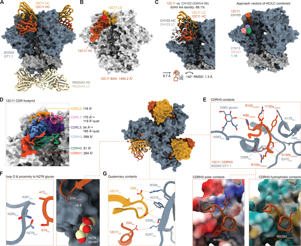
Eliciting potent and broadly neutralizing antibodies (bnAbs) is a major goal in HIV-1 vaccine development. Here, we describe how germline-targeting immunogen BG505 SOSIP germline trimer 1.1 (GT1.1), generated through structure-based design, engages a diverse range of VRC01-class bnAb precursors. A single immunization with GT1.1 expands CD4 binding site (CD4bs)-specific VRC01-class B cells in knock-in mice and drives VRC01-class maturation. In nonhuman primates (NHPs), GT1.1 primes CD4bs-specific neutralizing serum responses. Selected monoclonal antibodies (mAbs) isolated from GT1.1-immunized NHPs neutralize fully glycosylated BG505 virus. Two mAbs, 12C11 and 21N13, neutralize subsets of diverse heterologous neutralization-resistant viruses. High-resolution structures revealed that 21N13 targets the same conserved residues in the CD4bs as VRC01-class and CH235-class bnAbs despite its low sequence similarity (~40%), whereas mAb 12C11 binds predominantly through its heavy chain complementarity-determining region 3. These preclinical data underpin the ongoing evaluation of GT1.1 in a phase 1 clinical trial in healthy volunteers.


Conflict of interest statement

Competing interests

AC, PJK, JPM, IAW, ABW and RWS are inventors on a patent related to BG505 SOSIP Env trimers, and MMR and RWS are inventors on a patent related to germline-targeting HIV-1 Env trimers. SP is currently a full-time employee of Moderna, Inc. FDB has consultancy relationships with Adimab, Third Rock Ventures, and The EMBO Journal.

Figures

Figure 1.
Figure 1.. Design and antigenicity of BG505 SOSIP germline trimer 1.1 (GT1.1).
(A) Structural overlay of BG505 SOSIPv4.1-GT1 (PDB: 5W6D) and gl-3BNC60 (PDB: 5FEC). (B) Detail of the predicted interaction between GT1 E275 (left) and GT1.1 K275 and the CDRH3 residue D99 of gl-3BNC60. (C) Conservation of the negative CDRH3 charges in VRC01-class bnAbs. (D) Schematic linear representation of GT1.1 with glycan occupancy data. All amino acid mutations compared to BG505 SOSIP are indicated in black, whereas the blue E275K mutation defines GT1.1. The glycan icons represent the predominant type of glycan observed at that N-linked glycosylation site. (E) Surface plasmon resonance (SPR) of antibody binding to GT1 (top panels) and GT1.1 (bottom panels). The sensorgrams show the specific binding signal in response units (RUs) as a function of time. The x axes are truncated at half the dissociation time monitored (Fig. S1C). The KD1 values (nM) fitted by bivalent modeling (Table S1) are indicated on top of the panels. ND, not determined.
Figure 2.
Figure 2.. GT1.1 primes diverse VRC01-class precursors in a knock-in mouse model.
(A) Schematic showing residues of loop D of GT1, GT1.1 and GT1.2 interacting with gl-PGV20. Note that in contrast to N279D, N279A and N279K reduce VRC01-class binding and are used in knock-out reagents. (B) Schematic of the VRC01-class precursor mouse model (top panel) and the immunization schedule (bottom panel) (n = 3–4 per group). (C) Germinal center (GC) B cell frequency (top panel) and epitope-specific (CD4bs) GC B cell frequency (bottom panel) at day 8 and day 44 post-immunization. (D) Logo plot showing the CDRH3 sequence of knock-in BCRs isolated from each of the groups indicated. The donut plots on the right summarize the percentage of isolated knock-in BCRs with a negative charge at CDRH3 positions 98–100. (E) Total and VRC01-class amino acid mutations in the IGHV1–2 region for recovered knock-in BCR sequences for each immunization group. The staggered black line shows the expected level of VRC01-class mutations as expected to be introduced by random SHM in IGHV1–2 (21). On track VRC01-class mutations are defined as mutations that are shared with VRC01, PGV04, PGV20, VRC-CH31, 3BNC60 or 12A12 (21, 53). (F) Half-maximal effective concentrations (EC50s) as a measure for binding for mAbs recovered at 44 days post-immunization (DPI) against the indicated Envs. (G) Half-maximal inhibitory concentrations (IC50s) of the mAbs in (D) against the indicated pseudoviruses. (H) Bio-layer interferometry (BLI) sensorgrams (left panel) of isolated mAbs (grey) compared with gl-VRC01 (light green) and VRC01 (dark green) binding to GT1.1 and the binding at 600 s for various other Envs (right panel). Binding was assessed for 300 s, after which the sensors were moved to empty wells to allow dissociation measurement (dotted line). ELISA and neutralization data are representative of at least two independent experiments.
Figure 3.
Figure 3.. GT1.1 primes CD4bs antibodies in a non-human primate model.
(A) Schematic of the immunization regimen (n = 6 animals per group). (B) Mean ± SD half-maximal effective dose titers (ED50s) as a measure for binding to GT1.1 of each immunization group. The syringes indicate immunization time points. (C) Mean ± SD ED50s to BG505 SOSIP (left panel) and ConM (right panel). (D) Mean ± SD half-maximal inhibitory dose titers (ID50s) as a measure for neutralization of each immunization group to GT1.1 (left panel) and BG505.T332N (right panel). The grey boxes represent the minimum titer tested. (E) Mean ID50s of each animal to VRC01-class signature viruses 426c.TM and its CD4bs knockout mutant, 426c.TM.N279K. The grey boxes represent the minimum titer tested. ND, not determined. (F) The ratio of ID50 titers to 426c.TM and 426c.TM.N279K as in (E). The limits of the grey boxes represent a three-fold increase/decrease. (G) Electron microscopy-based polyclonal epitope mapping (EMPEM) for each immunization group. The polyclonal specificities for two representative animals per group are shown as segmented EM maps. The graph on the right shows positive detection of antibodies to a given epitope by each animal, with the numbers representing the ID50 titers to BG505.T332N. (H) Longitudinal EMPEM analysis for animal A12N115. ELISA and neutralization data shown are representative of at least two independent experiments.
Figure 4.
Figure 4.. Monoclonal antibodies neutralize fully glycosylated viruses through targeting of conserved (CD4bs) epitopes.
(A-B) Overview of the isolated mAbs (label on top) in binding (EC50, (A)) and neutralization (IC50 (B)). The animal IDs and booster immunogens these animals received are indicated in (B). The dotted lines connecting (A) and (B) depict binding and neutralization of a particular mAb. (A) EC50 titers of the isolated mAbs (label on top) to each indicated SOSIP protein (labels on left). (B) IC50 titers of the isolated mAbs as in (A) to each indicated pseudovirus. ND, not determined. The first three animals belong to the GT1.1/BG505 SOSIP group, the second three animals to the GT1.1/ConM SOSIP group. (C) Table showing the IC50 titers of the isolated mAbs (label on bottom) against the indicated heterologous pseudoviruses. The letter in parentheses represents the HIV-1 clade of a particular pseudovirus. (D) Top (left) and side (right) view of the 2.9 Å cryo-EM reconstruction of the GT1.1–21N13–21M20-RM20A3 complex. (E) Top (left) and side (right) view of the 2.8 Å cryo-EM reconstruction of the GT1.1–12C11-RM20A3 complex. HC, heavy chain; LC, light chain. Data shown in A-C are representative of at least two independent experiments.
Figure 5.
Figure 5.. nAb 21N13 contacts a conserved CD4bs epitope and accommodates CD4bs-adjacent glycans.
(A) Cryo-EM-derived atomic model of GT1.1 in complex with 21N13, 21M20 and RM20A3 Fabs. (B) Footprint and buried surface area (BSA) of 21N13 and 21M20 Fabs. The red shading indicates Env regions contacted by both heavy chain and light chain. (C) Superimposition of the GT1.1–21N13 structure with published structures of Env-VRC01 (PDB: 6V8X, left panel) and Env-1–18 (PDB: 6UDJ, right panel). The icons below the figure represent the mean displacement and rotation angle as well as the root mean square deviation (RMSD) of VRC01 or 1–18 compared to 21N13. (D) Detailed interactions of 21N13 with GT1.1 (top row) compared to VRC01 (middle row) and 1–18 (bottom row). The structures were aligned on Env fragments and the viewpoints are identical between structures. Dashed lines indicate listed distances. R58HC of 21N13 uses its side chain to form a salt bridge to the conserved D457Env. Furthermore, several CD4bs bnAbs such as 1–18, VRC03 and 3BNC117 evolved insertions in their CDRH1 or FWRH3 regions to contact adjacent Env protomers (11, 13, 50). While 21N13 does not bear an insertion, its negatively charged CDRH1 is positioned similarly to that of 1–18, thereby facilitating a contact with K207Env on the adjacent protomer (E) 4.7 Å crystal structure of 21N13 Fabs in complex with BG505 SOSIP and 35O22 Fab. (F) Comparison of the 21N13-GT1.1 cryo-EM structure (orange) with the 21N13-BG505 SOSIP crystal structure (blue/purple), as a whole Env+21N13 comparison (left panel) as well as a 21N13 Fab only comparison, both based on gp120 alignment (right panel). (G) Detailed interactions of 21N13 with BG505 SOSIP around N197 and N276 glycans as in (D).
Figure 6.
Figure 6.. nAb 12C11 binds a quaternary-dependent apical CD4bs epitope.
(A) Cryo-EM-derived atomic model of GT1.1 in complex with 12C11 and RM20A3 Fabs. (B) Footprint and buried surface area (BSA) of 12C11 Fab. The red shading indicates Env regions contacted by both heavy chain and light chain. (C) Superimposition of the GT1.1–12C11 structure with a published structure of Env-CH103 (PDB: 4JAN, left panel). The icons below the figure represent the mean displacement and rotation angle as well as the root mean square deviation (RMSD) of CH103 compared to 12C11. The right panel compares the approach vectors of NHP nAbs 21N13 (Fig. 5) and 12C11 with known CD4bs bnAbs CH103, VRC01 and 1–18. (D) Footprint of 12C11 CDRs onto the trimer. The numbers represent the buried surface area (BSA) contributed by each CDR. (E) Detailed interaction of the CDRH3-mediated contacts between GT1.1 and 12C11 (top panel) and an illustration of the polar CDRH3 contacts (bottom left panel) and the hydrophobic contacts (bottom right panel). (F) Inset showing the 12C11-loop D interaction (left panel) and the distance of the closest 12C11 residue to the N276 glycan (right panel). (G) Inset showing the quaternary interactions by 12C11 light chain (mustard) and 12C11 heavy chain (red) with the neighboring V3 region.

References

    1. Julg B, Liu P-T, Wagh K, Fischer WM, Abbink P, Mercado NB, Whitney JB, Nkolola JP, McMahan K, Tartaglia LJ, Borducchi EN, Khatiwada S, Kamath M, LeSuer JA, Seaman MS, Schmidt SD, Mascola JR, Burton DR, Korber BT, Barouch DH, Protection against a mixed SHIV challenge by a broadly neutralizing antibody cocktail. Sci. Transl. Med. 9, (2017). - PMC - PubMed
    1. Sok D, Burton DR, Recent progress in broadly neutralizing antibodies to HIV. Nat. Immunol. 19, 1179–1188 (2018). - PMC - PubMed
    1. Corey L, Gilbert PB, Juraska M, Montefiori DC, Morris L, Karuna ST, Edupuganti S, Mgodi NM, deCamp AC, Rudnicki E, Huang Y, Gonzales P, Cabello R, Orrell C, Lama JR, Laher F, Lazarus EM, Sanchez J, Frank I, Hinojosa J, Sobieszczyk ME, Marshall KE, Mukwekwerere PG, Makhema J, Baden LR, Mullins JI, Williamson C, Hural J, McElrath MJ, Bentley C, Takuva S, Gomez Lorenzo MM, Burns DN, Espy N, Randhawa AK, Kochar N, Piwowar-Manning E, Donnell DJ, Sista N, Andrew P, Kublin JG, Gray G, Ledgerwood JE, Mascola JR, Cohen MS, Two Randomized Trials of Neutralizing Antibodies to Prevent HIV-1 Acquisition. N. Engl. J. Med. 384, 1003–1014 (2021). - PMC - PubMed
    1. Hraber P, Seaman MS, Bailer RT, Mascola JR, Montefiori DC, Korber BT, Prevalence of broadly neutralizing antibody responses during chronic HIV-1 infection. AIDS 28, 163–169 (2014). - PMC - PubMed
    1. van Schooten J, van Gils MJ, HIV-1 immunogens and strategies to drive antibody responses towards neutralization breadth. Retrovirology 15, 74 (2018). - PMC - PubMed

Publication types