. 2024 Dec 24;8(24):6308-6320.
doi: 10.1182/bloodadvances.2024012599.

Local radiation enhances systemic CAR T-cell efficacy by augmenting antigen crosspresentation and T-cell infiltration

Affiliations

Local radiation enhances systemic CAR T-cell efficacy by augmenting antigen crosspresentation and T-cell infiltration

Nektarios Kostopoulos et al. Blood Adv. .

Abstract

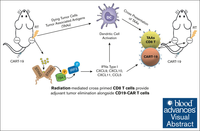
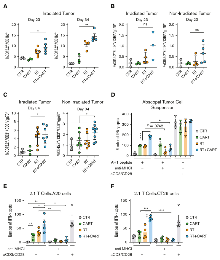
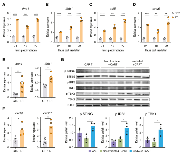
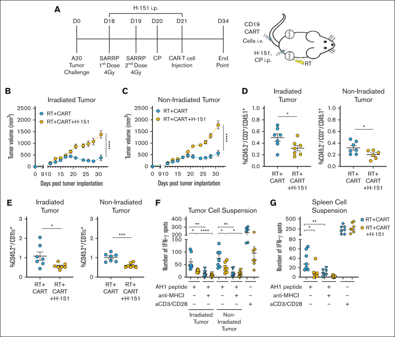
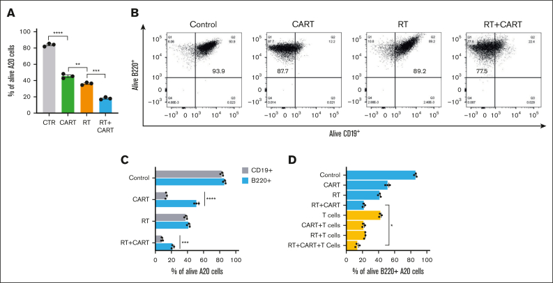
Chimeric antigen receptor (CAR) T-cell therapy targeting CD19 (CART-19) represents a significant advance in the treatment of patients with relapsed or refractory CD19+ B-cell lymphomas. However, a significant portion of patients either relapse or fail to respond. Moreover, many patients have symptomatic disease, requiring bridging radiation therapy (RT) during the period of CAR T-cell manufacturing. To investigate the impact of 1 to 2 fractions of low-dose RT on CART-19 treatment response, we developed a mouse model using A20 lymphoma cells for CART-19 therapy. We found that low-dose fractionated RT had a positive effect on generating abscopal systemic antitumor responses beyond the irradiated site. The combination of RT with CART-19 therapy resulted in additive effects on tumor growth in irradiated masses. Notably, a significant additional increase in antitumor effect was observed in nonirradiated tumors. Mechanistically, our results validate activation of the cyclic guanosine adenosine synthetase/stimulator of interferon genes pathway, tumor-associated antigen crosspriming, and elicitation of epitope spreading. Collectively, our findings suggest that RT may serve as an optimal priming and bridging modality for CAR T-cell therapy, overcoming treatment resistance and improving clinical outcomes in patients with CD19+ hematologic malignancies.


Conflict of interest statement

Conflict-of-interest disclosure: E.A.C. reports funding from Genmab, Genentech/Roche, AstraZeneca, CARGO, Juno Therapeutics, Novartis, Lymphoma Research Foundation, and Kite Pharma; and advisory board from AstraZeneca and Beigene. S.J.S. reports research funding from TG Therapeutics, Incyte, Adaptive Biotechnologies, Pharmacyclics, Merck, Genmab, Genentech/Roche, AstraZeneca, Cargo, AbbVie, and Lymphoma Research Foundation; consultancy fees and research funding from Genentech/Roche, Juno Therapeutics, and AbbVie; consultancy fees from Tessa Therapeutics, Loxo Oncology, BeiGene, Alimera Sciences, Acerta Pharma/AstraZeneca, and Nordic Nanovector; consultancy fees, honoraria, patents and royalties, and research funding from Novartis; consultancy fees, honoraria, and research funding from Celgene; and advisory board fees from AstraZeneca and BeiGene. The remaining authors declare no competing financial interests.

Figures

None
Graphical abstract
Figure 1.
Figure 1.
Enhanced abscopal antitumor effects in A20 lymphoma model with fractionated RT. (A) Working model: time line and schematic representation of in vivo A20 tumor–bearing mice treated with 2 × 4-Gy or 1 × 8-Gy RT in 1 of the 2 tumors. (B-C) A20 tumor growth from irradiated and nonirradiated abscopal tumor as followed by caliper measurements (n = 5-6 mice). (D) Gene ontology terms that are significantly enriched with an adjusted P value <.05 in the differentially expressed gene sets. Data represent irradiated tumors from the fractionated- or single-dose–treated groups. (E-F) Gene expression of granzyme B (gzmb) and perforin 1 (prf1) in irradiated and nonirradiated tumor 5 days after RT. (G-H) CD45+/CD3+ and CD45+/CD3+/CD8+ T-cell infiltration in irradiated and abscopal tumors 34 days after tumor implantation. Graphs show the mean ± standard error of the mean (SEM). ∗P < .01; ∗∗∗P < .001; ∗∗∗∗P < .0001. Data are representative of 3 independent experiments. CTR, control; ns, not significant; SARRP, Small Animal Radiation Research Platform.
Figure 2.
Figure 2.
Enhancing CART-19 cell infiltration and antitumor response through preceding RT in vivo. (A) Working model: time line and schematic representation of in vivo A20 tumor–bearing mice treated with RT followed by CART-19 cell injection. All mice of all groups were treated with the same cyclophosphamide (CP) lymphodepletion regimen. (B-C) A20 tumor growth from irradiated and abscopal tumor (n = 17-21). (D) Survival curve after treatment administration. (E) CD45.2+/CD3+ T-cell infiltration in irradiated and abscopal tumors. (F) CD45.2/CD3+/CD45.1+ T-cell infiltration in irradiated and abscopal tumor representing CART-19 cells. (G) CD45.2/CD3+/CD45.1+ T-cell infiltration in the spleen representing CART-19 cells. Graphs show the mean ± SEM. ∗P < .05; ∗∗P < .01; ∗∗∗P < .001; ∗∗∗∗P < .0001. Data are representative of 3 independent experiments. CTR, control.
Figure 3.
Figure 3.
RT/CART-19 combination treatment enhances crosspresentation of TAAs and T-cell responses to TAAs. (A) CD45.2+/CD11c+ DC infiltration in irradiated tumor on day 23 and day 34 after tumor challenge. (B-C) AH1-specific T-cell infiltration in irradiated and abscopal tumors on day 23 and day 34 after tumor challenge. (D) IFN-γ spots of an enzyme-linked immunospot (ELISPOT) assay after stimulation of tumor cell suspensions with AH1 peptide. (E) CD45.2+/CD3+ T cells isolated from treated mice stimulated with AH1 peptide and incubated with A20 tumor cells. (F) CD45.2+/CD3+ T cells isolated from treated mice stimulated with AH1 peptide and incubated with CT26 tumor cells. Graphs show the mean ± SEM. ∗P < .05; ∗∗P < .01; ∗∗∗P < .001; ∗∗∗∗P < .0001. CTR, control.
Figure 4.
Figure 4.
Enhanced tumor protection in a colorectal model through adoptive transfer of CD3+ T cells from mice treated with RT plus CART-19. (A) Working model: time line and schematic representation of in vivo A20 tumor–bearing mice treated with 2 × 4-Gy RT followed by CAR T cells and adoptive T-cell transfer to CT26 tumor–bearing mice. All mice of all groups were treated with the same CP lymphodepletion regimen. (B) AH1-specific T cells from donor mice after ex vivo expansion, before ACT. (C) CT26 tumor growth of recipient mice (n > 4). (D) CD45+/CD3+/CD8+ T-cell tumor infiltration. (E) CD45+/CD3+/CD8+/CD107+ T-cell tumor infiltration. (F) CD45+/CD3+/CD8+/AH1-specific T-cell tumor infiltration. (G) IFN-γ spots of an ELISPOT assay after AH1 peptide stimulation of splenocytes from mice that received ACT. Graphs show the mean ± SEM. ∗P < .05; ∗∗P < .01; ∗∗∗P < .001; ∗∗∗∗P < .0001. Data are representative of 2 independent experiments. ACT, Adoptive Cell Therapy.
Figure 5.
Figure 5.
RT-induced activation of the cGAS/STING pathway promotes IFN type I gene and chemokine expression upregulation in A20 tumor cells. (A-D) Expression of ifna1, ifnb1, ccl5, and cxcl9 after 24, 48, and 72 hours of RT in vitro. (E-F) Expression of ifna1, inb1, cxcl9, and cxcl11 48 hours post in vivo RT. (G) Activation of STING pathway in A20 tumors treated with CART-19 alone or combination of CART-19 and RT. Graphs show the mean ± SEM. ∗P < .05; ∗∗P < .01; ∗∗∗P < .001; ∗∗∗∗P < .0001. b-TUB, b-tubulin; RT, Radiation Therapy; p-IRF3, phosphorylated-Interferon Regulatory Factor 3; p-STING, phosphorylated-Stimulator of interferon genes; p-TBK1, phosphorylated-TANK-binding kinase 1.
Figure 6.
Figure 6.
RT enhances CAR T-cell therapy through STING activation. (A) Working model: time line and schematic representation of in vivo A20 tumor–bearing mice treated with RT followed by CART-19 cell injection with or without the STING antagonist H-151. All mice of all groups were treated with the same CP lymphodepletion regimen. (B-C) Tumor growth from irradiated and abscopal tumors (n = 8). (D) CD45.2/CD3+/CD45.1+ T-cell infiltration in irradiated and abscopal tumors, representing the CART-19 cells. (E) CD45.2+/CD11c+ DC infiltration in irradiated and abscopal tumors. (F-G) IFN-γ spots after overnight stimulation of tumor or spleen cell suspensions with AH1 peptide with or without anti–MHC I antibody. Graphs show the mean ± SEM. ∗P < .05; ∗∗P < .01; ∗∗∗P < .001; ∗∗∗∗P < .0001. Data are representative of 1 experiment.
Figure 7.
Figure 7.
Enhancing sensitivity of target cells to CTL function through RT: implications for CAR T-cell efficacy and antigen escape. (A) Percentage of A20 tumor cells alive after RT, CAR T-cell, and RT and CAR T-cell treatments. (B) Flow cytometry for B220 and CD19 markers after in vitro treatment of A20 tumor cells with RT, CAR T-cell, and RT and CAR T-cell treatments. (C) B220+ and CD19+ alive cells after treatments. (D) B220+ alive cells after treatment with RT, CAR T cells, and T cells isolated from CT26 tumor–bearing mouse adoptively transferred with T cells from RT plus CAR T-cell donor. Graphs show the mean ± SEM. ∗P < .05; ∗∗P < .01; ∗∗∗P < .001; ∗∗∗∗P < .0001. CTR, XXX.

References

    1. Park JH, Rivière I, Gonen M, et al. Long-term follow-up of CD19 CAR therapy in acute lymphoblastic leukemia. N Engl J Med. 2018;378(5):449–459. - PMC - PubMed
    1. Lee DW, Kochenderfer JN, Stetler-Stevenson M, et al. T cells expressing CD19 chimeric antigen receptors for acute lymphoblastic leukaemia in children and young adults: a phase 1 dose-escalation trial. Lancet. 2015;385(9967):517–528. - PMC - PubMed
    1. Maude SL, Frey N, Shaw PA, et al. Chimeric antigen receptor T cells for sustained remissions in leukemia. N Engl J Med. 2014;371(16):1507–1517. - PMC - PubMed
    1. Neelapu SS, Locke FL, Bartlett NL, et al. Axicabtagene ciloleucel CAR T-cell therapy in refractory large B-cell lymphoma. N Engl J Med. 2017;377(26):2531–2544. - PMC - PubMed
    1. Schuster SJ, Bishop MR, Tam CS, et al. Tisagenlecleucel in adult relapsed or refractory diffuse large B-cell lymphoma. N Engl J Med. 2019;380(1):45–56. - PubMed