. 2024 Aug 30;20(8):e1012504.
doi: 10.1371/journal.ppat.1012504. eCollection 2024 Aug.

Rab27a GTPase and its effector Myosin Va are host factors required for efficient Oropouche virus cell egress

Affiliations

Rab27a GTPase and its effector Myosin Va are host factors required for efficient Oropouche virus cell egress

Juan O Concha et al. PLoS Pathog. .

Abstract

Oropouche fever, a debilitating illness common in South America, is caused by Oropouche virus (OROV), an arbovirus. OROV belongs to the Peribunyaviridae family, a large group of RNA viruses. Little is known about the biology of Peribunyaviridae in host cells, especially assembly and egress processes. Our research reveals that the small GTPase Rab27a mediates intracellular transport of OROV induced compartments and viral release from infected cells. We show that Rab27a interacts with OROV glycoproteins and colocalizes with OROV during late phases of the infection cycle. Moreover, Rab27a activity is required for OROV trafficking to the cell periphery and efficient release of infectious particles. Consistently, depleting Rab27a's downstream effector, Myosin Va, or inhibiting actin polymerization also hinders OROV compartments targeting to the cell periphery and infectious viral particle egress. These data indicate that OROV hijacks Rab27a activity for intracellular transport and cell externalization. Understanding these crucial mechanisms of OROV's replication cycle may offer potential targets for therapeutic interventions and aid in controlling the spread of Oropouche fever.


Conflict of interest statement

The authors have declared that no competing interests exist.

Figures

Fig 1
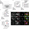
Fig 1. OROV replication cycle in HeLa cells.
(A) Growth kinetics of OROV. HeLa cells monolayers were inoculated with OROV (MOI = 4) and the cell-free supernatant was collected at the indicated hours post-infection (h.p.i) and clarified. Infectious virus titers released into the cell culture media were quantified in Vero cells by plaque assay and are expressed as PFU/ml. In parallel, plasma membrane permeability in OROV-infected cells was measured by trypan blue exclusion from three independent experiments. Data represent the mean ± SD at indicated h.p.i. N = 3 (B) Intracellular viral proteins analyzed by western blot. HeLa cells monolayers infected as in (A) were processed for immunoblot at the indicated times post-infection. Detection of viral proteins was performed using mouse anti-OROV antiserum that recognizes Gc glycoprotein and N protein. Polyclonal rabbit anti-GAPDH antibody were used as a loading control of cell lysate samples (C) Monolayers of HeLa cells grown on coverslips was inoculated with OROV (MOI = 4) and fixed at the indicated post-infection times. The presence and intracellular distribution of the virus were monitored by indirect immunofluorescence, staining the cells with the anti-OROV same antiserum used for (B), followed by staining with a secondary anti-mouse IgG conjugated to Alexa Fluor 488 (in green). Nuclei are stained with DAPI (in blue) Scale bar: 10 μm.
Fig 2
Fig 2. Rab27a knockdown impairs the release of OROV in HeLa cells.
(A) HeLa cells were silenced for Rab27a or Rab27b using siRNA and later inoculated with OROV (MOI = 4). After 24 h, cells were lysed and subjected to western blot analysis using mouse anti-OROV antiserum, rabbit anti-Rab27a, rabbit anti-Rab27b and rabbit anti-GAPDH (used as a loading control). (B) HeLa cells conditioned media was used for viral titer determination by plaque assay for each condition. Data are the mean ± SD of three independent experiments. p>0.05 was considered as not significant (one-way ANOVA followed by Tukey´s multiple comparisons test). (C) HeLa shRab27a and shControl cells were mock transfected or transfected with control siRNA or Rab27b siRNA, as indicated, and later inoculated with OROV (MOI = 4). After 24 h, cells were lysed and subjected to western blot analysis using antibodies against indicated proteins. (D) The clarified supernatant of each condition was used for viral titer determination by the plaque assay method. Data are the mean ± SD of three independent experiments. p>0.05 was considered as not significant (one-way ANOVA followed by Tukey´s multiple comparisons test).
Fig 3
Fig 3. Rab27a colocalizes with OROV in late stages of infection.
(A) HeLa shControl (upper panels) and shRab27a (lower panels) cells growing on coverslips were inoculated with OROV (MOI = 4) and fixed at 24 hours post-infection. Cells were permeabilized and stained with primary mouse anti-OROV, rabbit anti-Rab27a and sheep anti-TGN46 antibodies, followed by secondary anti-mouse IgG 488 (green), anti-rabbit IgG 594 (red) and anti-sheep IgG 647 (cyan). Nuclei are stained with DAPI (in blue). Scale bar = 10 μm. Insets represent the boxed areas. Scale bar = 2 μm. A maximum intensity projection image across the Z-stack for OROV staining is shown in each case. Scale bar = 10 μm. (B) Colocalization between TGN46 and OROV from the panel A, bars represent the mean ± SD of the Manders’ colocalization coefficient (at least 10 cells from three independent experiments). p>0.05 was considered as not significant (Unpaired t test).
Fig 4
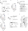
Fig 4. Overexpression of Rab27a mutants affects OROV intracellular distribution and release.
(A) HeLa cell lines with inducible expression of Rab27a WT, Rab27a Q78L or Rab27a T23N mutants, or HeLa parental cells, were infected with OROV (MOI = 4) and after 4 hours the cells were treated with 10 ng/ml of Doxycycline to induce the expression. After 24 h.p.i., cells were lysed and subjected to western blot analysis using mouse anti-OROV (which recognizes N and Gc proteins), rabbit anti-Rab27a and rabbit anti-GAPDH used as a loading control. (B-C) Densitometry quantification of the Gc and N signals relative to GAPDH, as shown in panel A (n = 3 independent experiments). p>0.05 was considered as not significant (one-way ANOVA followed by Dunnett’s multiple comparisons test). (D) The clarified cell culture media of each condition in (A) were used for PFU assay. Data are the mean ± SD of three independent experiments. p>0.05 was considered as not significant. (one-way ANOVA followed by Tukey´s multiple comparisons test). (E) HeLa cell lines with inducible expression of Rab27a WT, Rab27a Q78L or Rab27a T23N mutants, growing on cover slips were infected with OROV (MOI = 4) and after 4 hours the cells were treated with 10ng/ml of Doxycycline to induce the expression. After 24 h.p.i., the cells were fixed, permeabilized and incubated with anti-OROV (mouse), anti-Rab27a (rabbit) and anti-TGN46 (sheep) antibodies, followed anti-mouse IgG 488 (green), anti-rabbit IgG 594 (red) and anti-sheep IgG 647 (cyan) secondary antibodies. Nuclei are stained with DAPI (in blue). Scale bar = 10 μm. (F) Colocalization between TGN46 and OROV from (E), bars represent the mean ± SD of the Manders’ colocalization coefficient (at least 10 cells from three independent experiments). p>0.05 was considered as not significant (one-way ANOVA followed by Tukey´s multiple comparisons test).
Fig 5
Fig 5. Rab27a interacts with OROV proteins and associates with the viral particles.
(A) HeLa cells monolayers were infected with OROV (MOI = 4) and after 4 hours were transfected with plasmids expressing GFP-Rab27a and GFP as a control, 24 h.p.i. the cells were collected. The cells were lysed and subjected to affinity capture (IP) using GFP affinity matrices before western blot using mouse anti-OROV antiserum (that recognizes Gc and N proteins), rabbit anti-GFP and rabbit anti-GAPDH as a loading control. (B) Densitometry quantification of the amount of Gc immunoprecipitated by the GFP affinity matrices as shown in panel A (n = 3 independent experiments). p>0.05 was considered as not significant (Unpaired t test). (C) HEK293T cells monolayers were cotransfected with HA-Gn and HA-Gc plasmids and either GFP or GFP-Rab27a WT, 24 h after transfection, the cells were collected. The cells were lysed and subjected to affinity capture (IP) using GFP affinity matrices before western blot using rabbit anti-HA (to recognize Gc and Gn), mouse anti-GFP, and rabbit anti-actin as a loading control. (D-E) Densitometry quantification of the amount of Gc and Gn immunoprecipitated by the GFP affinity matrices as shown in panel C (n = 4 independent experiments). p>0.05 was considered as not significant (Unpaired t test). (F) HeLa cells monolayers were infected with OROV (MOI = 4) and after 4 hours were transfected with plasmids expressing GFP-Rab27a WT and GFP as a control, 24 hpi the cells and the supernatant were collected. The cells were lysed and subjected to western blot using the mouse anti-OROV, rabbit anti-GFP and rabbit anti-GAPDH as a loading control. The supernatant was ultracentrifuged (100000 x g for 90’ at 4°C) and subjected to western blot analysis using the same antibodies used for the cell lysate. (G) HeLa cells were infected with OROV (MOI = 4) or mock infected, 24 h.p.i. The cells and media were collected and virus particles were concentrated from the media by ultracentrifugation (100,000 × g for 90’ at 4°C). The lysates of cells and extracellular particles were analyzed by western blot using the antibodies indicated. AP1γ1 and CD9 were used as a cell cytoplasm contamination marker and as EVs marker respectively. p>0.05 was considered as not significant (Unpaired t test).
Fig 6
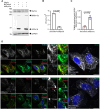
Fig 6. The Rab27a downstream effector Myosin Va is important for OROV trafficking and release.
(A) HeLa shControl and shMyoVa cells were infected with OROV (MOI = 4) or mock infected. After 24 h, cells were lysed and subjected to western blot analysis using mouse anti-OROV, rabbit anti-MyoVa and rabbit anti-GAPDH used as a loading control. (B) The cell culture media of each condition was used for PFU assay. Data are the mean ± SD of three independent experiments. p>0.05 was considered as not significant (Unpaired t test). (C) Colocalization between TGN46 and OROV from (D), bars represent the mean ± SD of the Manders’ colocalization coefficient (at least 5 cells from three independent experiments). p>0.05 was considered as not significant (Unpaired t test). (D) HeLa shControl (upper panels) and shMyoVa (lower panels) cells growing on coverslips were inoculated with OROV (MOI = 4) and fixed at 24 h.p.i. Cells were permeabilized and stained with primary anti-OROV (mouse), anti-MyoVa (Rabbit) and anti-TGN46 (sheep) antibodies, followed by secondary anti-mouse IgG 488 (green), anti-rabbit IgG 594 (red) and anti-sheep IgG 647 (cyan). Nuclei are stained with DAPI (in blue). Scale bar = 10 μm. Insets represent the boxed areas. Scale bar = 2 μm. A maximum intensity projection image across the Z-stack for OROV staining is shown in each case. Scale bar = 10 μm. p>0.05 was considered as not significant (Unpaired t test).
Fig 7
Fig 7. MyoVa KO Fibroblasts show alteration in the viral compartments’ morphology and a reduction in OROV release.
(A) Control (MyoVa WT) and MyoVa-null (MyoVa KO) human fibroblasts were infected with OROV (MOI = 4) or mock infected. After 24 h, cells were lysed and subjected to western blot analysis using mouse anti-OROV (which recognizes N and Gc proteins), rabbit anti-MyoVa and rabbit anti-GAPDH used as a loading control. (B-C) Densitometry quantification of the amount of Gc and N as shown in panel A (n = 3 independent experiments). p>0.05 was considered as not significant (Unpaired t test). (D) The clarified cell culture media of each condition (panel A) were used for viral titer determination by plaque assay. Data are the mean ± SD of three independent experiments. p>0.05 was considered as not significant (Unpaired t test). (E) MyoVa WT and MyoVa KO fibroblasts growing on cover slips were infected with OROV (MOI = 4). After 24 h, the cells were fixed, permeabilized and incubated with anti-OROV (mouse) and anti-TGN46 (sheep) antibodies, followed anti-mouse IgG 488 (green) and anti-sheep IgG 594 (red) secondary antibodies. Nuclei are stained with DAPI (in blue). Scale bar = 10 μm. Insets represent the boxed areas. Scale bar = 2 μm.
Fig 8
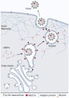
Fig 8. Proposed model for the Rab27a and Myosin Va mediated OROV cell egress.
OROV envelope glycoprotein complexes are synthesized in the endoplasmic reticulum and transported to the Golgi, where virus assembly occurs for most orthobunyaviruses. The GTPase Rab27a may associate with the limiting membrane of these viral-induced Golgi/TGN-derived structures via interaction with OROV glycoproteins. This interaction likely occurs before the envelopment process is complete, occasionally resulting in the incorporation of Rab27a into nascent virions. The presence of Rab27a engages effector proteins, including Myosin Va, which propels the large virion-containing vesicles along actin filaments to the cell periphery. This movement continues across the actin cortical network, ultimately reaching the plasma membrane. At the plasma membrane, exocytic membrane fusion occurs, leading to the release of viral particles from the cells. Image created with BioRender.com.

References

    1. Barbosa NS, Concha JO, daSilva LLP, Rezaei N. Bunyavirus. Encyclopedia of Infection and Immunity. Oxford: Elsevier; 2022. p. 207–18.
    1. Elliott RM. Molecular biology of the Bunyaviridae. J Gen Virol. 1990;71 (Pt 3):501–22. doi: 10.1099/0022-1317-71-3-501 . - DOI - PubMed
    1. Vernal S, Martini CCR, da Fonseca BAL. Oropouche Virus-Associated Aseptic Meningoencephalitis, Southeastern Brazil. Emerg Infect Dis. 2019;25(2):380–2. doi: 10.3201/eid2502.181189 ; PubMed Central et al.: PMC6346467. - DOI - PMC - PubMed
    1. Wesselmann KM, Postigo-Hidalgo I, Pezzi L, de Oliveira-Filho EF, Fischer C, de Lamballerie X, et al.. Emergence of Oropouche fever in Latin America: a narrative review. Lancet Infect Dis. 2024. Epub 20240125. doi: 10.1016/S1473-3099(23)00740-5 . - DOI - PubMed
    1. Pinheiro FP, Travassos da Rosa AP, Travassos da Rosa JF, Ishak R, Freitas RB, Gomes ML, et al.. Oropouche virus. I. A review of clinical, epidemiological, and ecological findings. Am J Trop Med Hyg. 1981;30(1):149–60. . - PubMed

MeSH terms

LinkOut - more resources