. 2024 Sep;300(9):107724.
doi: 10.1016/j.jbc.2024.107724. Epub 2024 Aug 28.

Nonstructural protein 4 of human norovirus self-assembles into various membrane-bridging multimers

Affiliations

Nonstructural protein 4 of human norovirus self-assembles into various membrane-bridging multimers

Adrien Royet et al. J Biol Chem. 2024 Sep.

Abstract

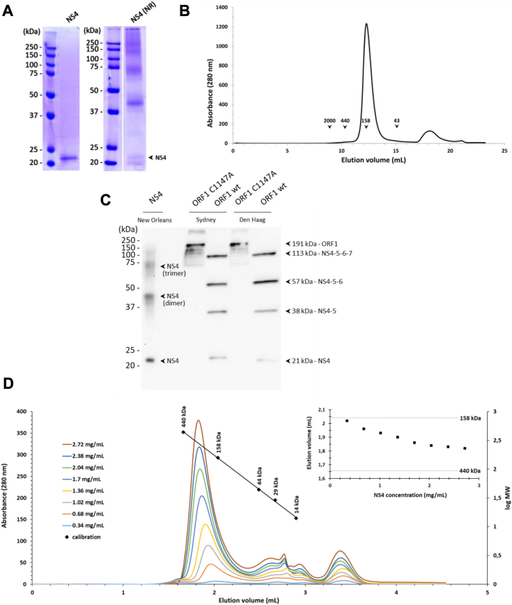
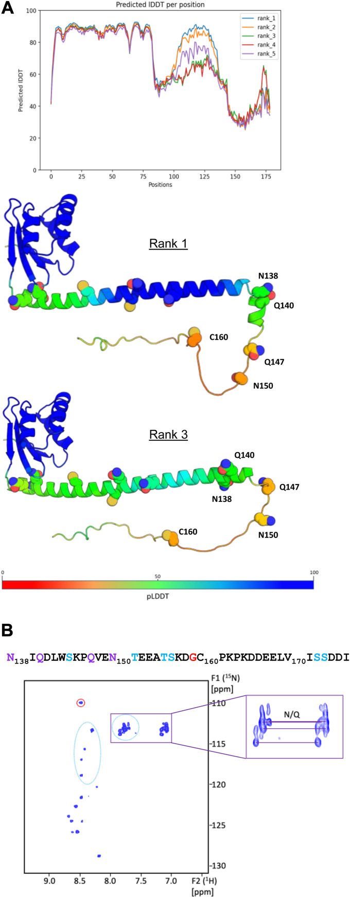
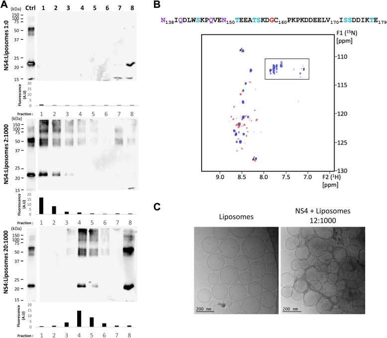
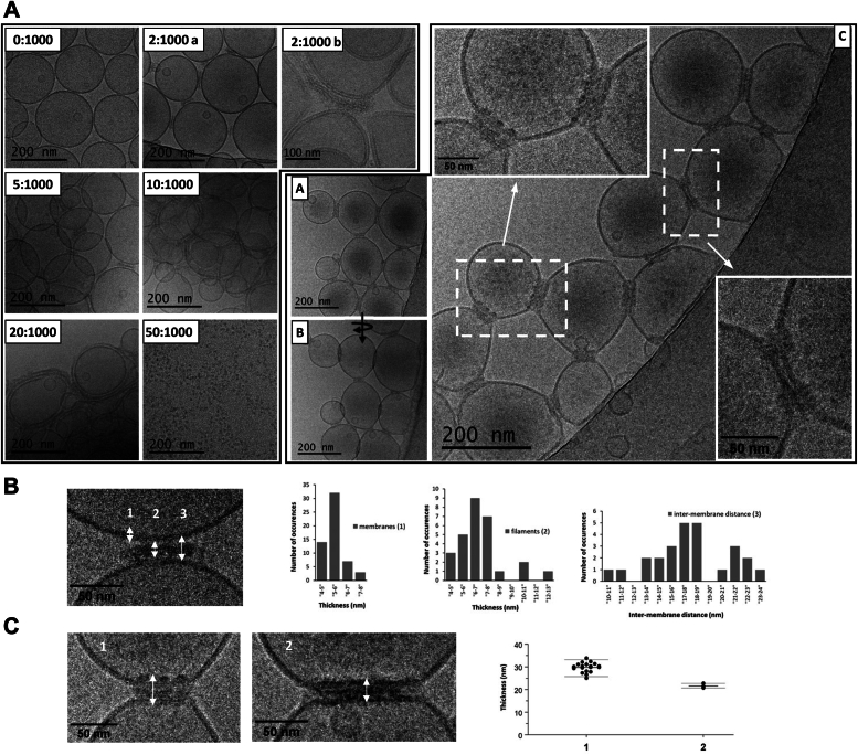
Single-stranded, positive-sense RNA ((+)RNA) viruses replicate their genomes in virus-induced intracellular membrane compartments. (+)RNA viruses dedicate a significant part of their small genomes (a few thousands to a few tens of thousands of bases) to the generation of these compartments by encoding membrane-interacting proteins and/or protein domains. Noroviruses are a very diverse genus of (+)RNA viruses including human and animal pathogens. Human noroviruses are the major cause of acute gastroenteritis worldwide, with genogroup II genotype 4 (GII.4) noroviruses accounting for the vast majority of infections. Three viral proteins encoded in the N terminus of the viral replication polyprotein direct intracellular membrane rearrangements associated with norovirus replication. Of these three, nonstructural protein 4 (NS4) seems to be the most important, although its exact functions in replication organelle formation are unknown. Here, we produce, purify, and characterize GII.4 NS4. AlphaFold modeling combined with experimental data refines and corrects our previous crude structural model of NS4. Using simple artificial liposomes, we report an extensive characterization of the membrane properties of NS4. We find that NS4 self-assembles and thereby bridges liposomes together. Cryo-EM, NMR, and membrane flotation show formation of several distinct NS4 assemblies, at least two of them bridging pairs of membranes together in different fashions. Noroviruses belong to (+)RNA viruses whose replication compartment is extruded from the target endomembrane and generates double-membrane vesicles. Our data establish that the 21-kDa GII.4 human norovirus NS4 can, in the absence of any other factor, recapitulate in tubo several features, including membrane apposition, that occur in such processes.

Keywords: cryo-EM; lipid–protein interaction; liposome; membrane; nonstructural protein; norovirus; plus-stranded RNA virus; protein assembly; viral protein; viral replication.


Conflict of interest statement

Conflict of interest The authors declare that they have no conflicts of interest with the contents of this article.

Figures

Figure 1
Figure 1
Biochemical analysis of purified recombinant NS4.A, 6His-tagged NS4 protein from GII.4 New Orleans variant was produced in bacteria and purified in presence of DDM. Protein sample was analyzed by SDS-PAGE in reducing (left) and nonreducing (NR, right) conditions. The arrow indicates the band corresponding to the expected molecular weight of denatured monomeric NS4. B, the elution profile of DDM-solubilized NS4 was analyzed by size-exclusion chromatography on a Superdex 200 10/300 GL column, and compared to standard globular molecular weight markers whose elution volumes are indicated. C, WT or C1147A mutated ORF1 from GII.4 Sydney or Den Haag variants were expressed by a wheat-germ cell-free expression system. Expression profiles were analyzed by Western blot and compared to pure 6His-tagged New Orleans NS4. Since we used anti-NS4 antibody, only fragments containing NS4 are detected. Arrows indicate the bands corresponding to uncleaved full-length C1147A ORF1 or processing intermediates generated after self-cleavage of WT ORF1. The expected sizes of the proteins were calculated by taking into account the presence of a Strep-tag at both N- and C-terminal extremities (15). D, elution profile of DDM-solubilized NS4 at different concentrations was analyzed on an analytical Superdex 200 5/150 column. NS4 samples at constant volume but decreasing concentrations were injected onto the column and resulting chromatograms were superimposed for comparison. The calibration curve of the column using molecular weight markers is indicated. The inset represents the elution volume of the main peak as a function of the input protein concentration. The elution volumes of 158 and 440 kDa molecular weight markers are indicated with a thin line. DDM, n-dodecyl-β-D-maltoside; GII.4, genogroup II genotype 4; NS, nonstructural.
Figure 2
Figure 2
Structural analysis of purified recombinant NS4.A, AlphaFold2 modeling of NS4 from the GII.4 New Orleans variant. Top, predicted LDDT score per residue for the five top-ranked models. Bottom, two models (ranks 1 and 3 from top to bottom) are represented as ribbons with alpha carbons colored according to the residue pLDDT score. The pLDDT scale is indicated, from the lowest (red, 0) to the highest confidence (blue, 100). Side chains for cysteines, asparagines, and glutamines in residues 86 to 179 are displayed as spheres. The most C-terminal cysteine (C160), asparagines (N138 and N150), and glutamines (Q140 and Q147) are labeled. B, 2D 1H-15N HSQC spectrum acquired with DDM-solubilized 15N-labeled NS4 is shown. The sequence corresponding to the ∼40-residue intrinsically disordered region is indicated, with Gly in red, Ser and Thr in cyan, and Asn and Gln in purple. The 15N-HN crosspeaks that could correspond to Gly/Ser/Thr residues are encircled on the spectrum using the same color code. Peaks connected by thin lines indicate possible correlations of Asn and Gln side chains -NH2 groups (zoomed in the inset). DDM, n-dodecyl-β-D-maltoside; GII.4, genogroup II genotype 4; HSQC, heteronuclear single quantum coherence; NS, nonstructural; pLDDT, predicted local-distance difference test.
Figure 3
Figure 3
Effects of NS4 interactions with liposomes.A, DDM-solubilized NS4 was incubated 10 min with PC-PS liposomes (ratios, 2:1000, 20:1000 NS4:lipids mol:mol) and subjected to a flotation assay on 0 to 20% sucrose gradients. After ultracentrifugation, 8 fractions were collected from top to bottom (lanes 1–8) and analyzed by Western blot using our anti-NS4 antibody. Control was NS4 without liposomes (ratio 1:0). One hundred nanograms of NS4 were loaded on each gel as an input control (lane Ctrl). In addition, the fluorescence of each fraction was measured to detect the location of fluorescent liposomes in the gradient; values are indicated below each corresponding Western blot membrane. B, 2D 1H-15N SOFAST-HMQC spectra acquired with DDM-solubilized NS4 incubated without (blue) and with liposomes (ratio 12:1000 NS4:lipids mol:mol) (red) are superimposed. Figure is annotated as in Fig. 2B. C, NMR sample of PC-PS liposomes incubated with DDM-solubilized NS4 was imaged by cryo-EM (right panel) and compared to liposomes without the protein (left panel). A representative region of each sample is shown. Scale bars are indicated. DDM, n-dodecyl-β-D-maltoside; NS, nonstructural; HMQC, hetero multiple quantum coherence; HSQC, heteronuclear single quantum coherence; PC, phosphatidylcholine; pLDDT, predicted local-distance difference test; PS, phosphatidylserine.
Figure 4
Figure 4
Imaging of NS4-clustered liposomes and associated NS4 assemblies. DDM-solubilized NS4 was incubated with PC-PS liposomes at multiple ratios and imaged by cryo-EM. A, left, representative images at the edges of liposome clusters for NS4:lipids ratios from 0:1000 (only NS4 buffer) to 20:1000 are shown. For 50:1000 liposome, clusters could not be included in a thin ice layer, showing on grids only objects that seem to be NS4 oligomers. 2:1000 b is an image from a separate experiment on a higher end microscope. Right, images from the grid indicated in red on Fig. S4B, at a ratio of 20:1000 NS4:lipids mol:mol (NS4 54 μM; lipids 2.6 mM). B, is the same location as (A) after a 30° tilt of the sample (indicated by an arrow). C, is a zoom on another region with the insets detailing the zipper-like structures bridging liposomes. B and C, quantification of intervening material between pairs of liposomes. See Experimental procedures and Fig. S4 for methodology. B, for 2:1000 micrographs, we measured membrane thickness as a control, filament thickness, and intermembrane distance. C, quantification of zippers and zipper-like objects (1 and 2, respectively) between bridged liposomes visible on the 20:1000 grid of Figure 4A, right. We measured thickness of the two distinct objects. DDM, n-dodecyl-β-D-maltoside; NS, nonstructural; PC, phosphatidylcholine; PS, phosphatidylserine.

References

    1. den Boon J.A., Diaz A., Ahlquist P. Cytoplasmic viral replication complexes. Cell Host Microbe. 2010;8:77–85. - PMC - PubMed
    1. Zhang Z., He G., Filipowicz N.A., Randall G., Belov G.A., Kopek B.G., et al. Host lipids in positive-strand RNA virus genome replication. Front. Microbiol. 2019;10:286. - PMC - PubMed
    1. Romero-Brey I., Bartenschlager R. Membranous replication factories induced by plus-strand RNA viruses. Viruses. 2014;6:2826–2857. - PMC - PubMed
    1. Knoops K., Kikkert M., Worm S.H., Zevenhoven-Dobbe J.C., van der Meer Y., Koster A.J., et al. SARS-coronavirus replication is supported by a reticulovesicular network of modified endoplasmic reticulum. PLoS Biol. 2008;6:e226. - PMC - PubMed
    1. Prasad V., Bartenschlager R. A snapshot of protein trafficking in SARS-CoV-2 infection. Biol. Cell. 2022;115 - PMC - PubMed

LinkOut - more resources