. 2024 Sep;300(9):107725.
doi: 10.1016/j.jbc.2024.107725. Epub 2024 Aug 28.

Interaction between the TBC1D24 TLDc domain and the KIBRA C2 domain is disrupted by two epilepsy-associated TBC1D24 missense variants

Affiliations

Interaction between the TBC1D24 TLDc domain and the KIBRA C2 domain is disrupted by two epilepsy-associated TBC1D24 missense variants

Risa Tona et al. J Biol Chem. 2024 Sep.

Abstract

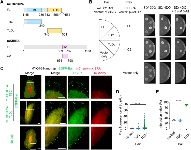
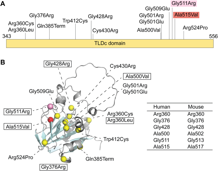
Mutations of human TBC1D24 are associated with deafness, epilepsy, or DOORS syndrome (deafness, onychodystrophy, osteodystrophy, cognitive disability, and seizures). The causal relationships between TBC1D24 variants and the different clinical phenotypes are not understood. Our hypothesis is that phenotypic heterogeneity of missense mutations of TBC1D24 results, in part, from perturbed binding of different protein partners. To discover novel protein partners of TBC1D24, we conducted yeast two-hybrid (Y2H) screen using mouse full-length TBC1D24 as bait. Kidney and brain protein (KIBRA), a scaffold protein encoded by Wwc1, was identified as a partner of TBC1D24. KIBRA functions in the Hippo signaling pathway and is important for human cognition and memory. The TBC1D24 TLDc domain binds to KIBRA full-length and to its C2 domain, confirmed by Y2H assays. No interaction was detected with Y2H assays between the KIBRA C2 domain and TLDc domains of NCOA7, MEAK7, and OXR1. Moreover, the C2 domains of other WWC family proteins do not interact with the TLDc domain of TBC1D24, demonstrating specificity. The mRNAs encoding TBC1D24 and KIBRA proteins in mouse are coexpressed at least in a subset of hippocampal cells indicating availability to interact in vivo. As two epilepsy-associated recessive variants (Gly511Arg and Ala515Val) in the TLDc domain of human TBC1D24 disrupt the interaction with the human KIBRA C2 domain, this study reveals a pathogenic mechanism of TBC1D24-associated epilepsy, linking the TBC1D24 and KIBRA pathways. The interaction of TBC1D24-KIBRA is physiologically meaningful and necessary to reduce the risk of epilepsy.

Keywords: C2-domain; KIBRA; TBC1D24; TLDc; WWC1; epilepsy; hippocampus; protein-protein interaction; synapse; yeast two-hybrid.


Conflict of interest statement

Conflict of interest The authors declare that they have no conflicts of interest with the contents of this article.

Figures

Figure 1
Figure 1
mTBC1D24 interacts with mKIBRA.A, diagrams of domain content of TBC1D24 full length (FL) and KIBRA FL mouse proteins. B, Y2H assays using Mus musculus (m) mTBC1D24 FL and mKIBRA FL proteins. Yeast cells expressing both bait and prey plasmids grow on SD/-2DO agar plate. When yeast have both prey and bait plasmids and prey and bait proteins interact, they grow on SD/-4DO and SD/-4DO + 3-AT (3-amino-1,2,4-triazole) agar plates. 3-AT is a competitive inhibitor of the HIS3 gene product increasing interaction stringency. C, representative images of NanoSPD 2.0 assay showing that the EGFP-TBC1D24–mCherry-mKIBRA complex binds to the MYO10-HMM-EGFP-Nanotrap and is transported by the MYO10 motor domain to tips of filopodia of a HeLa cell (top panels) showing yellow signals (red + green fluorescence). However, mCherry-KIBRA remains in the cell body in the absence of the EGFP-mTBC1D24 bait (bottom panels). Magnified images of white boxed areas of left images are shown on the right three right images. The scale bars represent 10 μm. Schematic of the NanoSPD 2.0 assay is shown in Fig. S1. D, intensity correlation analysis of data from C (right panel) shows an increase of mCherry-mKIBRA at filopodial tips in the presence of EGFP-mTBC1D24. Data are mean ± SD with each data point representing a single filopodium. E, spatial correlation analysis of images from panel C shows a significantly higher interaction index (t test) for EGFP-mTBC1D24 and mCherry-mKIBRA indicating an interaction between mTBC1D24 and mKIBRA. Statistical analyses were performed using 2-tailed t test for calculating an interaction index and a Mann-Whitney U test was used for intensity correlation analysis for comparing interaction of mTBC1D24 FL and mKIBRA FL with the no-bait negative control. Data are presented as mean ± SD with each data point representing the average interaction index from a single experiment. ∗∗∗∗p < 0.0001, ∗∗∗p < 0.001; all data are from three independent experimental determinations (n = 484–512 filopodia). 3-AT, 3-amino-1,2,4-triazole; HMM, heavy meromyosin; KIBRA, kidney and brain protein; mKIBRA, mouse KIBAR; NanoSPD, nanoscale pull-down; SD, synthetic defined; TBC, Tre2/Bub2/Cdc16; TBC1D24, TBC1 domain family member 24; Y2H, yeast two-hybrid.
Figure 2
Figure 2
The TLDc domain of mTBC1D24 interacts with the C2 domain of mKIBRA.A, protein diagrams of domain constructs of mTBC1D24 and mKIBRA, B, Y2H assays using mTBC1D24 TBC and TLDc domains and mKIBRA FL and C2 domain. C, representative images of a NanoSPD 2.0 assay showing that EGFP-TBC1D24 TLDc domain–mCherry-KIBRA C2 domain complex binds to the MYO10-HMM-EGFP-Nanotrap and is transported to tips of filopodia (top panels). However, mCherry-KIBRA C2 domain remains in the HeLa cell body in presence of the TBC domain of EGFP-TBC1D24 as bait (bottom panels). The scale bars represent 10 μm. D, NanoPSD 2.0 assay intensity correlation analysis of data from C shows an increase of mCherry-mKIBRA C2 domain at filopodial tips in the presence of EGFP-mTBC1D24 TLDc domain. Statistical analyses were performed using one-way ANOVA for calculating intensity correlation analysis for comparing mTBC1D24 TBC domain and TLDc domain and the no-bait negative control with the mKIBRA C2 domain. Data are mean ± SD with each data point representing a single filopodium. E, spatial correlation analysis of images from panel C (left panel) show a significantly higher interaction index (one-way ANOVA) for EGFP-mTBC1D24 TLDc domain and mCherry-mKIBRA C2 domain indicating an interaction between the TLDc domain of mTBC1D24 and the C2 domain of mKIBRA. Data are presented as mean ± SD with each data point representing the average interaction index from a single experiment. ∗∗∗∗p < 0.0001; all data are from three independent experiments (n = 527–591 filopodia total). FL, full length; HMM, heavy meromyosin; KIBRA, kidney and brain protein; mKIBRA, mouse KIBAR; NanoSPD, nanoscale pull-down; TBC, Tre2/Bub2/Cdc16; TBC1D24, TBC1 domain family member 24; TLDc, TBC/LysM-associated domain containing; Y2H, yeast two-hybrid.
Figure 3
Figure 3
mKIBRA FL and the mKIBRA C2 domain interact with the TLDc domain of mTBC1D24.A, Y2H assays using WT mKIBRA FL or mKIBRA FL with a Cys772Ala substitution as bait and WT mKIBRA FL or mKIBRA FL with a Cys772Ala substitution as prey. Yeast cotransformed with WT mKIBRA FL plus WT mKIBRA FL, WT mKIBRA FL plus mKIBRA FL with the Cys772Ala substitution, and mKIBRA FL with Cys772Ala substitution plus mKIBRA FL with Cys772Ala substitution grew on selection media. These data indicate that mKIBRA FL forms homodimers without using the intramolecular disulfide bond of Cys772 in the C2 domain. Y2H assay using the WT mKIBRA C2 domain or mKIBRA C2 domain with the Cys772Ala substitution as bait and WT mKIBRA C2 domain or mKIBRA C2 domain with the Cys772Ala substitution as prey showed that yeast did not grow in the presence of any combination of bait and prey. These data indicate that the C2 domain alone does not form a dimer in yeast. B, yeast cotransformed with mTBC1D24 FL or the TLDc domain as bait and mKIBRA FL or the C2 domain as prey grew on the selection media, indicating both mTBC1D24 FL and the TLDc domain interacts with mKIBRA FL and the C2 domain in yeast. KIBRA, kidney and brain protein; mKIBRA, mouse KIBAR; TBC, Tre2/Bub2/Cdc16; TBC1D24, TBC1 domain family member 24; TLDc, TBC/LysM-associated domain containing; Y2H, yeast two-hybrid; full-length.
Figure 4
Figure 4
Only the mTBC1D24 TLDc domain interacts with the mKIBRA C2 domain. Y2H assays using the TLDc domains of the five TLDc domain-containing proteins and mKIBRA C2 domain as bait and prey, respectively. Yeast cotransformed with mTLDC2 TLDc domain and mKIBRA C2 domain grew on selection media (section number 4). However, yeast cotransformed with mTLDC2 TLDc and the empty-prey vector also grew (section number 10) indicating that mTLDC2 TLDc domain has intrinsic autoactivation ability and cannot be tested in our Y2H assay. This autoactivation activity of mTLDC2 TLDc domain can be eliminated in the presence of 50 mM 3-AT (Fig. S3E) and does not show the interaction with the C2 domain of mKIBRA. 3-AT, 3-amino-1,2,4-triazole; KIBRA, kidney and brain protein; mKIBRA, mouse KIBAR; mTLDC2, mouse TLDC2; TBC1D24, TBC1 domain family member 24; TLDc, TBC/LysM-associated domain containing; Y2H, yeast two-hybrid.
Figure 5
Figure 5
mWWC2 C2 domain does not interact with mTBC1D24 TLDc domain.A, amino acid sequence alignment of C2 domains of mouse WWC family proteins. KIBRA (WWC1), RefSeq: NP_740749.1 and WWC2, RefSeq: NP_598552.2. Dark gray and light gray indicate identical and similar amino acids, respectively. B, Y2H assays using the TLDc domain of mTBC1D24 and the C2 domains of mKIBRA or mWWC2 as bait and prey, respectively. KIBRA, kidney and brain protein; mKIBRA, mouse KIBAR; TBC, Tre2/Bub2/Cdc16; TBC1D24, TBC1 domain family member 24; TLDc, TBC/LysM-associated domain containing; WWC1, WW and C2 domain containing 1; Y2H, yeast two-hybrid.
Figure 6
Figure 6
Tbc1d24 and Wwc1 (Kibra) mRNAs are expressed in the same cells of the mouse hippocampus.A, drawing of a coronal section of mouse brain and regions analyzed in this study. cornu ammonis (CA) 1 to 4 and dentate gyrus (DG) are labeled. B, in situ hybridization using RNAscope probes and coronal sections of the hippocampus from a C57BL/6J male WT mouse at postnatal day 66. Top panel shows Tbc1d24 mRNA (magenta, probe-Mm-Tbc1d24-C3) and Wwc1 (Kibra) mRNA (green, probe-Mm-Wwc1-03) expressed in hippocampal cells. DAPI (purple-blue-gray) stains nuclei. Lower panels show Z-stack of confocal images taken through the cell body including the nucleus and then compressed. Scale bars are 200 μm. C, in situ hybridization using Tbc1d24 and Wwc1 RNAscope probes combined with Neurin (NeuN) antibody. NeuN is a neural cell marker and is shown in white. Left column shows regions of hippocampus, and yellow boxes indicate areas from which magnified images were taken. Scale bars are 20 μm. The expression of Tbc1d24 mRNA (magenta) and Wwc1 mRNA (green) were observed in the same cells in CA1-3 and DG areas in the hippocampus. Z-stack images of high magnifications were analyzed using orthogonal projections. DAPI is shown in purple-blue-gray. Merged 1 shows combined images using Tbc1d24 and Kibra RNAscope probes and nuclei stained with DAPI. Merged two images show immunofluorescence for the antibody against NeuN. Yellow dotted lines emphasize the location of the plasma membrane of a cell. Scale bars are 5 μm. Additional independent experiments with male and female mice are shown in Fig. S5. DAPI, 4′,6-diamidino-2-phenylindole; KIBRA, kidney and brain protein; TBC, Tre2/Bub2/Cdc16; TBC1D24, TBC1 domain family member 24; WWC1, WW and C2 domain containing 1.
Figure 7
Figure 7
Epilepsy associated-missense variants in the TLDc domain of human TBC1D24. Fourteen epilepsy associated variants in the TLDc domain of hTBC1D24 are shown on a protein diagram of the TLDc domain of hTBC1D24 shown in (A) and AlphaFold protein predicted structure (entry: Q9ULP9) shown in (B). Residues of the TLDc domain of mTBC1D24 corresponding to hTBC1D24 variants in boxes were used for Y2H assays. Ala515Val (red highlight in A and red ball in B) entirely abrogates the interaction of hTBC1D24 with KIBRA in a Y2H assay (Fig. 8). Gly511Arg (pink highlight in A and pink ball in B) appears to reduce affinity strength of the interaction between hTBC1D24 and hKIBRA based on 3-AT Y2H titration assays (Fig. 9). Table in this figure shows human TBC1D24 amino acid residues and the corresponding mouse TBC1D24 residues used in Y2H assays. 3-AT, 3-amino-1,2,4-triazole; hKIBRA, human kidney and brain protein; TBC, Tre2/Bub2/Cdc16; TBC1D24, TBC1 domain family member 24; TLDc, TBC/LysM-associated domain containing; Y2H, yeast two-hybrid.
Figure 8
Figure 8
Epilepsy-associated Ala517Val amino acid substitution in the TLDc domain of mTBC1D24 abolishes the interaction with mKIBRA.A, Y2H assays using six mTBC1D24 substitution variants corresponding to reported epilepsy-associated variants of hTBC1D24. Ala517Val abolished the interaction between mTBC1D24 and mKIBRA, whereas the other substitutions do not prevent the interaction of mTBC1D24 and mKIBRA. B, Y2H assays using mTBC1D24 with a series of side chain substitutions of mTBC1D24 Ala517 residue corresponding to hTBC1D24 Ala515. KIBRA, kidney and brain protein; mKIBRA, mouse KIBAR; TBC, Tre2/Bub2/Cdc16; TBC1D24, TBC1 domain family member 24; TLDc, TBC/LysM-associated domain containing; Y2H, yeast two-hybrid.
Figure 9
Figure 9
hTBC1D24 TLDc domain interacts with hKIBRA C2 domain. Y2H assays show that hTBC1D24 TLDc domain binds to hKIBRA C2 domain. Yeast cotransformed with hTBC1D24 and hKIBRA grew on the SD/-3DO selection medium, but not on SD/-4DO selection medium which is more stringent than SD/-3DO. This indicates that the interaction between hTBC1D24 and hKIBRA is weaker compared to the interaction between mTBC1D24 and mKIBRA. hKIBRA, human kidney and brain protein; mKIBRA, mouse KIBAR; TBC, Tre2/Bub2/Cdc16; TBC1D24, TBC1 domain family member 24; SD, synthetic defined; TLDc, TBC/LysM-associated domain containing; Y2H, yeast two-hybrid.
Figure 10
Figure 10
Epilepsy-associated variants Gly511Arg and Ala515Val of hTBC1D24 disrupt the interaction with hKIBRA. Yeast growth was tested on selection medium with histidine (DO-2) or without histidine (DO-3) in presence of various concentrations of 3-AT of calibrated yeast cell concentrations. A, semiquantitative Y2H analyses of hTBC1D24 and hKIBRA. Ala515Val hTBC1D24 abolishes the interaction with hKIBRA, while the Gly511Arg hTBC1D24 disrupts the interaction but only with a higher concentration of 3-AT. B, semiquantitative Y2H analyses of mTBC1D24 and mKIBRA. mTBC1D24 Gly513 and Ala517 correspond to Gly511 and Ala515 of hTBC1D24, respectively. Ala517Val mTBC1D24 abolishes the interaction with mKIBRA in all conditions tested. However, Gly513Arg mTBC1D24 allows for an interaction with mKIBRA unlike hTBC1D24 Gly511Arg. 3-AT, 3-amino-1,2,4-triazole; TBC, Tre2/Bub2/Cdc16; hKIBRA, human kidney and brain protein; hTBC1D24, human TBC1D24; mKIBRA, mouse KIBAR; TBC1D24, TBC1 domain family member 24; Y2H, yeast two-hybrid.

References

    1. Fukuda M. TBC proteins: GAPs for mammalian small GTPase Rab? Biosci. Rep. 2011;31:159–168. - PubMed
    1. Arnaud-Arnould M., Tauziet M., Moncorge O., Goujon C., Blaise M. Crystal structure of the TLDc domain of human NCOA7-AS. Acta Crystallogr. F Struct. Biol. Commun. 2021;77:230–237. - PMC - PubMed
    1. Finelli M.J., Oliver P.L. TLDc proteins: new players in the oxidative stress response and neurological disease. Mamm. Genome. 2017;28:395–406. - PMC - PubMed
    1. Azaiez H., Booth K.T., Bu F., Huygen P., Shibata S.B., Shearer A.E., et al. TBC1D24 mutation causes autosomal-dominant nonsyndromic hearing loss. Hum. Mutat. 2014;35:819–823. - PMC - PubMed
    1. Rehman A.U., Santos-Cortez R.L., Morell R.J., Drummond M.C., Ito T., Lee K., et al. Mutations in TBC1D24, a gene associated with epilepsy, also cause nonsyndromic deafness DFNB86. Am. J. Hum. Genet. 2014;94:144–152. - PMC - PubMed

MeSH terms

Substances

Supplementary concepts