. 2024 Oct 15:201:106653.
doi: 10.1016/j.nbd.2024.106653. Epub 2024 Aug 28.

Neurovascular mechanisms of cognitive aging: Sex-related differences in the average progression of arteriosclerosis, white matter atrophy, and cognitive decline

Affiliations

Neurovascular mechanisms of cognitive aging: Sex-related differences in the average progression of arteriosclerosis, white matter atrophy, and cognitive decline

Daniel C Bowie et al. Neurobiol Dis. .

Abstract

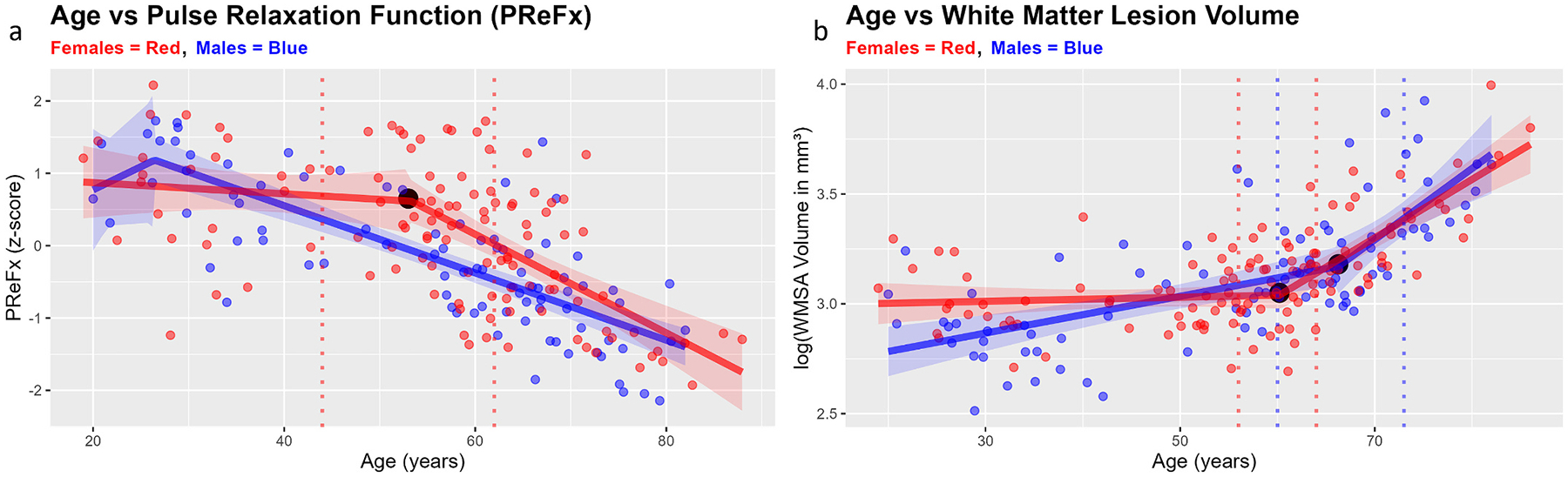
Arterial stiffness (arteriosclerosis) has been linked to heightened risks for cognitive decline, and ultimately for Alzheimer's disease and other forms of dementia. Importantly, neurovascular outcomes generally vary according to one's biological sex. Here, capitalizing on a large sample of participants with neuroimaging and behavioral data (N = 203, age range = 18-87 years), we aimed to provide support for a hierarchical model of neurocognitive aging, which links age-related declines in cerebrovascular health to the rate of cognitive decline via a series of intervening variables, such as white matter integrity. By applying a novel piecewise regression approach to our cross-sectional sample to support Granger-like temporal inferences, we show that, on average, a precipitous decline in cerebral arterial elasticity (measured with diffuse optical imaging of the cerebral arterial pulse; pulse-DOT) precedes an acceleration in the development of white matter lesions by nearly a decade, with women protected from these deleterious effects until approximately age 50, the average onset of menopause. By employing multiple-mediator path analyses while controlling for sex, we show that age may impair cognition via the sequential indirect effects of arteriosclerosis and white matter atrophy on fluid, but not crystallized, abilities. Importantly, we replicate these results using pulse pressure, an independent index of arterial health, thereby providing converging evidence for the central role of arteriosclerosis as an accelerating factor in normal and pathological aging and identifying robust sex-related differences in the progression of cerebral arteriosclerosis and white matter degradation.

Keywords: Arteriosclerosis; Cerebral arterial pulse based on diffused optical tomography (pulse-DOT); Cerebrovascular health; Cognitive aging; Sex differences; White matter lesions.


Conflict of interest statement

Declaration of competing interest None of the authors of this article has any financial or other conflicts of interest regarding this work.

Figures

Fig. 1.
Fig. 1.
Hypothesized arterial dysfunction pathway to cognitive decline (measures). Greyed out panels indicate variables for which we do not have measures of in the present paper. Evidence suggests that cardiorespiratory fitness (CRF) may moderate the relationship between age and arterial health, thereby providing a point of intervention (via regular aerobic exercise) to prevent or slow the age-related progression of arteriosclerosis. Female sex hormones such estradiol may also confer protective effects to the cerebrovasculature prior to menopausal onset (≈age 50), after which production of estrogens declines precipitiously.
Fig. 2.
Fig. 2.
Scatterplots with piecewise linear regression lines superimposed (shaded areas indicate 95 % confidence interval bands). Participants are colour-coded by their self-reported sex (females = RED, males = BLUE). The black dot in each plot indicates the ‘best’ breakpoint estimate for each piecewise relationship, or the age at which the difference in slopes between the two regression lines is largest—these breakpoints are statistically significant after employing a Holm-Bonferroni correction. The vertical dashed lines represent the lower and upper limits of the 95 % confidence intervals for each estimate. The breakpoint estimates for (a) cerebral arterial health and (b) cerebral white matter atrophy differ by approximately 8 years, with partially nonoverlapping confidence intervals. Together, this suggests that, on average, declines in cerebrovascular health precede the accelerated formation of white matter lesions. WMSA = white matter signal abnormalities.
Fig. 3.
Fig. 3.
Scatterplots with piecewise linear regression lines superimposed (shaded areas indicated 95 % confidence interval bands). Participants are colour-coded by their self-reported sex (females = RED, males = BLUE). The black dot in each plot indicates the ‘best’ breakpoint estimate for each piecewise relationship, or the age at which the difference in slopes between the two regression lines is largest—these breakpoints are statistically significant after employing a Holm-Bonferroni correction. The vertical dashed lines represent the lower and upper limits of the 95 % confidence intervals for each estimate. The breakpoint estimates for (a) fluid cognition and (b) crystallized or verbal cognition are seemingly attributable to different physiological phenomena, due to their relatively large discrepancy. The accelerated decline in fluid cognition seemingly occurs within the interval during which we observe accelerated declines in cerebrovascular health and white matter health in our data, which is consistent with prior evidence indicating that fluid abilities are more susceptible to neurobiological deterioration than crystallized cognition.
Fig. 4.
Fig. 4.
Scatterplots with separate piecewise linear regression lines for females and males superimposed (red and blue shaded areas indicated 95 % confidence interval bands for females and males, respectively). The black dot(s) in each plot indicates the ‘best’ breakpoint estimate for each piecewise relationship, or the age at which the difference in slopes between the two regression lines is largest—these breakpoints are statistically significant after employing a Holm-Bonferroni correction. The red and blue vertical dashed lines represent the lower and upper limits of the 95 % confidence intervals for each estimate for females and males, respectively. The breakpoint estimates for (a) PReFx are only significant for females at age 53, roughly coinciding with the onset of menopause and a drop in estrogen production. Males’ cerebrovascular health seemingly declines steadily throughout the lifespan. Males and females both exhibited statistically significant breakpoints for (b) white matter lesion volume, with females’ occurring at 60 and males’ occurring later at age 66. Contrary to males, however, females’ white matter health does not significantly decrease until age 60, presumably due to the loss of the vasoprotective effects of estradiol after age 50, which then manifest as white matter lesions years later. These results are consistent with the hypothesis that cerebrovascular declines contribute to the formation of white matter lesions.
Fig. 5.
Fig. 5.
Scatterplots with separate piecewise linear regression lines for females and males superimposed (red and blue shaded areas indicated 95 % confidence interval bands for females and males, respectively). The black dot(s) in each plot indicates the ‘best’ breakpoint estimate for each piecewise relationship, or the age at which the difference in slopes between the two regression lines is largest—these breakpoints are statistically significant after employing a Holm-Bonferroni correction. The red and blue vertical dashed lines represent the lower and upper limits of the 95 % confidence intervals for each estimate for females and males, respectively. The breakpoint estimates for (a) fluid cognition are only significant for females at age 58, which is within the presumed interval during which vascular health declines and white matter lesion development accelerates. Males’ fluid abilities seemingly decline steadily throughout the lifespan. Only females exhibited a statistically significant breakpoint for (b) crystallized or verbal cognition, occurring at 65. This suggests that crystallized cognition may be less vulnerable to declines in cerebrovascular health and cerebral matter atrophy.
Fig. 6.
Fig. 6.
Serial mediation diagram examining the effect of age on fluid cognition via two intervening variables, PReFx (a pulse-DOT parameter measuring cerebral arterial stiffness) and T1w white matter hypointensities (an index of white matter lesions or WMSAs). Self-reported sex is controlled for in these analyses but is not included in the diagram to maintain visual clarity. The two-mediator sequential indirect effect is significant, which is consistent with the hypothesis that age-related declines in cerebral arterial health contribute to the formation of white matter lesions and negatively impact fluid cognition. Red font/arrows indicate paths implicated in the multiple-mediator indirect effect. *p < .05, **p < .01, ***p < .001.
Fig. 7.
Fig. 7.
Serial mediation diagram examining the effect of age on crystallized cognition via two intervening variables, PReFx (a pulse-DOT parameter measuring cerebral arterial stiffness) and T1w white matter hypointensities (an index of white matter lesions or WMSAs). Self-reported sex is controlled for in these analyses but is not included in the diagram to maintain visual clarity. The two-mediator sequential indirect effect is not significant, which is consistent with the hypothesis that age-related declines in cerebral arterial health contribute to the formation of white matter lesions but do not severely impact crystallized cognition. *p < .05, **p < .01, ***p < .001.

Update of

References

    1. Abdelkarim D, Zhao Y, Turner MP, Sivakolundu DK, Lu H, Rypma B, 2019. A neural-vascular complex of age-related changes in the human brain: anatomy, physiology, and implications for neurocognitive aging. Neurosci. Biobehav. Rev 107, 927–944. 10.1016/j.neubiorev.2019.09.005. - DOI - PubMed
    1. Beck AT, Steer RA, Brown GK, 1996. Manual for the Beck Depression Inventory-II.
    1. Buckner RL, Head D, Parker J, Fotenos AF, Marcus D, Morris JC, Snyder AZ, 2004. A unified approach for morphometric and functional data analysis in young, old, and demented adults using automated atlas-based head size normalization: reliability and validation against manual measurement of total intracranial volume. NeuroImage 23 (2), 724–738. 10.1016/j.neuroimage.2004.06.018. - DOI - PubMed
    1. Burt VL, Harris T, 1994. The third National Health and nutrition examination survey: contributing data on aging and health. Gerontologist 34 (4), 486–490. 10.1093/geront/34.4.486. - DOI - PubMed
    1. Calamante F, Thomas DL, Pell GS, Wiersma J, Turner R, 1999. Measuring cerebral blood flow using magnetic resonance imaging techniques. J. Cereb. Blood Flow Metab 19 (7), 701–735. 10.1097/00004647-199907000-00001. - DOI - PubMed

Publication types

MeSH terms