. 2024 Aug 30;15(1):7481.
doi: 10.1038/s41467-024-51273-1.

Phosphoribosyl modification of poly-ubiquitin chains at the Legionella-containing vacuole prohibiting autophagy adaptor recognition

Affiliations

Phosphoribosyl modification of poly-ubiquitin chains at the Legionella-containing vacuole prohibiting autophagy adaptor recognition

Min Wan et al. Nat Commun. .

Abstract

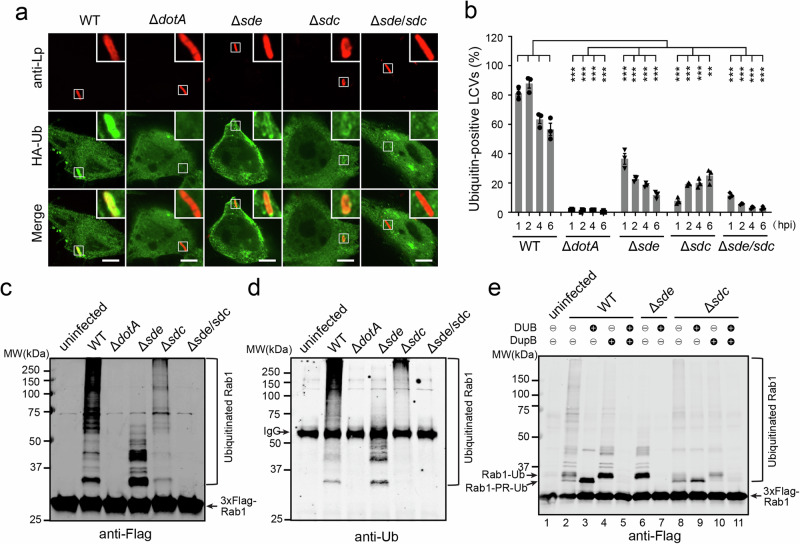
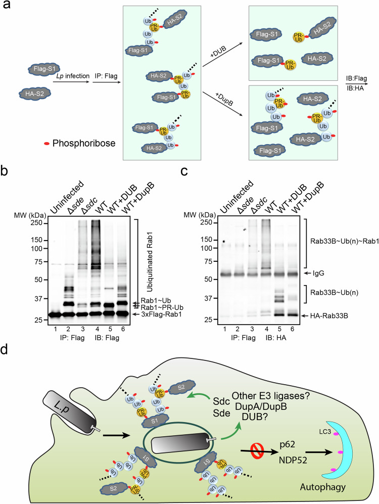
Ubiquitination is a posttranslational modification in eukaryotes that plays a significant role in the infection of intracellular microbial pathogens, such as Legionella pneumophila. While the Legionella-containing vacuole (LCV) is coated with ubiquitin (Ub), it avoids recognition by autophagy adaptors. Here, we report that the Sdc and Sde families of effectors work together to build ubiquitinated species around the LCV. The Sdc effectors catalyze canonical polyubiquitination directly on host targets or on phosphoribosyl-Ub conjugated to host targets by Sde. Remarkably, Ub moieties within poly-Ub chains are either modified with a phosphoribosyl group by PDE domain-containing effectors or covalently attached to other host substrates via Sde-mediated phosphoribosyl-ubiquitination. Furthermore, these modifications prevent the recognition by Ub adaptors and therefore exclude host autophagy adaptors from the LCV. In this work, we shed light on the nature of the poly-ubiquitinated species present at the surface of the LCV and provide a molecular mechanism for the avoidance of autophagy adaptors by the Ub-decorated LCV.


Conflict of interest statement

The authors declare no competing interests.

Figures

Fig. 1
Fig. 1. The sde and sdc families of effectors are required for the ubiquitination of host targets and the enrichment of ubiquitinated species at the LCV.
a Representative confocal images showing the Ub signals (green) at the LCV in HEK293T cells challenged with specified Legionella strains (red) at 1 hpi. Scale Bar: 5 μm. b Quantitative analysis of Ub-positive LCVs in cells infected with the indicated Legionella strains. Data were shown as means ± SEM of three independent experiments. At least 45 randomly selected LCVs were counted for each condition. Statistical comparisons between two groups were conducted using unpaired two-tailed Student’s t-tests. The P values (1 h, 2 h, 4 h, 6 h) are: <0.0001, <0.0001, 0.0001, 0.0002 (ΔdotA vs WT); 0.0005, <0.0001, <0.0001, 0.0005 (Δsde vs WT); <0.0001, <0.0001, <0.0001, 0.0002 (Δsdc vs WT); <0.0001, <0.0001, 0.0002, 0.0035 (Δsde/sdc vs WT). **p < 0.01; ***p < 0.001. c Western-blot analysis of Rab1 ubiquitination. HEK293T cells were transfected with plasmids expressing FcγRII and 3xFlag-Rab1 and then infected with the indicated Legionella strains for 1 h. Cell lysates were prepared and 3xFlag-Rab1 was immunoprecipitated using anti-Flag resins. Precipitated materials were analyzed by Western blot using anti-Flag or (d) anti-Ub antibodies. e Western-blot analysis 3xFlag-Rab1 after immunoprecipitation from cells challenged with indicated Legionella strains followed by DUB and/or DupB treatment. Source data for b is shown in Source Data. Data shown in (c), (d), and (e) are one representative experiment of three independent experiments. Uncropped blots are shown in Source Data.
Fig. 2
Fig. 2. Sde and Sdc facilitate the recruitment of their host targets to the LCV.
a Representative confocal images show the localization of EGFP-Rab1 (green) in HEK293T cells challenged with specified Legionella strains (red) for 1 h. Scale Bar: 5 μm. b Quantitative analysis of Rab1-positive LCVs in cells infected with the indicated Legionella strains. Data were shown as means ± SEM of three independent experiments. At least 60 randomly selected LCVs were counted for each condition. Statistical comparisons between two groups were conducted using unpaired two-tailed Student’s t-tests. The P values are: <0.0001 (ΔdotA vs WT); <0.0001 (Δsde vs WT); 0.0015 (Δsdc vs WT); <0.0001 (Δsde/sdc vs WT). **p < 0.01; ***p < 0.001. c Representative confocal images show the recruitment of an ER-marker protein Sec61β (green) at the LCV (red). Scale Bar: 5 μm. d Quantitative analysis of Sec61β-positive LCVs. Data were shown as means ± SEM of three independent experiments. At least 60 randomly selected LCVs were counted for each condition. Statistical comparisons between two groups were conducted using unpaired two-tailed Student’s t-tests. The P values are: 0.0007 (ΔdotA vs WT); 0.0011 (Δsde vs WT); 0.0082 (Δsdc vs WT); 0.001 (Δsde/sdc vs WT). **p < 0.01; ***p < 0.001. Source data for (b) and (d) is shown in Source Data.
Fig. 3
Fig. 3. Sde and Sdc synthesize mixed ubiquitin chains on their host targets.
a A schematic presentation of the mixed Ub chains on a substrate and the predicted outcomes after DUB or DupB cleavage. b Immunoblot image of in vitro ubiquitinated Rab33B after the cleavage by DUB and/or DupB. Purified recombinant 4xFlag-Rab33B was first ubiquitinated by SidC and SdeA enzymes in vitro and then immobilized on anti-Flag resins followed by the cleavage with DUB and/or DupB. The cleaved products released in the supernatant (Sup) and remained on the beads (Beads) were analyzed by SDS-PAGE followed by anti-Ub Western blot. c Immunoblot analysis of Rab33B prepared from infected cells after the cleavage by DUB and/or DupB. HEK293T cells expressing FCγRII and 4xFlag-Rab33B were infected with wild-type Legionella strain for 2 h. 4xFlag-Rab33B was enriched by anti-Flag resins, followed by the treatment with DUB and/or DupB. The cleaved products that were released in the supernatant (Sup) and that remained on the beads (Beads) were analyzed by SDS-PAGE followed by anti-Ub Western blot. Data shown in (b) and (c) are one representative experiment of three independent experiments. Uncropped blots are shown in Source Data.
Fig. 4
Fig. 4. Sde suppresses the recruitment of p62 to the LCV by disrupting the physical interaction between p62 and Ub.
a Representative confocal images show the localization of mCherry-p62 (red) in HEK293T cells challenged with specified Legionella strains (green) for 1 h. Scale Bar: 5 μm. b Quantitative analysis of p62-positive LCVs in cells infected with the indicated Legionella strains. Data were shown as means ± SEM of three independent experiments. At least 33 randomly selected LCVs were counted for each condition. Statistical comparisons between two groups were conducted using unpaired two-tailed Student’s t-tests. The P values (1 h, 2 h, 4 h) are: 0.0547, 0.0651, 0.1212 (ΔdotA vs WT); 0.0473, 0.0937, 0.318 (Δsdc vs WT); 0.0005, 0.0304, 0.1779 (Δsde vs WT); 0.0004, 0.008, 0.0065, (Δsde+pSdeA vs Δsde); 0.0204, 0.1862, 0.1343 (Δsde+pSdeAEE/AA); 0.0008, 0.0126, 0.0243 (Δsde+pSdeAH277A). NS = nonsignificance; *p < 0.05; **p < 0.01; ***p < 0.001. c Co-immunoprecipitation assay of the interaction between p62 and ubiquitinated Rab1. HEK293T cells expressing FcγRII, 3xFlag-Rab1, and HA-p62 were infected with the indicated Legionella strains for 1 h. 3xFlag-Rab1 was enriched by anti-Flag immunoprecipitation. Immunoprecipitated materials were analyzed by anti-Flag (Rab1) and anti-HA (p62) Western blot. d The amount of HA-p62 bound to Rab1 was normalized to the value of Δsde group. Data were shown as means ± SEM of three independent experiments. Statistical comparisons between two groups was conducted using unpaired two-tailed Student’s t-tests. The P values are: 0.8082 (ΔdotA vs WT); 0.7847 (Δsdc vs WT); <0.0001 (Δsde vs WT); <0.0001 (Δsde+pSdeA vs Δsde); 0.0108 (Δsde+ pSdeAEE/AA); <0.0001 (Δsde+ pSdeAH277A). NS nonsignificance; *p < 0.05; ***p < 0.001. e Ribbon diagrams of the UBA-Ub complex (PDB ID: 2g3q) and the modeled UBA-PR-Ub complex. The UBA domain is colored in blue and the Ub is in yellow. The PR group attached to Ub R42 is shown in red spheres. Note the steric collision between the UBA domain and the PR group. This figure was generated using PyMol. Source data for (b) and (d) is shown in Source Data. Data shown in (c) is one representative experiment of three independent experiments. Uncropped blots are shown in Source Data.
Fig. 5
Fig. 5. Sde modifies poly-Ub chains by phosphoribosylation.
a In vitro modification of K63-linked poly-Ub chains by SdeA. Mono-Ub or K63-linked poly-Ub chains were incubated with the catalytic core of SdeA and NAD+. The products were analyzed by SDS-PAGE followed by staining with Coomassie blue (left panel) or Pro-Q diamond (right panel). b Schematic diagram of the poly-Ub chains attached to the substrate and experimental flow of biochemical and Mass spectrometry analysis of the poly-Ub chains. c Biochemical analysis of Ub modification by Sde in vitro. Recombinant 4x-Flag-Rab33B was ubiquitinated in vitro by the catalytic domain of SidC or by both SidC and SdeA. Ubiquitinated Rab33B was immobilized on anti-Flag beads and treated with a canonical DUB. The samples before and after the DUB treatment were analyzed by SDS-PAGE followed by anti-Ub Western blot (top panel) or by Pro-Q diamond stain (bottom panel). d LC-MS/MS spectrum of a Ub peptide from an in vitro ubiquitinated sample showing the phosphoribosyl modification at Ub R42. Recombinant 4x-Flag Rab33B was ubiquitinated in vitro by SidC and SdeA and then enriched by anti-Flag beads. The resulting bound proteins were treated with a purified DUB, and the Ub molecules released from the cleavage were analyzed by LC-MS/MS. e Biochemical analysis of Ub modification by Sde in vivo. HEK293T cells expressing FCγRII and 4xFlag-Rab33B were infected with WT or Δsde strain for 2 h. 4xFlag-Rab33B was immunoprecipitated by anti-Flag resins and then treated with a canonical DUB. The samples before and after the DUB treatment were analyzed by SDS-PAGE followed by anti-Ub Western blot (top panel) or by Pro-Q diamond stain (bottom panel). Samples prepared from cells infected with the Δsde strain were loaded with 1 fold (1x) or 6 folds (6x) of the amount of the sample from the WT infection. f LC-MS/MS spectrum of a Ub peptide from a sample prepared from Legionella infected cells showing the phosphoribosyl modification at Ub R42. Samples were prepared similarly as in (e). Data shown in (a), (c), and (e) are one representative experiment of three independent experiments. Uncropped gels and blots are shown in Source Data.
Fig. 6
Fig. 6. Both DupA and DupB play a role in the processing of ADPR-Ub to PR-Ub.
a Schematic representation of DupA and DupB modification on free poly-Ub chains. b K63-linked poly-Ub chains were first modified by purified SdeAH277A and subsequently treated with DupA or DupB. Conversion of ADPR-Ub to PR-Ub following DupA or DupB treatment was analyzed by SDS-PAGE followed by Coomassie blue (left panel) or Pro-Q diamond stain (right panel). c Schematic representation of DupA and DupB modification on a poly-ubiquitinated substrate. d 4x-Flag Rab33B was poly-ubiquitinated by recombinant SidC and modified by SdeAH277A. Ubiquitinated Rab33B was immobilized on anti-Flag beads and then treated with DupA or DupB. The samples were then treated with a canonical DUB to release mono-Ub. Samples before and after DUB cleavage were analyzed by SDA-PAGE followed by anti-Ub Western blot (top panel) or by Pro-Q diamond stain (bottom panel). Data shown in (b) and (d)are one representative experiment of three independent experiments. Uncropped gels and blots are shown in Source Data.
Fig. 7
Fig. 7. Cross-linking of multiple Sdc and Sde targets by canonical and PR-ubiquitination.
a Schematic model of two host substrates that are cross-linked by canonical and PR-ubiquitination and the experimental flow of verification. In this model, a poly-Ub chain attached to one substrate (either through canonical or PR-ubiquitination), and the Ub moieties within the Ub chain can be used to modify a second substrate via PR-ubiquitination. The cleavage by a canonical DUB will result in unmodified and mono- or multi-mono PR-ubiquitinated products, while the cleavage by DupB will yield unmodified and ubiquitinated (with a variable length) products. b, c Biochemical analysis of substrate crossing-linking in Legionella infection. HEK293T cells expressing FCγRII, 3xFlag-Rab1, and HA-Rab33B were infected with indicated Legionella strains for 2 h. 3xFlag-Rab1 was immunoprecipitated by anti-Flag resins and then treated with a canonical DUB or DupB. The samples before and after the treatment were analyzed by SDS-PAGE followed by anti-Flag (b) or anti-HA Western blot (c). d A schematic model of unconventional poly-Ub chains at the LCV serving as a scaffold for cross-linking multiple host targets and preventing host autophagy detection. Data shown in (b) and (c) are one representative experiment of three independent experiments. Uncropped blots are shown in Source Data.

Update of

References

    1. Cuervo, A. M. Autophagy: in sickness and in health. Trends Cell Biol.14, 70–77 (2004). 10.1016/j.tcb.2003.12.002 - DOI - PubMed
    1. Shintani, T. & Klionsky, D. J. Autophagy in health and disease: a double-edged sword. Science306, 990–995 (2004). 10.1126/science.1099993 - DOI - PMC - PubMed
    1. Yang, Z. & Klionsky, D. J. Mammalian autophagy: core molecular machinery and signaling regulation. Curr. Opin. Cell Biol.22, 124–131 (2010). 10.1016/j.ceb.2009.11.014 - DOI - PMC - PubMed
    1. Lin, M. G. & Hurley, J. H. Structure and function of the ULK1 complex in autophagy. Curr. Opin. Cell Biol.39, 61–68 (2016). 10.1016/j.ceb.2016.02.010 - DOI - PMC - PubMed
    1. Nakatogawa, H., Ichimura, Y. & Ohsumi, Y. Atg8, a ubiquitin-like protein required for autophagosome formation, mediates membrane tethering and hemifusion. Cell130, 165–178 (2007). 10.1016/j.cell.2007.05.021 - DOI - PubMed

LinkOut - more resources