. 2024 Aug 30;15(8):637.
doi: 10.1038/s41419-024-07029-2.

TGF-β signaling promotes eosinophil activation in inflammatory responses

Affiliations

TGF-β signaling promotes eosinophil activation in inflammatory responses

Chen Zhu et al. Cell Death Dis. .

Abstract

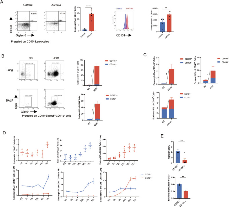
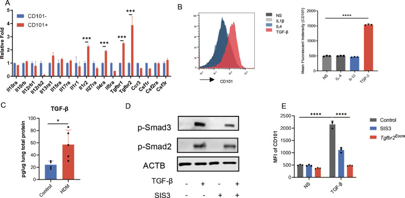
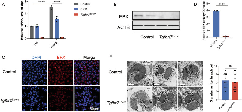
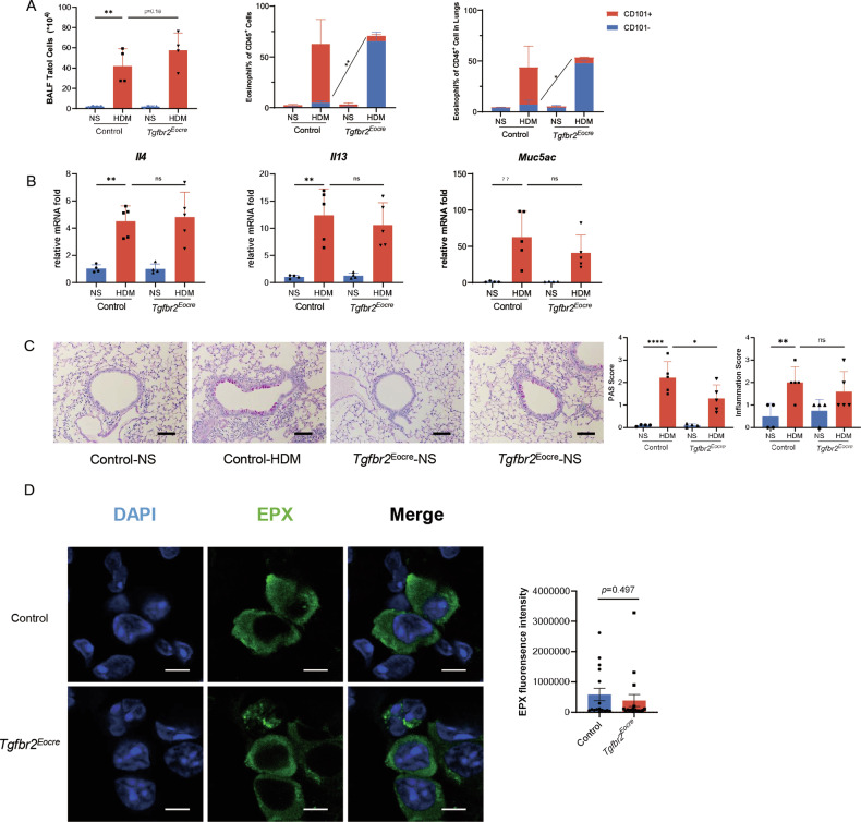
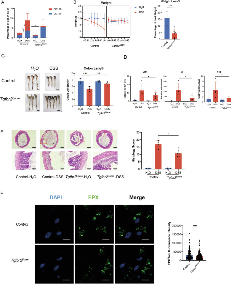
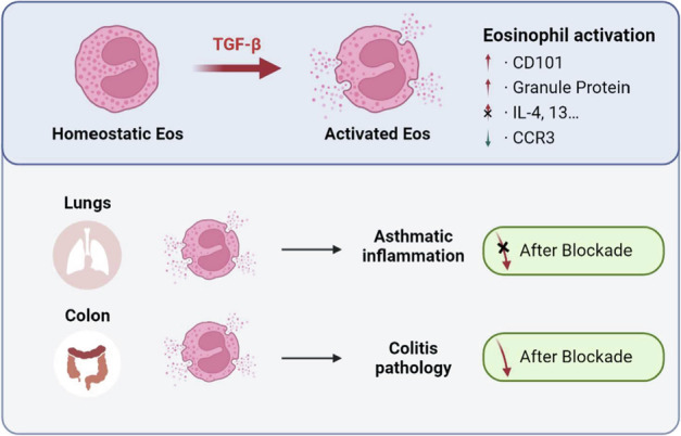
Eosinophils, traditionally associated with allergic phenomena, play a pivotal role in inflammatory responses. Despite accumulating evidence suggesting their pro-inflammatory function upon activation, the underlying mechanisms governing eosinophil activation remain incompletely characterized. In this study, we investigate the local activation of pulmonary and colon eosinophils within the inflammatory microenvironment. Leveraging transcriptional sequencing, we identify TGF-β as a putative regulator of eosinophil activation, leading to the secretion of granule proteins, including peroxidase. Genetic deletion of TGF-β receptors on eosinophils resulted in the inhibition of peroxidase synthesis, affirming the significance of TGF-β signaling in eosinophil activation. Using models of HDM-induced asthma and DSS-induced colitis, we demonstrate the indispensability of TGF-β-driven eosinophil activation in both disease contexts. Notably, while TGF-β signaling did not significantly influence asthmatic inflammation, its knockout conferred protection against experimental colitis. This study delineates a distinct pattern of eosinophil activation within inflammatory responses, highlighting the pivotal role of TGF-β signaling in regulating eosinophil behavior. These findings deepen our comprehension of eosinophil-related pathophysiology and may pave the way for targeted therapeutic approaches in allergic and inflammatory diseases.


Conflict of interest statement

The authors declare no competing interests.

Figures

Fig. 1
Fig. 1. Increased infiltration of locally activated Eos in various inflammatory responses.
A The proportion of Eos in peripheral blood of asthmatic patients, and CD101 expression (as mean fluorescence intensity) in circulating Eos in asthmatic patients. B Percentage of Eos subsets in inflammatory cells in BALF and lung homogenates. BALF cells were harvested 24 h after last exposure in HDM-induced mouse model. Eos were labeled as live CD45+SiglecF+CD11c cells. n = 4. C Percentage of Eos subsets in lung homogenates in chitin and papain induced mouse models. n = 4. D Dynamic changes (6 h, 12 h, 24 h, 48 h, 72 h from last exposure) of Eos in bone marrow, peripheral blood and lung tissue after last exposure in HDM model. E Migrated rate of Eos in transwell migration assay, and Ccr3 mRNA level of different Eos subsets. Sample size is indicated as individual plots in column graphs. Data are triplicate by individual experiments except A. **p < 0.01; ***p < 0.001; ****p < 0.0001.
Fig. 2
Fig. 2. TGF-β signaling pathway promoted Eos activation.
A Differential expression of receptors of CD101−/+ eosinophils according to the result of RNAseq. n = 3. B The expression of CD101 in stimulated Eos from NJ.1638 mice. Left, representative image of flow cytometry. Right, mean fluorescence intensity of Eos treated with 10 ng/ml IL-4, 20 ng/ml IL-1β and 10 ng/ml TGF-β. C TGF-β concentration in lung homogenates of HDM model. D Expression of Smads signaling pathway in Eos after TGF-β stimulation. E Mean fluorescence intensity of CD101 in Eos. Eos were treated with 5 μM SIS3 intervention, or purified from NJ.1638×Eocre-Tgfbr2 f/f mouse (which was hybrid by NJ.1638 and Eocre-Tgfbr2 f/f mouse, an Eos abundant and Tgfbr2 specific knockout in Eos mouse strain). Sample size is indicated as individual plots in column graphs. Data are triplicated by individual experiments except A. *p < 0.05; ***p < 0.001; ****p < 0.0001.
Fig. 3
Fig. 3. TGF-β promoted Eos activation by synthesizing Eos peroxidase.
A Relative mRNA expression of Epx in Eos from NJ.1638×Eocre-Tgfbr2f/f mouse. B Blots of EPX expression in Eos. C Immunofluorescence images of EPX expression in Tgfbr2 knockout Eos. Scale bar, 50 μm. D Relative Eos peroxidase activity in Tgfbr2 knockout Eos. E Left, Representative images on electron microscopy of Eos. Scale bar, 2 μm. Right, granules content in Eos. Sample size is indicated as individual plots in column graphs. Data are triplicate by individual experiments. ****p < 0.0001. ns not significant.
Fig. 4
Fig. 4. Inhibition of the TGF-β signaling pathway prevented Eos activation but did not suppress asthmatic inflammation.
A Cellularity in HDM-induced asthma model. Left, total cells count in BALF. Middle, Eos subsets percentage in BALF cells. Right, Eos subsets percentage in lung homogenates. n = 4. B Relative mRNA expression of lung inflammatory cytokines Il4, Il13 and Muc5ac. C Airway inflammation evaluation in asthmatic model. Left, representative images of lung histology. Scale bar, 200μm. Middle, PAS score of asthmatic mice. Right, inflammation score of asthmatic mice. D Immunofluorescence of lung tissue of HDM model. Left, representative image of immunofluorescence. EPX, green. DAPI, blue. Right, statistics of fluorescence intensity of EPX. Scale bar, 5 μm. Sample size is indicated as individual plots in column graphs. Data are triplicate by individual experiments. *p < 0.05; **p < 0.01; ****p < 0.0001. ns not significant.
Fig. 5
Fig. 5. TGF-β signaling pathway blockade suppressed Eos activation and inflammation in colitis mice.
A Different Eos subset percentage in colon of DSS-induced colitis mice. n = 3. B Weight loss in experimental colitis. Left, dynamic weight change of colitis mice after DSS exposure. n = 4. Right, percentage of weight loss after the establishment of DSS-induce colitis model. C Colon length shortening in experimental colitis. Left, representative images of mice colon. Scale bar, 1 cm. Right, Colon length of experimental colitis mice. D Relative mRNA expression of IL-1β, IL-6 and IL-10. E Histology of experimental colitis. Left, representative images of colitis tissue. Right, inflammatory score of colitis. F Immunofluorescence of foci in colon tissue of DSS model. Left, representative image of immunofluorescence. EPX, green. DAPI, blue. Right, statistics of fluorescence intensity of EPX foci. Scale bar, 5 μm. Sample size is indicated as individual plots in column graphs. Data are triplicated by individual experiments. *p < 0.05; **p < 0.01; ***p < 0.001. ns not significant.
Fig. 6
Fig. 6. Summary.
Our research suggested that TGF-β promote Eos local activation, represented as increased CD101 and granule protein. In asthma, blocking TGF-β induced Eos activation did not alleviate airway inflammation. But in colitis, blocking TGF-β could inhibit inflammatory response in colon.

References

    1. Klion AD, Ackerman SJ, Bochner BS. Contributions of eosinophils to human health and disease. Annu Rev Pathol. 2020;15:179–209. 10.1146/annurev-pathmechdis-012419-032756 - DOI - PMC - PubMed
    1. Lee JJ, Dimina D, Macias MP, Ochkur SI, McGarry MP, O’Neill KR, et al. Defining a link with asthma in mice congenitally deficient in eosinophils. Science. 2004;305:1773–6. 10.1126/science.1099472 - DOI - PubMed
    1. Ignacio A, Shah K, Bernier-Latmani J, Koller Y, Coakley G, Moyat M, et al. Small intestinal resident eosinophils maintain gut homeostasis following microbial colonization. Immunity. 2022;55:1250–67.e12. 10.1016/j.immuni.2022.05.014 - DOI - PubMed
    1. Sugawara R, Lee EJ, Jang MS, Jeun EJ, Hong CP, Kim JH, et al. Small intestinal eosinophils regulate Th17 cells by producing IL-1 receptor antagonist. J Exp Med. 2016;213:555–67. 10.1084/jem.20141388 - DOI - PMC - PubMed
    1. Masterson JC, McNamee EN, Fillon SA, Hosford L, Harris R, Fernando SD, et al. Eosinophil-mediated signalling attenuates inflammatory responses in experimental colitis. Gut. 2015;64:1236–47. 10.1136/gutjnl-2014-306998 - DOI - PMC - PubMed

Substances