. 2024 Aug 30;15(1):7547.
doi: 10.1038/s41467-024-51959-6.

Mapping the immunopeptidome of seven SARS-CoV-2 antigens across common HLA haplotypes

Affiliations

Mapping the immunopeptidome of seven SARS-CoV-2 antigens across common HLA haplotypes

Asolina Braun et al. Nat Commun. .

Abstract

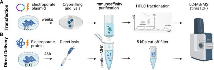
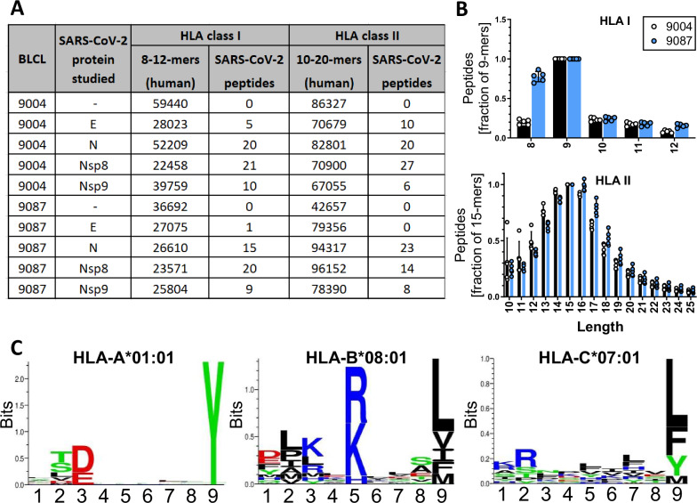
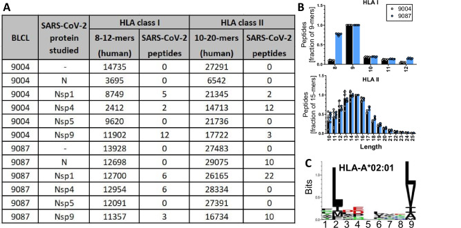
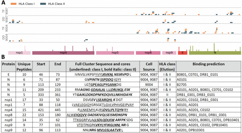
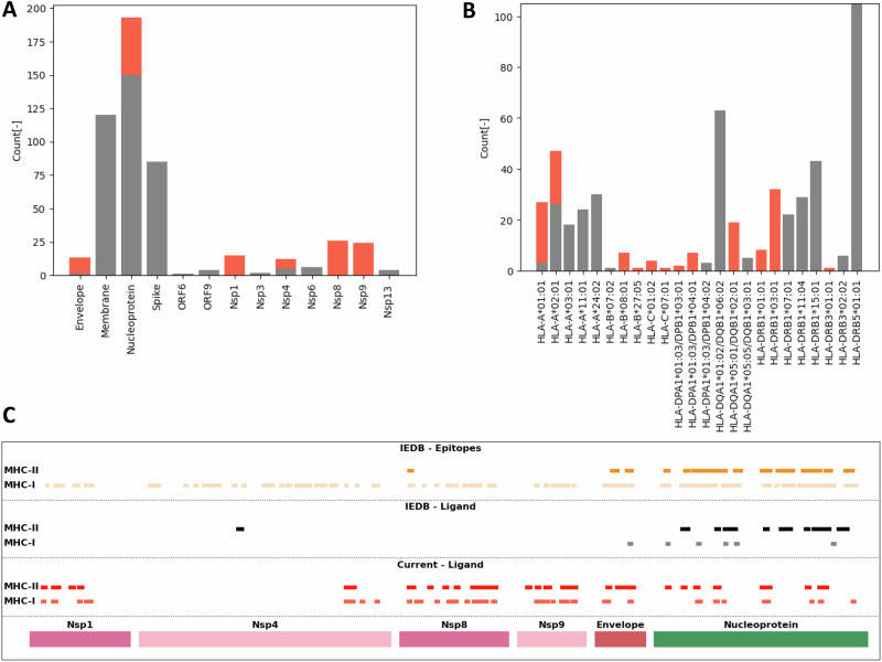
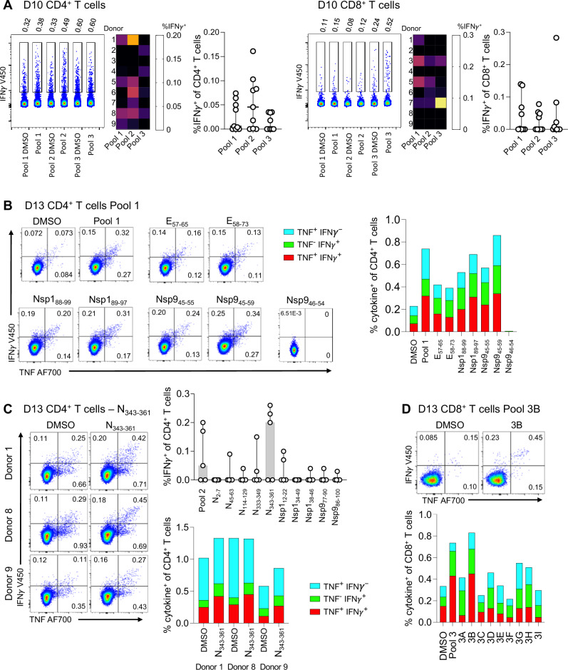
Most COVID-19 vaccines elicit immunity against the SARS-CoV-2 Spike protein. However, Spike protein mutations in emerging strains and immune evasion by the SARS-CoV-2 virus demonstrates the need to develop more broadly targeting vaccines. To facilitate this, we use mass spectrometry to identify immunopeptides derived from seven relatively conserved structural and non-structural SARS-CoV-2 proteins (N, E, Nsp1/4/5/8/9). We use two different B-lymphoblastoid cell lines to map Human Leukocyte Antigen (HLA) class I and class II immunopeptidomes covering some of the prevalent HLA types across the global human population. We employ DNA plasmid transfection and direct antigen delivery approaches to sample different antigens and find 248 unique HLA class I and HLA class II bound peptides with 71 derived from N, 12 from E, 28 from Nsp1, 19 from Nsp4, 73 from Nsp8 and 45 peptides derived from Nsp9. Over half of the viral peptides are unpublished. T cell reactivity tested against 56 of the detected peptides shows CD8+ and CD4+ T cell responses against several peptides from the N, E, and Nsp9 proteins. Results from this study will aid the development of next-generation COVID vaccines targeting epitopes from across a number of SARS-CoV-2 proteins.


Conflict of interest statement

NT, EJL, GP, MSK, JVK, and ABS are employed by Evaxion Biotech A/S which holds IP for identifying neoepitopes and personalized immunotherapy. AWP is on the scientific advisory board of Evaxion Biotech A/S. AWP is on the advisory board of Bioinformatics Solutions (Canada), and Grey Wolf Therapeutics (UK) and is a co-founder of Resseptor Therapeutics (Australia). The remaining authors declare no competing interests.

Figures

Fig. 1
Fig. 1. Workflow for the generation of SARS-CoV-2-derived HLA I and HLA II immunopeptidome data.
A Upper panel depicts the large-scale workflow used for the stable transfection and processing of B lymphoblastoid cell lines (BLCL) cells using EF1α-GoI-pIRES-DsRed plasmids containing SARS-CoV-2 genes. Cell pellets (4 x 10⁸ to 1 x 10⁹) were homogenised using a cryomill, lysed, and peptide-HLA complexes immunoaffinity purified using first the W6/32 antibody (HLA class I) and subsequently a mix of SPV-L3, LB3.1, B7/21 antibodies (HLA class II). Peptides were separated from HLA using reverse-phase high-performance liquid chromatography (HPLC). B The lower panel shows the workflow schematic for small-scale protein antigen direct delivery experiments. SARS-CoV-2-derived proteins were electroporated into BLCL and 0.9 ×10⁸ to 1.6 × 10⁸ cells collected 48 h later. Cell pellets were subjected to direct lysis and immunoaffinity purification. Peptides were separated from HLA using 5 kilodalton (kDa) molecular weight cut-off filters. All samples were analysed using liquid chromatography-tandem mass spectrometry (LC-MS/MS) and PASEF® on TimsTOF Pro. MHC: Major histocompatibility complex. Created with BioRender.com released under a Creative Commons Attribution-NonCommercial-NoDerivs 4.0 International license.
Fig. 2
Fig. 2. Immunopeptidomic profile of class I and class II eluted peptides presented by stably transfected BLCL.
Samples were analysed using PEAKS Online using a 5% FDR cut-off. A Relevant eluted peptide numbers, headers in bold. B Mean length distribution of class I and class II eluted peptides, SD is shown, based on all n = 5 cell lines in A; 9087: black bars, 9004: blue bars. C Representative Gibbs Cluster peptide motif analysis of 9-mers from the HLA class I elution of 9087_E is shown. Attribution of individual clusters to known HLA class I alleles expressed in 9087 is shown above each cluster, clusters are based on 1130-5414 peptides. Analysis using Immunolyser. Nsp: non-structural protein, BLCL: B lymphoblastoid cell line, HLA: human leukocyte antigen. Source data are provided as a Source Data file.
Fig. 3
Fig. 3. Immunopeptidomic profile of class I and class II eluted peptides presented by BLCL 48 h after direct delivery of protein antigen.
Samples were analysed using PEAKS Online using a 5% FDR cut-off. A Relevant eluted peptide numbers, headers in bold. B Mean length distribution of HLA I and HLA II eluted peptides. SD is shown, based on all n = 5 cell lines in A; 9087: black bars, 9004: blue bars. C Representative Gibbs Cluster peptide motif analysis of 9-mers from an HLA class I elution of 9004 with delivery of Nucleoprotein is shown. HLA-A*02:01-derived peptides dominate the HLA class I immunopeptidome, cluster size is 2602 peptides. Analysis using Immunolyser. Nsp non-structural protein, BLCL B lymphoblastoid cell line, HLA human leukocyte antigen. Source data are provided as a Source Data file.
Fig. 4
Fig. 4. Distribution of HLA-I and HLA-II presented protein regions within SARS-CoV-2 derived investigated proteins.
A SARS-CoV-2 derived peptides found across stable transfected (TF) B lymphoblastoid cell lines (BLCL) and direct delivery (DD) antigen approaches and their alignment to source proteins. Peaks represent mutations in variants of concern. B Table of antigen presentation hotspots. Peptide sequences derived from human leukocyte antigen (HLA) class I elutions are underlined, peptide sequences derived from HLA class II elutions are shown in bold italic. Nsp: non-structural protein, Source data are provided as a Source Data file.
Fig. 5
Fig. 5. Comparison of data generated in the current study and data available in IEDB.
Ligands identified by immunopeptidomics in the current study were compared to those deposited in the Immune Epitope Database (IEDB) for SARS-CoV-2 reading frames (A) and HLA restriction (B). The same data represented as an alignment to source proteins shows the ligand coverage of proteins analysed in the current study and the ligand/epitope coverage from the data annotated in IEDB, divided by MHC Class I or II restriction (C). IEDB epitopes were restricted to ELISPOT, intracellular staining and multimer staining assay types. Nsp: non-structural protein, HLA human leukocyte antigen, MHC Major histocompatibility complex. Source data are provided as a Source Data file.
Fig. 6
Fig. 6. CD4+ and CD8 + T cell reactivity towards detected SARS-CoV-2 peptides.
A Representative concatenated FACS plots of IFNγ expression in Donor 1 CD4+ and Donor 7 CD8 + T cells in response to peptide pools 1, 2, 3 (left). Frequency of IFNγ + CD4+ and CD8 + T cells across donors following stimulation with peptide pools (background DMSO subtracted). Percentage IFNγ+ of CD4+ and CD8 + T cells depicted as a heatmap (middle) and median with 95% CI is shown (n = 9 donors, mean with SD; right). B Representative FACS plots of CD4 + T cell IFNy+ response (left) of Donor 3. Frequency of IFNγ + TNF + CD4 + T cells after stimulation with individual pool 1 SARS-CoV-2 peptides (right). C Representative FACS plots for CD4 + T cell response to N343-361 (left). Frequency of IFNγ + CD4 + T cells stimulated with pool 2 individual peptides (background DMSO subtracted, n = 5 donors, mean with SD; top). Frequency of IFNγ + TNF + CD4 + T cells after stimulation with DMSO and N343-361 (bottom). D Representative FACS plot of IFNγ + CD8 + T cells (top). IFNγ + TNF+ expression in response to stimulation with peptide pool 3 subpools (bottom). D10/D13 day 10/13, CD cluster of differentiation, IFN interferon, TNF Tumor necrosis factor, DMSO dimethyl sulfoxidy, Nsp non-structural protein. Source data are provided as a Source Data file.

References

    1. Plante, J. A. et al. Spike mutation D614G alters SARS-CoV-2 fitness. Nature592, 116–121 (2021). 10.1038/s41586-020-2895-3 - DOI - PMC - PubMed
    1. Jangra, S. et al. SARS-CoV-2 spike E484K mutation reduces antibody neutralisation. Lancet Microbe2, e283–e284 (2021). 10.1016/S2666-5247(21)00068-9 - DOI - PMC - PubMed
    1. Low, J. S. et al. Clonal analysis of immunodominance and cross-reactivity of the CD4 T cell response to SARS-CoV-2. Science372, 1336–1341 (2021). 10.1126/science.abg8985 - DOI - PMC - PubMed
    1. Mallajosyula, V. et al. CD8(+) T cells specific for conserved coronavirus epitopes correlate with milder disease in COVID-19 patients. Sci. Immunol.6, eabg5669 (2021). 10.1126/sciimmunol.abg5669 - DOI - PMC - PubMed
    1. Augusto, D. G. et al. A common allele of HLA is associated with asymptomatic SARS-CoV-2 infection. Nature620, 128–136 (2023). - PMC - PubMed

MeSH terms