. 2024 Aug 30;15(1):7555.
doi: 10.1038/s41467-024-51793-w.

Molecular recognition of an odorant by the murine trace amine-associated receptor TAAR7f

Affiliations

Molecular recognition of an odorant by the murine trace amine-associated receptor TAAR7f

Anastasiia Gusach et al. Nat Commun. .

Abstract

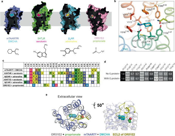
There are two main families of G protein-coupled receptors that detect odours in humans, the odorant receptors (ORs) and the trace amine-associated receptors (TAARs). Their amino acid sequences are distinct, with the TAARs being most similar to the aminergic receptors such as those activated by adrenaline, serotonin, dopamine and histamine. To elucidate the structural determinants of ligand recognition by TAARs, we have determined the cryo-EM structure of a murine receptor, mTAAR7f, coupled to the heterotrimeric G protein Gs and bound to the odorant N,N-dimethylcyclohexylamine (DMCHA) to an overall resolution of 2.9 Å. DMCHA is bound in a hydrophobic orthosteric binding site primarily through van der Waals interactions and a strong charge-charge interaction between the tertiary amine of the ligand and an aspartic acid residue. This site is distinct and non-overlapping with the binding site for the odorant propionate in the odorant receptor OR51E2. The structure, in combination with mutagenesis data and molecular dynamics simulations suggests that the activation of the receptor follows a similar pathway to that of the β-adrenoceptors, with the significant difference that DMCHA interacts directly with one of the main activation microswitch residues, Trp6.48.


Conflict of interest statement

The authors declare the following competing interests: C.G.T. is a shareholder, consultant and member of the Scientific Advisory Board of Sosei Heptares. Unique materials described in this paper are freely available upon reasonable request. The remaining authors declare no competing interests.

Figures

Fig. 1
Fig. 1. Overall structure of the mTAAR7f-Gs complex.
a Cryo-EM density of the entire complex. b Cartoon of the mTAAR7f-Gs complex (ribbon representation) with bound DMCHA (pale blue) and CHS (pale brown) shown as spheres. The inset shows density for DMCHA (pale blue) and surrounding residues (purple) in mesh. The view is from the extracellular surface and is 90° orthogonal to the receptor cartoon viewed in the membrane plane. c The three most populated ligand binding poses derived from MD simulations conducted either in the absence or presence of the G protein (ligand orientation from the cryo-EM structure is shown in grey) are shown in stick representation. d Differences in ligand RMSD from five distinct MD simulations either with or without G protein are depicted. The error bars represent the SD and a two-sided t-test showed no statistical difference (ns; p = 0.94) between the mean ligand RMSDs.
Fig. 2
Fig. 2. The mTAAR7f orthosteric binding site and comparison to other receptors.
a Sliced surface representation of the OBS of DMCHA-bound mTAAR7f, serotonin-bound 5HT4R, adrenaline-bound β2AR and propionate-bound OR51E2; ligand atoms are depicted as spheres and the ligand structures are shown at the bottom of the panel. b Binding pose of DMCHA and details of ligand-receptor interactions. Amino acid residues ≤3.9 Å from the ligand are shown with polar interactions depicted as dashed lines. c Amino acid residues in the OBS within 3.9 Å of the ligand of mTAAR7f, aminergic receptors and an odorant receptor, OR51E2: (PDB IDs; h5HT4R, 7XT8; β1AR, 7JJO; β2AR, 4LDO; h5HT1DR, 7E32; OR51E2, 8F76). Superscripts refer to the Ballesteros–Weinstein naming convention. d Frequency of ligand contacts as determined during MD simulations. e The relative positions of the OBS in OR51E2 and mTAAR7f are shown after superposition of the receptors. Ligands (propionate and DMCHA) are shown as spheres.
Fig. 3
Fig. 3. G protein recruitment to mTAAR7f.
a BRET ratios measured for increasing concentrations of the agonist DMCHA for the wild-type mTAAR7f (WT) and 11 mutants, and normalised with respect to cell surface expression levels. Three to six experiments were performed independently (see Supplementary Table 2 for details) with single measurements per experiment and error bars represent the SD. b Comparison of values for Emax. c Comparison of values for EC50. d Cell surface expression data. In (bd), error bars represent the SD with the statistical significance compared to the wild type receptor determined using a two-sided Welch’s t-test (****p < 0.0001; **p 0.001 to 0.01; *p 0.01 to 0.05; ns not significant, p ≥ 0.05). The number of independent experiments and numerical values for the mean, errors and p values are given in Supplementary Table 2. e Numerical values determined from data plotted in (b) and (c).
Fig. 4
Fig. 4. Interactions between mini-Gs and mTAAR7f.
a Cartoon of the mTAAR7f-Gs complex with an inset highlighting interactions between the α5 helix of mini-Gs and mTAAR7f (distance cut-off ≤3.9 Å). b Comparison of amino acid contacts (distance cut-off ≤3.9 Å) made by the α-subunit of Gs and mTAAR7f, h5HT4R (PDB 7XT8), hβ2AR (PDB 3SN6) and OR51E2 (PDB 8F76); blue, polar contacts; grey, van der Waals contacts.
Fig. 5
Fig. 5. Activation switches in mTAAR7f and β2AR.
a Conformational changes in functional motifs are depicted in an alignment of the inactive state structure of hβ2AR (yellow, carazolol-bound, PDB 2RH1), an active state structure of hβ2AR (orange, BI-167107-bound, PDB 3SN6) and the mTAAR7f structure (purple). b Increase in the TM5 bulge in β2AR upon the transition from an inactive state (left panel, yellow, PDB 2RH1) to the active state (right panel, orange, PDB 4LDO). Both structures are aligned with the active structure of mTAAR7f-Gs-DMCHA (purple). Hydrogen bonds between the receptors and their corresponding ligands are shown as dashed lines. c Alignment of amino acid residues in the bulge region of aminergic GPCR representatives with mTAAR7f, hTAAR9 and hTAAR1. One amino acid in the bulge region is absent in mTAAR7f and hTAAR9.
Fig. 6
Fig. 6. Comparison between structures of mTAAR7f, mTAAR9 and hTAAR1.
a Superposition of the agonist-bound G-protein-coupled structures of mTAAR7f (blue), mTAAR9 (magenta; PDB ID 8ITF), hTAAR1 (pale blue; PDB ID 8WCB) and hTAAR1 (green; PDB ID 8UHB). b Overlay of ligands derived from receptor alignments in (a). c Comparison of side chain positions in the receptor alignments.

Update of

References

    1. Firestein, S. How the olfactory system makes sense of scents. Nature413, 211–218 (2001). 10.1038/35093026 - DOI - PubMed
    1. Serizawa, S. et al. Negative feedback regulation ensures the one receptor-one olfactory neuron rule in mouse. Science302, 2088–2094 (2003). 10.1126/science.1089122 - DOI - PubMed
    1. Gainetdinov, R. R., Hoener, M. C. & Berry, M. D. Trace amines and their receptors. Pharmacol. Rev.70, 549–620 (2018). 10.1124/pr.117.015305 - DOI - PubMed
    1. Li, Q. & Liberles, S. D. Odor sensing by trace amine-associated receptors. in Chemosensory Transduction (eds. Zufall, F. & Munger, S. D.) 67–80 (Elsevier, 2016).
    1. Rutigliano, G., Accorroni, A. & Zucchi, R. The case for TAAR1 as a modulator of central nervous system function. Front. Pharmacol.8, 987 (2018). 10.3389/fphar.2017.00987 - DOI - PMC - PubMed

LinkOut - more resources