. 2024 Nov;38(11):2355-2365.
doi: 10.1038/s41375-024-02391-8. Epub 2024 Aug 30.

IL-10R inhibition reprograms tumor-associated macrophages and reverses drug resistance in multiple myeloma

Affiliations

IL-10R inhibition reprograms tumor-associated macrophages and reverses drug resistance in multiple myeloma

Jennifer Sun et al. Leukemia. 2024 Nov.

Abstract

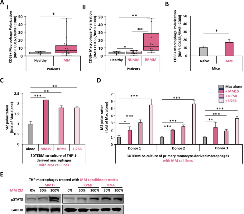
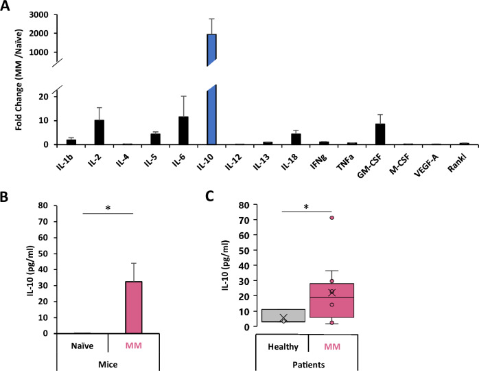
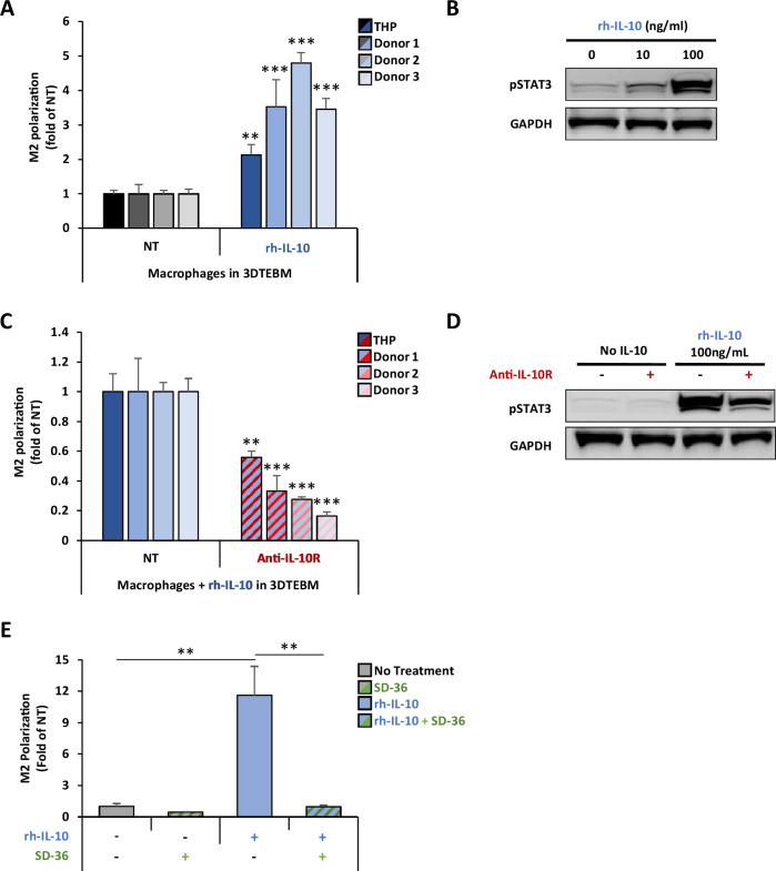
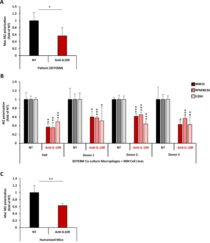
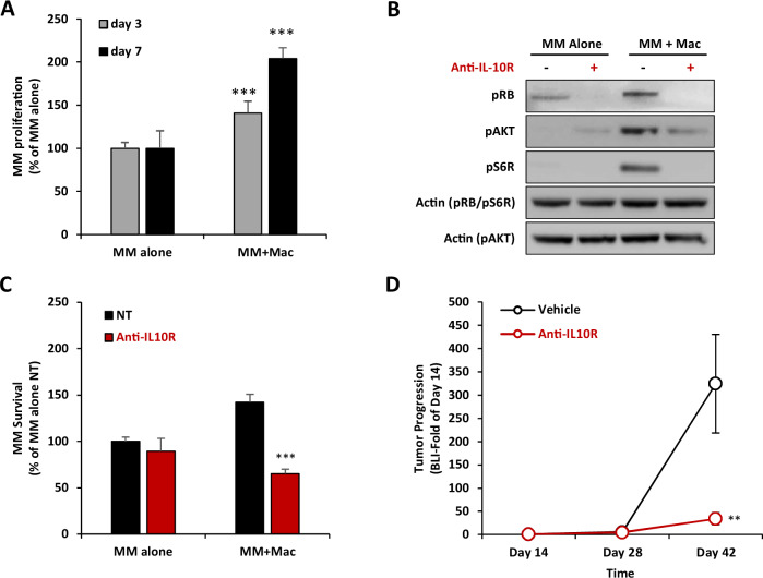
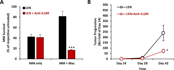
Multiple myeloma (MM) is the cancer of plasma cells within the bone marrow and remains incurable. Tumor-associated macrophages (TAMs) within the tumor microenvironment often display a pro-tumor phenotype and correlate with tumor proliferation, survival, and therapy resistance. IL-10 is a key immunosuppressive cytokine that leads to recruitment and development of TAMs. In this study, we investigated the role of IL-10 in MM TAM development as well as the therapeutic application of IL-10/IL-10R/STAT3 signaling inhibition. We demonstrated that IL-10 is overexpressed in MM BM and mediates M2-like polarization of TAMs in patient BM, 3D co-cultures in vitro, and mouse models. In turn, TAMs promote MM proliferation and drug resistance, both in vitro and in vivo. Moreover, inhibition of IL-10/IL-10R/STAT3 axis using a blocking IL-10R monoclonal antibody and STAT3 protein degrader/PROTAC prevented M2 polarization of TAMs and the consequent TAM-induced proliferation of MM, and re-sensitized MM to therapy, in vitro and in vivo. Therefore, our findings suggest that inhibition of IL-10/IL-10R/STAT3 axis is a novel therapeutic strategy with monotherapy efficacy and can be further combined with current anti-MM therapy, such as immunomodulatory drugs, to overcome drug resistance. Future investigation is warranted to evaluate the potential of such therapy in MM patients.


Conflict of interest statement

Dr. Azab is the founder and owner of Cellatrix LLC that has an exclusive license for the 3DTEBM technology described in part of the experiments in this paper; however, Cellatrix has no contribution to this study. Other authors state no conflicts of interest.

Figures

Fig. 1
Fig. 1. MM induces M2 polarization in macrophages in patients, in vivo, and in vitro.
A Effect of MM on macrophage phenotype in human BM. Macrophage polarization, in BM macrophages in (i) healthy (n = 5) or MM (n = 20) subjects, was represented as M2/M1 ratio. (ii) Macrophage polarization stratifying for newly diagnosed MM (NDMM, n = 10) or relapsed refractory MM (RRMM, n = 10) patients. B Effect of MM on macrophage phenotype in a humanized CD34 + NCG mouse model. Macrophages polarization in naïve (n = 3) or MM-inoculated (n = 3) humanized CD34 + NCG mice. Macrophage polarization was tested by flow cytometry and represented as M2/M1 ratio. C Effect of MM co-culture on THP-1-derived macrophage M2 polarization in vitro. THP-1-derived macrophages were cultured alone, or co-cultured with MM.1S, RPMI8266, or U266 cells at 1:1 ratio in the 3D tissue-engineered bone marrow (3DTEBM) for 3 days. Macrophage polarization was tested by flow cytometry and represented as M2/M1 ratio. D Effect of MM co-culture on human primary macrophage polarization in vitro. Human PBMC-derived macrophages (from 3 independent donors) were cultured alone, or co-cultured with MM.1S, RPMI8266, or U266 cells at 1:1 ratio in the 3DTEBM for 3 days. Macrophage polarization was tested by flow cytometry and represented as M2/M1 ratio. E Effect of MM conditioned media on STAT3 signaling in macrophages. THP-1-derived macrophages were cultured for 30 min with increasing concentrations of conditioned media which was used from MM cell lines (MM.1S, RPMI8266, or U266) culture for 24 h (CM). Macrophages were lysed and levels of pSTAT3 were detected by western blotting. (Bars = Average ± SD *p < 0.05; **p < 0.01; ***p < 0.001).
Fig. 2
Fig. 2. Multiple myeloma produces high levels of IL-10.
A Cytokine screening of the BM supernatant of MM-bearing vs naïve mice. Cytokine levels in BM supernatants of naïve (n = 5) and MM-inoculated (n = 5) KaLwRij immunocompetent mice was measured by Luminex. Cytokine levels MM bearing mice are represented as fold-change of naïve mice. B Levels of IL-10 in the BM supernatant of MM-bearing vs naïve mice. IL-10 levels in BM supernatants of naïve (n = 5) and MM-inoculated (n = 5) KaLwRij immunocompetent mice was measured by Luminex. IL-10 levels we presented as pg/ml. C IL-10 concentrations in BM supernatants of healthy donors (n = 3) and MM patients (n = 10), measured using enzyme-linked immunosorbent assay (ELISA) and presented as pg/ml. (Bars: Average ± SEM, *p < 0.05).
Fig. 3
Fig. 3. IL-10 induces M2 polarization in TAMs through activation of STAT3 signaling.
A Effect of IL-10 on macrophage M2 polarization in vitro. THP-1-derived and primary monocyte-derived macrophages (from 3 independent donors) were treated with 100 ng/ml rhIL-10 in the 3DTEBM for 3 days. Macrophage polarization was tested by flow cytometry and represented as M2/M1 ratio, and a fold-change of non-treated condition (NT). B Effect of IL-10 on STAT3 signaling in macrophages. THP-1-derived macrophages were treated with rhIL-10 (0, 10, 100 ng/ml) for 30 min, and STAT3 activation was measured by western blotting. C Effect of anti-IL-10R mAb on macrophage polarization in vitro. THP-1-derived and primary monocyte-derived macrophages (from 3 independent donors) treated with rhIL-10 (100 ng/ml) in combination with anti-IL-10R mAb (5 µg/ml) in the 3DTEBM for 3 days. Macrophage polarization was tested by flow cytometry and represented as M2/M1 ratio, and a fold-change of non-treated condition (NT). D Effect of anti-IL-10R inhibition on pSTAT3 activation in THP-1-derived macrophages. THP-1-derived macrophages were pretreated with or without anti-IL-10R mAb (5 µg/ml) for 30 min before addition of rhIL-10 (100 ng/ml), and STAT3 activation was measured by western blotting. E Effect of selective cellular STAT3 protein degradation on macrophage polarization. THP-1-derived macrophages were treated with the STAT3 proteolysis targeting chimera (PROTAC) molecule SD-36 (2.5uM) for 24 h. Then, rh-IL-10 (100 ng/ml) was added for 3 additional days. Macrophage polarization was tested by flow cytometry and represented as M2/M1 ratio, and a fold-change of non-treated condition (NT). (Bars = Average ± SD, **p < 0.01).
Fig. 4
Fig. 4. IL-10R inhibition reverses MM-induced TAM M2 polarization.
A Effect of anti-IL-10R mAb on patient BM macrophage polarization ex vivo. BM aspirates from 4 MM patients were cultured in the 3DTEBM model, and treated with or without anti-IL-10R mAb (5 µg/ml) for 3 days. Macrophage polarization was tested by flow cytometry and represented as M2/M1 ratio, and a fold-change of non-treated condition (NT). B Effect of anti-IL-10R mAb on macrophage polarization in vitro. THP-1-derived macrophages and primary monocyte-derived macrophages (from 3 independent donors) were co-cultured with MM1S, RPMI8266, or U266 cells (1:1 ratio) and treated with or without anti-IL-10R mAb (5µg/ml) for 3 days. Macrophage polarization was tested by flow cytometry and represented as M2/M1 ratio, and a fold-change of non-treated condition (NT). C Effect of anti-IL-10R mAb on macrophage polarization in a humanized mice model, in vivo. huCD34 NCG mice with human MM tumors were treated with (n = 3) or without (n = 3) anti-IL-10R mAb (5 mg/kg, i.p., twice a week) for 2 weeks. BM was harvested, and macrophage polarization was tested by flow cytometry and represented as M2/M1 ratio, and as fold-change of non-treated condition (NT). (Bars = Average ± SD, *p < 0.05; **p < 0.01; ***p < 0.001;).
Fig. 5
Fig. 5. IL-10R inhibition reverse TAM-induced MM proliferation.
A Effect of macrophages on MM proliferation. MM.1S-CBR-GFP cells were cultured alone or co-cultured at 1:1 ratio with THP-1-derived macrophages for 3 or 7 days in 3DTEBM. MM proliferation is determined by GFP cell count, presented as % of MM alone. Statistical significance is compared between the co-culture condition and the respective monoculture condition of each day. B Effect of IL-10R inhibition on TAM-induced MM proliferation and cell cycle signaling. THP-1-derived macrophages were pretreated with or without of anti-IL-10R mAb (5µg/ml) for 4 h, and MM.1S cells were cultured alone or with macrophages at 1:1 ratio for 24 h. MM.1S cells were then detached from macrophages, lyzed, and proteins were analyzed by western for proliferation (pAKT and S6R) and cell cycle signaling (pRB). Actin was blotted as the loading control. C Effect of IL10R inhibition on TAM-induced MM proliferation. MM.1S-CBR-GFP cells were cultured alone or co-cultured at 1:1 ratio with THP-1-derived macrophages in 3DTEBM and treated with or without anti-IL-10R mAb (5 µg/ml) for 3 days. MM survival is determined by GFP cell count by flow cytometry, presented as % of MM monoculture NT condition. Statistical significance is compared between anti-IL-10R mAb and NT. D Effect of anti-IL-10R mAb on in vivo MM tumor progression. NSG mice (n = 14) were inoculated with MM.1S-CBR-GFP and injected with human PBMC-derived macrophages. Treatment started at day 14 post-inoculation, mice were randomly divided into two groups which were treated with vehicle control (n = 7), or anti-IL-10R mAb (5 mg/kg, i.p, twice/week, n = 7). Tumor progression was measured at days 14, 28, and 42 by BLI, and represented as fold of day 14. Statistical significance is compared between anti-IL-10R mAb and NT. (Bars = Average ± SD, **p < 0.01; ***p < 0.001).
Fig. 6
Fig. 6. Anti-IL10R mAb reverses MM drug resistance to lenalidomide in vitro and in vivo.
A Effect of IL-10R inhibition on TAM-induced MM drug resistance to lenalidomide (LEN), in vitro. MM.1S-CBR-GFP cells were cultured alone or at 1:1 ratio with THP-1-derived macrophages in 3DTEBM and treated with or without LEN (1 uM), with or without anti-IL-10R mAb (5 µg/ml) for 3 days. MM survival was determined by GFP cell count by flow cytometry, represented as % of untreated MM respective mono- or co-culture. Statistical significance is compared between the combination and LEN-only conditions. B Effect of IL-10R inhibition on TAM-induced MM drug resistance to lenalidomide (LEN), in vivo. NSG mice (n = 14) were inoculated with MM.1S-CBR-GFP and injected with human PBMC-derived macrophages. Treatment started at day 14 post-inoculation) for all cohort (n = 14), mice were randomly divided into two groups which were treated with LEN alone (4mg/kg, Per Os, Daily, n = 7), or a combination of LEN with anti-IL10R mAb (2.5 mg/kg, i.p., twice/week, n = 7). Tumor progression was measured at days 14, 28 and 42 by BLI, and represented as fold of day 14. Statistical significance is compared between LEN alone and the combination of LEN + anti-IL-10R mAb. (Bars= Average ±SD,*: p < 0.05; ***: p < 0.001).

References

    1. van de Donk NWCJ, Pawlyn C, Yong KL. Multiple myeloma. Lancet. 2021;397:410–27. - PubMed
    1. Azab AK, Runnels JM, Pitsillides C, Moreau A-S, Azab F, Leleu X, et al. CXCR4 inhibitor AMD3100 disrupts the interaction of multiple myeloma cells with the bone marrow microenvironment and enhances their sensitivity to therapy. Blood. 2009;113:4341–51. - PMC - PubMed
    1. Kawano Y, Moschetta M, Manier S, Glavey S, Görgün GT, Roccaro AM, et al. Targeting the bone marrow microenvironment in multiple myeloma. Immunological Rev. 2015;263:160–72. - PubMed
    1. Uckun FM. Dual targeting of multiple myeloma stem cells and myeloid-derived suppressor cells for treatment of chemotherapy-resistant multiple myeloma. Front Oncol. 2021;11. - PMC - PubMed
    1. Shah UA, Mailankody S. Emerging immunotherapies in multiple myeloma. Bmj. 2020. - PubMed

MeSH terms