. 2024 Sep 15;38(17):e70021.
doi: 10.1096/fj.202400198R.

Promotion of endoplasmic reticulum retrotranslocation by overexpression of E3 ubiquitin-protein ligase synoviolin 1 reduces endoplasmic reticulum stress and preserves cone photoreceptors in cyclic nucleotide-gated channel deficiency

Affiliations

Promotion of endoplasmic reticulum retrotranslocation by overexpression of E3 ubiquitin-protein ligase synoviolin 1 reduces endoplasmic reticulum stress and preserves cone photoreceptors in cyclic nucleotide-gated channel deficiency

Fan Yang et al. FASEB J. .

Abstract

Cone photoreceptor cyclic nucleotide-gated (CNG) channels play an essential role in phototransduction and cellular Ca2+ homeostasis. Mutations in genes encoding the channel subunits CNGA3 and CNGB3 are associated with achromatopsia, progressive cone dystrophy, and early-onset macular degeneration. Cone loss in patients with achromatopsia and cone dystrophy associated with CNG channel mutations has been documented by optical coherence tomography and in mouse models of CNG channel deficiency. Cone death in CNG channel-deficient retinas involves endoplasmic reticulum (ER) stress-associated apoptosis, dysregulation of cellular/ER Ca2+ homeostasis, impaired protein folding/processing, and impaired ER-associated degradation (ERAD). The E3 ubiquitin-protein ligase synoviolin 1 (SYVN1) is the primary component of the SYVN1/SEL1L ER retrotranslocon responsible for ERAD. Previous studies have shown that manipulations that protect cones and reduce ER stress/cone death in CNG channel deficiency, such as increasing ER Ca2+ preservation or treatment with an ER chaperone, increase the expression of SYVN1 and other components of the ER retrotranslocon. The present work investigated the effects of SYVN1 overexpression. Intraocular injection of AAV5-IRBP/GNAT2-Syvn1 resulted in overexpression of SYVN1 in cones of CNG channel-deficient mice. Following treatment, cone density in Cnga3-/- mice was significantly increased, compared with untreated controls, outer segment localization of cone opsin was improved, and ER stress/apoptotic cell death was reduced. Overexpression of SYVN1 also led to increased expression levels of the retrotranslocon components, degradation in ER protein 1 (DERL1), ERAD E3 ligase adaptor subunit (SEL1L), and homocysteine inducible ER protein with ubiquitin-like domain 1 (HERPUD1). Moreover, overexpression of SYVN1 likely enhanced protein ubiquitination/proteasome degradation in CNG channel-deficient retinas. This study demonstrates the role of SYVN1/ERAD in cone preservation in CNG channel deficiency and supports the strategy of promoting ERAD for cone protection.

Keywords: CNG channel; ER stress; ERAD; SYVN1; cone photoreceptors; retinal degeneration.


Conflict of interest statement

Disclosures

All authors declare that they have no conflict of interest.

Figures
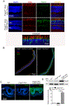
Figure 1.
Figure 1.. SYVN1 was overexpressed in cones of CNG channel-deficient mice after intraocular injection with AAV5-IRBP/GNAT2-Syvn1.
A-B. Cnga3−/− mice were injected with AAV5-IRBP/GNAT2-Syvn1 at P5 and analyzed at 4 months for retinal expression/cone localization of SYVN1 and PNA by immunofluorescence labeling. A. Shown are representative confocal images of SYVN1 and PNA labeling in the mouse retina. ONL, outer nuclear layer; INL, inner nuclear layer. B. Shown are representative virtual slide scanner images of SYVN1 and PNA labeling in the mouse retina. C-D. Cnga3−/−/Nrl−/− mice were injected at P5 with AAV5-IRBP/GNAT2-Syvn1 and analyzed at P40 for SYVN1 expression by immunofluorescence labeling and immunoblotting. C. Shown are representative confocal images of SYVN1 labeling in the mouse retina. ONL, outer nuclear layer; INL, inner nuclear layer. D. Shown are representative immunoblotting images and the corresponding quantitative analysis. Data are presented as mean ± SEM of 3-4 assays using retinas prepared from 8-12 mice (***p < 0.001).
Figure 2.
Figure 2.. Treatment with AAV-Syvn1 increased cone survival in CNG channel-deficient mice.
A. Cnga3−/− mice received AAV5-IRBP/GNAT2-Syvn1 at P5 and were analyzed at 4 months for cone survival by immunolabeling. Shown are representative confocal images of PNA labeling on retinal cross sections and corresponding quantitative analysis. Data are presented as mean ± SEM (n=5-11 mice, ***p < 0.001).
Figure 3.
Figure 3.. Treatment with AAV-Syvn1 reduced cone death in Cnga3−/−/Nrl−/− mice.
Cnga3−/−/Nrl−/− mice received AAV5-IRBP/GNAT2-Syvn1 at P5 and were analyzed at P40 for cone death by TUNEL-labeling. Shown are representative confocal images of TUNEL-positive cells and the corresponding quantitative analysis. Data are presented as mean ± SEM (n=6-10 mice, *p < 0.05, **p < 0.01).
Figure 4.
Figure 4.. Treatment with AAV-Syvn1 increased outer segment localization of M-opsin in Cnga3−/− mice.
Cnga3−/− mice received AAV5-IRBP/GNAT2-Syvn1 at P5 and were analyzed at 4 months for M-opsin localization by immunofluorescence labeling. Shown are representative confocal images of M-opsin labeling on retinal cross sections and the corresponding quantitative analysis. ONL, outer nuclear layer; INL, inner nuclear layer. OS, outer segment; IS, inner segment; OPL, outer plexiform layer. Data are presented as mean ± SEM (n=5-8 mice, ***p < 0.001).
Figure 5.
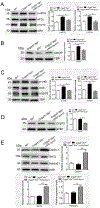
Figure 5.. Treatment with AAV-Syvn1 reduced ER stress and ER stress downstream signaling in Cnga3−/−/Nrl−/− mice and increased expression levels of ER retrotranslocation proteins.
Cnga3−/−/Nrl−/− mice received AAV5-IRBP/GNAT2-Syvn1 at P5, and retinas were analyzed at P40 by immunoblotting. A-D. Shown are representative immunoblotting images and the corresponding quantitative analysis for expressions of phospho-eIF2α (A), phospho-IRE1α (A), CHOP (B), phospho-CREB (C), CREB3l3 (C), and Grp75 (D). E. Shown are representative immunoblotting images and the corresponding quantitative analysis for expressions of DERL1, SEL1L, and HERPUD1. Data are presented as mean ± SEM of 3-4 assays using retinas prepared from 8-12 mice (*p < 0.05, **p < 0.01).
Figure 6.
Figure 6.. Effects of treatment with AAV-Syvn1 on the ubiquitin-proteasome activity in Cnga3−/−/Nrl−/− mice.
Cnga3−/−/Nrl−/− mice received AAV5-IRBP/GNAT2-Syvn1 at P5 and were analyzed at P40 for expressions of ubiquitin (A) and p53 (B) by immunoblotting. Shown are representative immunoblotting images and the corresponding quantitative analysis. Data are presented as mean ± SEM of 3-4 assays using retinas prepared from 8-12 mice (*p < 0.05).
Figure 7.
Figure 7.. The role of ER retrotranslocation/ERAD in cone protection in CNG channel deficiency.
In CNG channel deficiency, reduced cytosolic Ca2+ level stimulates ER Ca2+ channels, leading to increased Ca2+ release from the ER and a compensatory reduction of ER Ca2+ store/impaired ER Ca2+ homeostasis. Moreover, reduced cytosolic Ca2+ level also stimulates cGMP/PKG signaling, which is a strong inducer for the ER Ca2+ channels, impairing ER Ca2+ store. Impaired ER Ca2+ store in turn inhibits ER protein processing/protein folding, presumably via the regulation of the Ca2+-dependent ER chaperone proteins, leading to ER stress/cell death. Thus, suppressing ER Ca2+ channel activity improves protein trafficking in cones and reduces ER stress/cell death. The previous findings and results from the present study support that ER retrotranslocation/ERAD in cone photoreceptors is regulated at least by the following three factors: 1) ER Ca2+ store/homeostasis, 2) function of ER chaperones, and 3) expression levels of the retrotranslocon components. Increased Ca2+ store in the ER, enhanced protein folding/processing, and enhanced expression of the ER retrotranslocon components will facilitate ER retrotranslocation/ERAD and relieve ER stress, leading to preservation of photoreceptors.

References

    1. Kohl S, Baumann B, Broghammer M, Jagle H, Sieving P, Kellner U, Spegal R, Anastasi M, Zrenner E, Sharpe LT, and Wissinger B (2000) Mutations in the CNGB3 gene encoding the beta-subunit of the cone photoreceptor cGMP-gated channel are responsible for achromatopsia (ACHM3) linked to chromosome 8q21. Hum Mol Genet 9, 2107–2116 - PubMed
    1. Thiadens AA, Roosing S, Collin RW, van Moll-Ramirez N, van Lith-Verhoeven JJ, van Schooneveld MJ, den Hollander AI, van den Born LI, Hoyng CB, Cremers FP, and Klaver CC Comprehensive analysis of the achromatopsia genes CNGA3 and CNGB3 in progressive cone dystrophy. Ophthalmology 117, 825–830 e821 - PubMed
    1. Wissinger B, Gamer D, Jagle H, Giorda R, Marx T, Mayer S, Tippmann S, Broghammer M, Jurklies B, Rosenberg T, Jacobson SG, Sener EC, Tatlipinar S, Hoyng CB, Castellan C, Bitoun P, Andreasson S, Rudolph G, Kellner U, Lorenz B, Wolff G, Verellen-Dumoulin C, Schwartz M, Cremers FP, Apfelstedt-Sylla E, Zrenner E, Salati R, Sharpe LT, and Kohl S (2001) CNGA3 mutations in hereditary cone photoreceptor disorders. Am J Hum Genet 69, 722–737 - PMC - PubMed
    1. Thiadens AA, Slingerland NW, Roosing S, van Schooneveld MJ, van Lith-Verhoeven JJ, van Moll-Ramirez N, van den Born LI, Hoyng CB, Cremers FP, and Klaver CC (2009) Genetic etiology and clinical consequences of complete and incomplete achromatopsia. Ophthalmology 116, 1984–1989 e1981 - PubMed
    1. Nishiguchi KM, Sandberg MA, Gorji N, Berson EL, and Dryja TP (2005) Cone cGMP-gated channel mutations and clinical findings in patients with achromatopsia, macular degeneration, and other hereditary cone diseases. Hum Mutat 25, 248–258 - PubMed

MeSH terms

Substances

LinkOut - more resources