. 2025 Mar 7;46(10):926-939.
doi: 10.1093/eurheartj/ehae584.

Malondialdehyde-specific natural IgM inhibit NETosis triggered by culprit site-derived extracellular vesicles from myocardial infarction patients

Affiliations

Malondialdehyde-specific natural IgM inhibit NETosis triggered by culprit site-derived extracellular vesicles from myocardial infarction patients

Anna S Ondracek et al. Eur Heart J. .

Abstract

Background and aims: Neutrophil extracellular traps (NETs) trigger atherothrombosis during acute myocardial infarction (AMI), but mechanisms of induction remain unclear. Levels of extracellular vesicles (EV) carrying oxidation-specific epitopes (OSE), which are targeted by specific natural immunoglobulin M (IgM), are increased at the culprit site in AMI. This study investigated EV as inducers of NETosis and assessed the inhibitory effect of natural anti-OSE-IgM in this process.

Methods: Blood from the culprit and peripheral site of ST-segment elevation myocardial infarction (STEMI) patients (n = 28) was collected, and myocardial function assessed by cardiac magnetic resonance imaging (cMRI) 4 ± 2 days and 195 ± 15 days post-AMI. Extracellular vesicles were isolated from patient plasma and cell culture supernatants for neutrophil stimulation in vitro and in vivo, in the presence of a malondialdehyde (MDA)-specific IgM or an isotype control. NETosis and neutrophil functions were assessed via enzyme-linked immunosorbent assay and fluorescence microscopy. Pharmacological inhibitors were used to map signalling pathways. Neutrophil extracellular trap markers and anti-OSE-IgM were measured by ELISA.

Results: CD45+ MDA+ EV and NET markers were elevated at the culprit site. Extracellular vesicles induced neutrophil activation and NET formation via TLR4 and PAD4, and mice injected with EV showed increased NETosis. Malondialdehyde-specific IgM levels were inversely associated with citH3 in STEMI patient blood. An MDA-specific IgM inhibited EV-induced NET release in vitro and in vivo. CD45+ MDA+ EV concentrations inversely correlated with left ventricular ejection fraction post-AMI.

Conclusions: Culprit site-derived EV induce NETosis, while MDA-specific natural IgM inhibit this effect, potentially impacting outcome after AMI.

Keywords: Acute myocardial infarction; Extracellular vesicles; Natural IgM; Neutrophil extracellular traps; Oxidation-specific epitopes.


Figures

Structured Graphical Abstract
Different subsets of extracellular vesicles (EV) are released at the culprit site of ST-segment elevation myocardial infarction (STEMI), and a high percentage of leukocyte-derived EV carry oxidation-specific epitopes (OSE) such as malondialdehyde (MDA) epitopes (red). Culprit site EV from STEMI patients (MI-EV) can activate neutrophils and trigger peptidyl-arginine deiminase 4 (PAD4)–dependent formation of neutrophil extracellular traps (NETs) via toll-like receptor 4 (TLR4) signalling. The presence of the MDA-specific IgM antibody LR04 reduces MI–EV-induced NETosis. ST-segment elevation myocardial infarction patients with high levels of MDA-specific IgM, which have the capacity to balance activity of pro-inflammatory NETogenic EV, present with preserved left ventricular ejection fraction (LVEF).
Figure 1
Figure 1
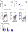
Concentration and percentage of CD45+ extracellular vesicles carrying malondialdehyde epitopes are elevated at the culprit site. Citrullinated histone H3 (citH3), malondialdehyde-specific immunoglobulin M, and extracellular vesicles were measured in culprit site and peripheral site plasma of acute myocardial infarction patients. Annexin V positivity and a size range of .2–1.1 µm were used to define large extracellular vesicles. Anti-CD45 and malondialdehyde-specific LR04 antibodies were used to define leukocyte origin and MDA+ extracellular vesicles. (A) Correlation of plasma levels of citH3 with malondialdehyde-specific immunoglobulin M in the periphery and (B) at the culprit sites; Spearman signed-rank correlations. Comparison between the periphery and culprit site of (C) annexin V–positive CD45+ events/µL, (D) Annexin V–positive CD45+ malondialdehyde-positive events/µL, and (E) percentage of malondialdehyde-positive extracellular vesicles gated from annexin V–positive CD45+ events; Wilcoxon matched-pairs signed-rank tests. (F) Percentages of malondialdehyde-carrying extracellular vesicles derived from leukocytes (CD45+), platelets (CD41a+), and endothelial cells (CD144+) were compared at the culprit site and in the periphery using a mixed model *P < .05, **P < .01, *** P < .001, ****P < .0001. Light-shaded dots represent female patients. RLU, relative light units
Figure 2
Figure 2
Extracellular vesicles induce neutrophil activation and neutrophil extracellular trap formation. (A–F) Neutrophils of healthy donors were stimulated either with extracellular vesicles or vehicle control, and supernatants were screened for the release of (A) deoxyribonucleic acid–myeloperoxidase complexes as neutrophil extracellular trap surrogate markers after 3 h, (B) neutrophil elastase after 3 h, (C) interleukin-8 after 3 h, (D) degranulation of myeloperoxidase after 30 min, (E) degranulation of neutrophil gelatinase–associated lipocalin after 30 min, and (F) reactive oxygen species production after 15 min. (G and H) Neutrophils of healthy donors were stimulated with myocardial infarction–extracellular vesicles, and formed neutrophil extracellular traps were stained with anti-deoxyribonucleic acid–histone (yellow) and anti-myeloperoxidase (red) antibodies. DAPI staining (blue) was used to visualize DNA/cell nuclei. (G) Representative pictures of neutrophil extracellular traps formed upon stimulation with vehicle (top) and culprit site myocardial infarction–extracellular vesicles (bottom), which were (H) quantified by normalizing the merged area to total DAPI area; paired t-tests, (E) data were log-transformed before statistical comparison, *P < .05, **P < .01, ***P < .001, ****P < .0001. MPO, myeloperoxidase; NE, neutrophil elastase; NGAL, neutrophil gelatinase–associated lipocalin; ROS, reactive oxygen species
Figure 3
Figure 3
Extracellular vesicle–induced neutrophil extracellular trap formation is dependent on TLR4, p38, and PAD4. Neutrophils of healthy donors were pre-treated with inhibitors of (A) TLR4 (TAK-242, 10 µM), (B) PAD4 (GSK484, 10 µM), (C) p38 (SB203580, 10 µM), (D) PKC (Go6979, 2 µM), (E) MEK (PD98059, 40 µM), and (F) NADPH oxidase (diphenyleniodonium chloride, 20 µM) or vehicle control for 20 min before stimulation with extracellular vesicles, PMA (125 nM), and ionomycin (4 µM) for 3 h to assess neutrophil extracellular trap formation by deoxyribonucleic acid–myeloperoxidase complexes; (G) matrix summarizing A–F, Wilcoxon matched-pairs signed-rank tests. (H–J) The involvement of TLR4 and PAD4 was confirmed using coronary myocardial infarction–extracellular vesicles on primary neutrophils. (H) Neutrophil extracellular traps were visualized by immunofluorescence microscopy and were defined as merge of chromatin and myeloperoxidase. Neutrophil extracellular trap area was normalized to DAPI area and presented as fold to the respective vehicle control for (I) TLR4 inhibition and (J) PAD4 inhibition; paired t-tests. (K) Potential signalling pathway of extracellular vesicle–induced neutrophil extracellular trap formation. *P < .05, **P < .01
Figure 4
Figure 4
Malondialdehyde-specific immunoglobulin M LR04 attenuate extracellular vesicle–induced neutrophil extracellular trap formation. (A) Neutrophils isolated from healthy donors were stimulated with extracellular vesicles released by lipopolysaccharide-activated THP-1 monocytic cells, either in the presence of malondialdehyde-specific immunoglobulin M LR04 or isotype control (25 µg/mL and 12.5 µg/mL). Deoxyribonucleic acid–myeloperoxidase complexes in the supernatant were measured as indicator of neutrophil extracellular trap formation. Repeated measures analysis of variance. (B and C) Neutrophils isolated from healthy donors were stimulated with myocardial infarction–extracellular vesicles pooled from culprit site plasma of six patients, either in the presence of LR04 or an isotype control (25 µg/mL). Neutrophil extracellular traps were visualized by immunofluorescence microscopy and were defined as merge of chromatin and myeloperoxidase. Neutrophil extracellular trap area was normalized to DAPI area and presented as fold of the respective vehicle control; repeated measures analysis of variance. *P < .05
Figure 5
Figure 5
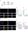
Malondialdehyde-specific immunoglobulin M LR04 attenuates myocardial infarction–extracellular vesicle–induced neutrophil extracellular trap formation in vivo. (A) Mouse treatment protocol. Baseline blood was drawn to determine malondialdehyde-specific immunoglobulin M levels 4 days prior to any injections. Upon sacrifice 3 h after injections, cytospins of haemolysed whole blood were prepared and analysed by immunofluorescence microscopy. (B and C) Representative pictures of cytospins of a mouse injected with (B) vehicle or (C) myocardial infarction–extracellular vesicles, respectively. (D and H) Ly6G+ (green) nuclei were identified, and the percentage of citH3+ (yellow) neutrophils was quantified. (E and I) An overlay of DNA and citH3 was quantified as NETs. (F and G) Representative cytospin pictures of a mouse treated with (F) myocardial infarction–extracellular vesicles and isotype or (G) myocardial infarction–extracellular vesicles and LR04. Red arrows indicate NETs, white arrows citH3+ neutrophils. Mann–Whitney U test in (D, E, and H), unpaired t-test in (I); * P < .05.
Figure 6
Figure 6
Association of 72-h left ventricular ejection fraction with malondialdehyde-positive extracellular vesicles. Spearman signed-rank correlations of cardiac magnetic resonance imaging–derived left ventricular ejection fraction (%) at 72 h with culprit site (A) malondialdehyde-positive extracellular vesicles and (B) CD45+ malondialdehyde-positive extracellular vesicles. (C) Receiver operating characteristic analysis of CD45+ malondialdehyde-positive extracellular vesicles from the culprit site (dots) and the periphery (empty circles) predicting reduced left ventricular ejection fraction (<40%). (D) Spearman signed-rank correlation of cardiac magnetic resonance imaging–derived left ventricular ejection fraction (%) at 72 h with the ratio of culprit site CD45+ malondialdehyde-positive extracellular vesicles and malondialdehyde–immunoglobulin M. Light-shaded dots represent female patients. A, area

References

    1. Cannon B. Cardiovascular disease: biochemistry to behaviour. Nature 2013;493:S2–3. 10.1038/493S2a - DOI - PubMed
    1. Heron M. Deaths: leading causes for 2018. Natl Vital Stat Rep 2021;70:1–115. 10.15620/cdc:104186 - DOI - PubMed
    1. Libby P. The changing landscape of atherosclerosis. Nature 2021;592:524–33. 10.1038/s41586-021-03392-8 - DOI - PubMed
    1. Distelmaier K, Adlbrecht C, Jakowitsch J, Winkler S, Dunkler D, Gerner C, et al. . Local complement activation triggers neutrophil recruitment to the site of thrombus formation in acute myocardial infarction. Thromb Haemost 2009;102:564–72. 10.1160/TH09-02-0103 - DOI - PubMed
    1. Distelmaier K, Winter M-P, Dragschitz F, Redwan B, Mangold A, Gleiss A, et al. . Prognostic value of culprit site neutrophils in acute coronary syndrome. Eur J Clin Invest 2014;44:257–65. 10.1111/eci.12228 - DOI - PubMed