. 2025 Mar 6;148(3):941-954.
doi: 10.1093/brain/awae278.

Brain-penetrant complement inhibition mitigates neurodegeneration in an Alzheimer's disease mouse model

Affiliations

Brain-penetrant complement inhibition mitigates neurodegeneration in an Alzheimer's disease mouse model

Wioleta M Zelek et al. Brain. .

Abstract

Complement activation is implicated in driving brain inflammation, self-cell damage and progression of injury in Alzheimer's disease and other neurodegenerative diseases. Here, we investigate the impact of brain delivery of a complement-blocking antibody on neurodegeneration in an Alzheimer's mouse model. We engineered a brain-penetrant recombinant antibody targeting the pro-inflammatory membrane attack complex. Systemic administration of this antibody in APPNL-G-F mice reduced brain levels of complement activation products, demonstrating successful brain entry and target engagement. Prolonged treatment decreased synapse loss, amyloid burden and brain inflammatory cytokine levels, concomitant with cognitive improvement compared to controls. These results underscore the potential of brain-penetrant complement-inhibiting drugs as promising therapeutics, targeting downstream of amyloid plaques in Alzheimer's disease.

Keywords: blood–brain barrier; drug delivery; mouse model; neuroinflammation; therapy.


Conflict of interest statement

The authors report competing interests.

Figures
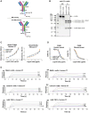
Figure 1
Figure 1
Generation and characterization of the brain penetrant anti-C7 recombinant monoclonal antibodies (r-mAbs). (A) Cartoon representing the design of the Nb62-r-mAb (top) and control-r-mAb (bottom). Complementary determining regions (CDRs) from mAb 73D1 were grafted onto a mouse IgG2a framework modified (D265A) to ablate Fc receptor binding. Nanobodies, Nb62 against low-affinity transferrin receptor (TfR) for the test, anti-GFP for the control, were expressed at the carboxy terminus of one heavy chain. ALFA-tag and 6-His tag were included in each construct for use in detection and purification. (B) Sodium dodecyl sulfate-polyacrylamide gel electrophoresis (SDS-PAGE) of Nb62-r-mAb and control-r-mAb. Proteins were purified on protein G then run on 7.5% acrylamide gels, either non-reduced (NR) or reduced with 5% β-mercaptoethanol (R). Separated proteins were stained with Coomassie Blue. NR = ∼150 kDa MW intact r-mAb; R = 55 kDa MW r-mAb heavy chain, 25 kDa MW r-mAb light chain and ∼70 kDa MW r-mAb heavy chain plus nanobody. (C) Direct ELISA. Human and mouse C7 were immobilized on wells then native mAb, Nb62-r-mAb or control-r-mAb added in a dilution series (0–10 µg/ml). Bound antibody was detected using labelled anti-mouse IgG. Binding curves for the three antibodies were superimposed and showed strong binding to both human and mouse C7. The assays were repeated three times with comparable results. The error bars are standard errors of duplicates. (D) Classical pathway haemolytic assays (CH50). Nb62-r-mAb and control-r-mAb were tested for inhibition of complement-mediated lysis in human and mouse serum. Both r-mAb inhibited mouse and human serum-mediated haemolysis as effectively as the native parent mAb 73D1. The experiments were repeated three times with comparable results. The error bars are standard errors of duplicates. (E) Surface plasmon resonance (SPR) was used to determine the binding of Nb62-r-mAb and control-r-mAb to human and mouse C7. Human or mouse C7 was immobilized directly onto a CM5 sensor chip, and the relevant r-mAb or native mAb 73D1 was flowed over the chip. Sensorgrams were collected and dissociation constants (KD) calculated using the Langmuir 1:1 binding model (Table 1). Representative sensorgrams are shown with fitted data in black (n = 3).
Figure 2
Figure 2
In vivo testing of the recombinant monoclonal antibodies (r-mAbs) for delivery to the brain. Entry of Nb62-r-mAb and control-r-mAb into brain was tested in 8-week-old C7-deficient mice. (A) r-mAb in total brain homogenate (TBH) was detected by sandwich ELISA and expressed as ng/mg protein. Nb62-r-mAb was detected in TBH at 2, 4 and 24 h time points, whereas control-r-mAb was not detected at any time point. (B) Western blotting confirmed the presence of Nb62-r-mAb but not control-r-Mab in TBH. (C) r-mAb levels in serum were measured by ELISA; both Nb62-r-mAb and control-r-mAb were detectable at each time point but significantly less Nb62-r-mAb was detected at 4 and 24 h (P = 0.0044 and P < 0.0001, respectively), suggesting rapid clearance. (D) Distribution of the r-mAbs in other organs at 2 h was tested in tissue lysates using ELISA; distribution patterns were different with the Nb62-r-mAb higher in muscle, brain and eye. *P < 0.05, **P < 0.01. All assays were repeated three times with comparable results. The error bars are standard errors of triplicates. Unpaired two-tailed t-test was used for the group comparison. Error bars correspond to the standard error of the mean.
Figure 3
Figure 3
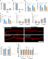
Impact of 1-week treatment with Nb62-recombinant monoclonal antibody (r-mAb) on brain parameters in APPNL-G-F mice. APPNL-G-F mice were treated with anti-C7 mAb plus either Nb62-r-mAb or control-r-mAb over 7 days; systemic inhibition of complement, inhibition of complement activation in brain, brain inflammation, and neurodegeneration were assessed. (A and B) Classical pathway haemolysis (CH50) assays confirm that systemic complement was inhibited in APPNL-G-F mice aged 5–6 months (control-r-mAb: n = 7, Nb62-r-mAb: n = 8) or 11–13 months (control-r-mAb: n = 5, Nb62-r-mAb: n = 6) over 1 week of systemic administration of 73D1 mAb and either r-mAb. (C and D) Sandwich ELISAs detecting mouse C3 fragments (C3b/iC3b/C3c; C) and terminal complement complex (TCC; D) in total brain homogenate (TBH). Both C3 fragments and TCC levels were significantly lower in Nb62-r-mAb-treated mice at either age. Error bars are standard errors for each dataset. Groups were compared using an unpaired two-tailed t-test. (E) Sandwich ELISA to measure levels of amyloid-β (Aβ) in tissue bound protein (TBP) demonstrating significantly decreased Aβ in TBP of Nb62-r-mAb-treated APPNL-G-F mice compared to controls in both age sets. (F and G) Immunostaining of Aβ plaques in hippocampus and cortex using anti-Aβ antibodies: 6E10 (F) and 4G8 (G) showed no significant difference in plaque coverage between Nb62-r-mAb- and control-r-mAb-treated APPNL-G-F mice at either age. (H) Representative confocal images of DiOIistics-labelled CA1 hippocampal dendritic segments in 5–6-month-old and 11–13-month-old APPNL-G-F mice treated with Nb62-r-mAb or control-r-mAb. Scale bar = 5 µm. (I and J) DiOIistics-labelled dendritic spines were analysed in prefixed coronal brain slices. Overall spine density, analysed from dendritic segments of at least 30 µm, was higher in Nb62-r-mAb-treated mice compared to controls at both ages, significant only in the 11–13-month set. Analysis of spine subtypes showed that the number of thin spines was significantly increased in Nb62-r-mAb-treated groups in both age sets. Unpaired two-tailed t-test was used to compare spine densities between groups. Error bars correspond to the standard error of the mean.
Figure 4
Figure 4
Impact of 3months of treatment with Nb62-r-mAb on brain parameters in APPNL-G-F mice. APPNL-G-F mice were treated with anti-C7 mAb plus either Nb62-r-mAb (n = 12) or control-r-mAb (n = 12) over 3 months; systemic complement activity, complement activation in brain, brain inflammation, neurodegeneration and cognition were assessed. (A) Classical pathway haemolytic assays (CH50) demonstrated that systemic complement was inhibited in the Nb62-r-mAb and control-r-mAb-treated mice over the 3-month time course. (B and C) Levels of C3 fragments (C3b/iC3b/C3c; B) and terminal complement complex (TCC; C) in the total brain homogenate (TBH) were significantly reduced at end point in Nb62-r-mAb-treated APPNL-G-F mice compared to controls. (D and E) Levels of cytokines: IL-1α and IL1-β in TBH at end point were significantly decreased in Nb62-r-mAb-treated APPNL-G-F mice compared to controls. (F) Levels of amyloid-β (Aβ) in tissue bound protein (TBP) were significantly decreased at end point in Nb62-r-mAb-treated APPNL-G-F mice compared to controls. (GI) Aβ plaques in hippocampus and cortex were either stained with the plaque stain X34 (G) or immunostained with anti-Aβ antibodies 6E10 (H) and 4G8 (I); analysis revealed no significant differences in plaque coverage with any of these stains at end point between the Nb62-r-mAb- and control-r-mAb-treated APPNL-G-F mice. (J) Representative confocal images of DiOIistics labelled CA1 hippocampal dendritic segments from APPNL-G-F mice treated with Nb62-r-mAb or control-r-mAb. Scale bar = 5 µm. (K and L) Quantification of DiOIistics-labelled dendritic spines in prefixed coronal brain slices. (K) APPNL-G-F mice treated with Nb62-r-mAb showed significantly increased overall spine density compared to control-r-mAb-treated mice. Analysis of spine subtypes showed that the numbers of thin and mushroom spines were significantly increased in Nb62-r-mAb-treated groups, most significantly for thin spines. (M) Representative images of Bassoon (green) and PSD95 (red) immunoreactive synaptic puncta in the stratum radium of Nb62-r-mAb- and control-r-mAb-treated APPNL-G-F mice at end point; scale bar = 5 µm. (N) Synaptic puncta stained with Bassoon or PSD95 were quantified (region of interest, 20 µm × 20 µm, 12 per mouse) using Imaris Spot function; puncta were increased in Nb62-r-mAb-treated mice compared with controls for both stains but significantly only for PSD95. (OQ) Comparison of Nb62-r-mAb-treated and control-r-mAb-treated APPNL-G-F mice in behavioural tests. (O) In the burrowing test, Nb62-r-mAb-treated mice burrowed significantly more of the gravel compared to controls. (P) In the Open Field (OF) test, Nb62-r-mAb mice spent significantly more time exploring the central area of the box. (Q) In the Novel Object Recognition (NOR) test, Nb62-r-mAb-treated mice spent significantly more time exploring the novel object. Each point represents one animal in these analyses. For all quantitative analyses, an unpaired two-tailed t-test was used to compare the two groups. Error bars correspond to standard error of the mean, and P-values are included where appropriate.

References

    1. Pardo-Moreno T, González-Acedo A, Rivas-Domínguez A, et al. Therapeutic approach to Alzheimer's disease: Current treatments and new perspectives. Pharmaceutics. 2022;14:1117. - PMC - PubMed
    1. Gupta GL, Samant NP. Current druggable targets for therapeutic control of Alzheimer's disease. Contemp Clin Trials. 2021;109:106549. - PubMed
    1. Imbimbo BP, Watling M. What have we learned from past failures of investigational drugs for Alzheimer's disease? Expert Opin Investig Drugs. 2021;30:1175–1182. - PubMed
    1. Karran E, De Strooper B. The amyloid hypothesis in Alzheimer disease: New insights from new therapeutics. Nat Rev Drug Discov. 2022;21:306–318. - PubMed
    1. Valiukas Z, Ephraim R, Tangalakis K, et al. Immunotherapies for Alzheimer's disease-A review. Vaccines (Basel). 2022;10:1527. - PMC - PubMed

MeSH terms

Substances