. 2024 Dec;23(6):2341-2359.
doi: 10.1007/s12311-024-01736-4. Epub 2024 Aug 31.

Postmortem Fatty Acid Abnormalities in the Cerebellum of Patients with Essential Tremor

Affiliations

Postmortem Fatty Acid Abnormalities in the Cerebellum of Patients with Essential Tremor

Mélissa Simard et al. Cerebellum. 2024 Dec.

Abstract

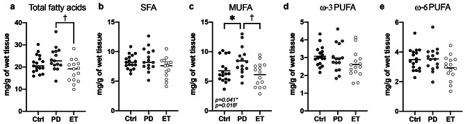
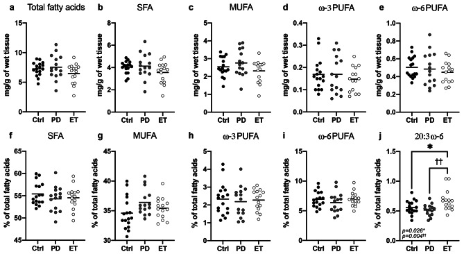
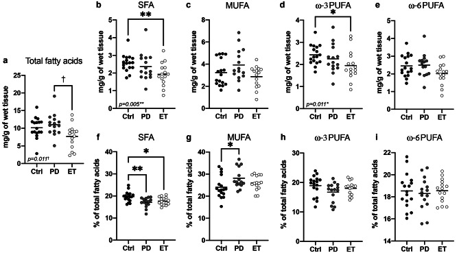
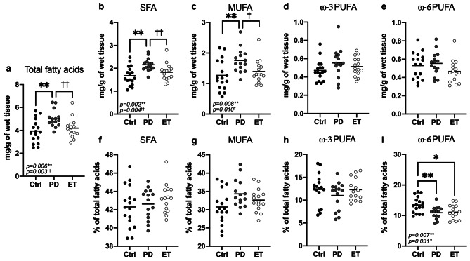
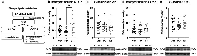
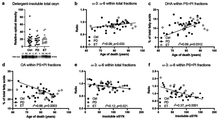
Fatty acids play many critical roles in brain function but have not been investigated in essential tremor (ET), a frequent movement disorder suspected to involve cerebellar dysfunction. Here, we report a postmortem comparative analysis of fatty acid profiles by gas chromatography in the cerebellar cortex from ET patients (n = 15), Parkinson's disease (PD) patients (n = 15) and Controls (n = 17). Phosphatidylcholine (PC), phosphatidylethanolamine (PE) and phosphatidylinositol (PI)/ phosphatidylserine (PS) were separated by thin-layer chromatography and analyzed separately. First, the total amounts of fatty acids retrieved from the cerebellar cortex were lower in ET patients compared with PD patients, including monounsaturated (MUFA) and polyunsaturated fatty acids (PUFA). The diagnosis of ET was associated with lower cerebellar levels of saturated fatty acids (SFA) and PUFA (DHA and ARA) in the PE fraction specifically, but with a higher relative content of dihomo-γ-linolenic acid (DGLA; 20:3 ω-6) in the PC fraction. In contrast, a diagnosis of PD was associated with higher absolute concentrations of SFA, MUFA and ω-6 PUFA in the PI + PS fractions. However, relative PI + PS contents of ω-6 PUFA were lower in both PD and ET patients. Finally, linear regression analyses showed that the ω-3:ω-6 PUFA ratio was positively associated with age of death, but inversely associated with insoluble α-synuclein. Although it remains unclear how these FA changes in the cerebellum are implicated in ET or PD pathophysiology, they may be related to an ongoing neurodegenerative process or to dietary intake differences. The present findings provide a window of opportunity for lipid-based therapeutic nutritional intervention.

Keywords: Essential tremor (ET); Fatty acids; Glycerophospholipids; Omega-3 polyunsaturated fatty acids (ω-3 PUFA).


Conflict of interest statement

Declarations. Ethics Approval and Consent to Participate: All human brain samples included in the present study were from patients or a family member who had filled out a consent form. Competing Interests: The authors declare no competing interests.

Figures

Fig. 1
Fig. 1
Total sum of fatty acids (absolute levels) within all phospholipid fractions in the cerebellar cortex of control subjects (n = 17), PD (n = 15) and ET (n = 15) patients. Levels of fatty acids were determined by GC-FID. Values are means ± S.E.M. expressed in absolute mass of wet tissue (mg/g of wet tissue). Statistical comparisons were performed using Kruskal-Wallis tests followed by nonparametric Wilcoxon post-hoc tests (*p < 0.05 comparing PD or ET patients to Controls; p < 0.05 comparing ET patients to PD ones). Abbreviations: ARA, arachidonic acid; Ctrl, control; DHA, docosahexaenoic acid; GC-FID, gas chromatography coupled to flame ionization detection; ET, essential tremor; MUFA, monounsaturated fatty acids; OA, oleic acid; PD, Parkinson’s disease; PUFA, polyunsaturated fatty acids; SFA, saturated fatty acids
Fig. 2
Fig. 2
Main fatty acids in the PC fraction extracted from the cerebellar cortex of subjects grouped according to the clinical diagnosis, controls (n = 17), PD (n = 15) and ET (n = 15) patients. (a-e) Absolute mass of wet tissue (mg/g of wet tissue). (f-j) Relative levels expressed in % of total fatty acids. Levels of fatty acids were determined by GC-FID and values are means ± S.E.M. Statistical comparisons were performed using Kruskal-Wallis tests followed by nonparametric Wilcoxon post-hoc tests (*p < 0.05 comparing PD or ET patients to Controls; p < 0.05; ††p < 0.01 comparing ET patients to PD ones). Abbreviations: Ctrl, control; GC-FID, gas chromatography coupled to flame ionization detection; ET, essential tremor; MUFA, monounsaturated fatty acids; PC, phosphatidylcholine; PD, Parkinson’s disease; PUFA, polyunsaturated fatty acids; SFA, saturated fatty acids
Fig. 3
Fig. 3
Main fatty acids in the PE fraction extracted from the cerebellar cortex of control subjects (n = 17), PD (n = 15) and ET (n = 15) patients. Levels of fatty acids were determined by GC-FID. Values are means ± S.E.M. expressed (a-e) in absolute mass of wet tissue (mg/g of wet tissue) or (f-i) in % of total fatty acids. Statistical comparisons were performed using Kruskal-Wallis tests followed by nonparametric Wilcoxon post-hoc tests, (*p < 0.05; **p < 0.01 comparing PD or ET patients to Controls; p < 0.05 comparing ET patients to PD ones). Abbreviations: Ctrl, control; GC-FID, gas chromatography coupled to flame ionization detection; ET, essential tremor; MUFA, monounsaturated fatty acids; PD, Parkinson’s disease; PE, phosphatidylethanolamine; PUFA, polyunsaturated fatty acids; SFA, saturated fatty acids
Fig. 4
Fig. 4
Main fatty acids in PS + PI fractions extracted from the cerebellar cortex of control subjects (n = 17), PD patients (n = 15) and ET patients (n = 15). Levels of fatty acids were determined by GC-FID. Values are means ± S.E.M. expressed (a-e) in absolute mass of wet tissue (mg/g of wet tissue) or (f-i) in % of total fatty acids. Statistical comparisons were performed using Kruskal-Wallis tests followed by nonparametric Wilcoxon post-hoc tests, (*p < 0.05; **p < 0.01 comparing PD or ET patients to Controls; p < 0.05; ††p < 0.01 comparing ET patients to PD ones). Abbreviations: Ctrl, control; GC-FID, gas chromatography coupled to flame ionization detection; ET, essential tremor; MUFA, monounsaturated fatty acids; PD, Parkinson’s disease; PI, phosphatidylinositol; PS, phosphatidylserine; PUFA, polyunsaturated fatty acids; SFA, saturated fatty acids
Fig. 5
Fig. 5
Expression of enzymes involved in ARA metabolism in cerebellar cortex of individuals with PD (n = 16) or ET (n = 15) in comparison with Controls (n = 19). (a) A schematic overview of ARA metabolism by 5-LOX and COX-2 after release from phospholipids. (b) Levels of 5-LOX, (c) cPLA2 and (d-e) COX-2 enzymes in (b, d) detergent-soluble and (c, e) TBS-soluble fractions. Levels of all proteins were determined by Western immunoblotting. Data are relative optical density values. Representative photo examples illustrate consecutive bands with actin shown as a loading control. Horizontal lines indicate mean ± SEM. Statistical comparisons were performed using one-way Kruskal-Wallis tests followed by nonparametric Wilcoxon post-hoc tests. Abbreviations: 5-LOX, 5-lipoxygenase; ARA, arachidonic acid; Ctrl, control; COX-2, cyclooxygenase 2; cPLA2, cytosolic phospholipase A 2; ET, essential tremor; PC, phosphatidylcholine; PGD2, prostaglandin D2; PGJ2, prostaglandin J2; PD, Parkinson’s disease; PE, phosphatidylethanolamine; PI, phosphatidylinositol; PS, phosphatidylserine; TBS, tris-buffered saline water-soluble
Fig. 6
Fig. 6
Linear regression analyses assessing age of death and insoluble phosphorylated α-synuclein to fatty acids. (a) Levels of total αsyn in the detergent-insoluble fraction were determined by Western immunoblotting and the normalization was done over the cerebellar cortex weight. Data are means ± S.E.M. of the relative optical density. Representative photo example illustrates bands for total αsyn. (b-f) Levels of fatty acids were determined by GC-FID and subjects were grouped according to (b-d) age of death or (e-f) insoluble monomeric α-syn (19 kDa) levels. Values are means ± S.E.M. Coefficient of determination (r2), p < 0.05. Abbreviations: αsyn, α-synuclein; Ctrl, control; DHA, docosahexaenoic acid; ET, essential tremor; GC-FID, gas chromatography coupled to flame ionization detection; pα-synuclein, phosphorylated α-synuclein; PD, Parkinson’s disease; PE, phosphatidylethanolamine; PI, phosphatidylinositol; PS, phosphatidylserine; PUFA, polyunsaturated fatty acids

Similar articles

References

    1. Louis ED, Ottman R. How many people in the USA have essential tremor? Deriving a population estimate based on epidemiological data. Tremor Other Hyperkinet Mov (N Y). 2014;4:259. - PMC - PubMed
    1. Louis ED, McCreary M. How common is essential Tremor? Update on the Worldwide Prevalence of essential tremor. Tremor Other Hyperkinet Mov (N Y). 2021;11:28. - PMC - PubMed
    1. Thenganatt MA, Louis ED. Distinguishing essential tremor from Parkinson’s disease: bedside tests and laboratory evaluations. Expert Rev Neurother. 2012;12(6):687–96. - PMC - PubMed
    1. Louis ED, Faust PL. Essential tremor pathology: neurodegeneration and reorganization of neuronal connections. Nat Rev Neurol. 2020. - PubMed
    1. Louis ED, Faust PL, Vonsattel JPG, Honig LS, Rajput A, Robinson CA, et al. Neuropathological changes in essential tremor: 33 cases compared with 21 controls. Brain. 2007;130(12):3297–307. - PubMed

LinkOut - more resources