. 2024 Sep 24;43(9):114665.
doi: 10.1016/j.celrep.2024.114665. Epub 2024 Aug 29.

Trigeminal innervation and tactile responses in mouse tongue

Affiliations

Trigeminal innervation and tactile responses in mouse tongue

Linghua Zhang et al. Cell Rep. .

Abstract

The neural basis of tongue mechanosensation remains largely mysterious despite the tongue's high tactile acuity, sensitivity, and relevance to ethologically important functions. We studied terminal morphologies and tactile responses of lingual afferents from the trigeminal ganglion. Fungiform papillae, the taste-bud-holding structures in the tongue, were convergently innervated by multiple Piezo2+ trigeminal afferents, whereas single trigeminal afferents branched into multiple adjacent filiform papillae. In vivo single-unit recordings from the trigeminal ganglion revealed lingual low-threshold mechanoreceptors (LTMRs) with distinct tactile properties ranging from intermediately adapting (IA) to rapidly adapting (RA). The receptive fields of these LTMRs were mostly less than 0.1 mm2 and concentrated at the tip of the tongue, resembling the distribution of fungiform papillae. Our results indicate that fungiform papillae are mechanosensory structures and suggest a simple model that links functional and anatomical properties of tactile sensory neurons in the tongue.

Keywords: CP: Neuroscience; LTMR; mechanoreceptor; somatosensation; taste bud; tongue; touch; trigeminal; trigeminal ganglion.


Conflict of interest statement

Declaration of interests The authors declare no competing interests.

Figures

Figure 1.
Figure 1.. Trigeminal afferents innervate both filiform and fungiform papillae and have the highest density at the tongue tip
(A and B) Dorsal views of the tongue showing DiI-labeled trigeminal axons in P1 (A) and P10 (B) mice. Arrows: filiform; triangles: fungiform. To reveal the fluorescent signals in the filiform papillae in (B), those in the fungiform papillae are displayed saturated. Scale bars: 500 μm for (A) and 100 μm for (B). (C and D) Longitudinal views of the tongue showing DiI-labeled trigeminal axons in fungiform (C) and filiform (D) papillae. Arrow in (C): axons terminating in extragemmal regions of a fungiform papilla. Scale bar: 20 μm. See also Figure S1. (E) The fluorescence intensity along the A-P axis across DiI-labeled tongue surfaces at different ages. (F) Schematic of the innervation patterns of TG neurons and GG neurons in filiform and fungiform papillae.
Figure 2.
Figure 2.. Trigeminal afferents asymmetrically innervate the extragemmal region of fungiform papillae and exhibit a ring-like termination pattern surrounding the taste pore, similar to Piezo2+ afferents
(A) Maximum intensity projection of the confocal z-stack in Figure 1B showing the ring-like innervation pattern of trigeminal afferents in fungiform papillae. Scale bar: 100 μm. (B) Optical sections of DiI-labeled trigeminal afferents in a fungiform papilla from a top-down view. The sections are labeled in an order from the deeper connective tissue (section 1) to the apical epithelium (section 8). Yellow dashed lines: taste bud regions. Scale bar: 25 μm. See also Video S1. (C) Fluorescence intensity of DiI in the ring region of fungiform papillae at each side of the tongue (top and bottom) on a linear scale (gray concentric circles). The dark blue line represents the mean intensity across fungiform papillae, while the light blue shading indicates the bootstrap 95% confidence interval. n = 26 and 27 papillae from 4 (2 P12 and 2 P21) mice for top and bottom, respectively. (D) Dorsal view of the tongue in a Piezo2-EGFP-IRES-Cre/+;Snap25LSL-EGFP/+ mouse and its Piezo2-EGFP-IRES-Cre/+; Snap25+/+ littermate. Scale bar: 500 μm. (E) Piezo2-expressing afferents innervate both fungiform and filiform papillae, and form a ring-like pattern (arrows) by innervating the extragemmal region of fungiform papillae. Scale bar: 100 μm.
Figure 3.
Figure 3.. Mouse filiform papillae are innervated by NFH+ myelinated free nerve endings
(A) Top: in the tongue of Plp1-EGFP/+ mice, no corpuscular end organs, but rather only myelinated free nerve endings, can be identified in the connective tissue core of filiform papillae. Bottom: in the glabrous skin, in contrast, Meissner corpuscles can be found in the dermal papillae. DAPI: cell nucleus. EGFP and S100β: (terminal) Schwann cells. Asterisks: cell bodies of Schwann cells. Arrows: cell processes or myelin from Schwann cells. See also Figure S2. (B and C) In PirtCre/+;R26LSL-tdTom/+;Plp1-EGFP/+ and Vglut2Cre/+;R26LSL-tdTom/+;Plp1-EGFP/+ mice, tdTom-labeled, myelinated afferents that colocalized with EGFP can be seen in both filiform and fungiform papillae. (B) Asterisks: taste bud cells in and around fungiform papillae; triangles: the myelin was lost at the nerve terminal in myelinated afferents; thin arrows: unmyelinated afferents. (C) Asterisks: muscle; arrowhead: intragemmal afferents; thick arrows: extragemmal afferents; thin arrows: unmyelinated afferents. (D) SplitCre labeled unmyelinated afferents in both filiform and fungiform papillae (arrows). Scale bars in (A)–(D): 20 μm. See also Figure S3 and Table S1.
Figure 4.
Figure 4.. Single-unit recordings reveal lingual LTMRs ranging from RA to IA with varied tactile response properties
(A) The recording setup. Green: lingual nerve. See also Figure S4. (B) Neural activity of single lingual LTMR units recorded in the TG in response to indentation at their RFs on the surface of the tongue. Neural traces with no spikes were cropped to make the onsets and offsets of indentation aligned for each column. An example SA-LTMR innervating the whisker pad was included for comparison. (C) The RFs of lingual LTMRs mapped by von Frey hairs (n = 25 from 19 mice; 16 IA-LTMRs in red, 9 RA-LTMRs in blue, and 1 unit that cannot be categorized into IA/RA in gray). (D) The AI and average firing duration of IA/RA-LTMRs for 3.9 mN stimulation. Top: schematic showing the calculation of AIt using a peristimulus time histogram (PSTH) generated from a Poisson distribution. (E) PSTHs (bin width: 50 ms, n = 5 events) and interspike intervals (ISIs, bin width: 10 ms) of the three example lingual units in (B) in response to 3.9 mN. See also Figure S5. (F) Cumulative firing rate over total firing rate for IA/RA-LTMRs during 3.9 mN stimulation. Traces longer than 4 s were cropped. Arrow: an RA-LTMR with extremely low firing rates (LZ2203 Pos 6 Unit 3 in Figure S5B). (G) Average firing rate of IA/RA-LTMRs during 3.9 mN stimulation (mean ± SD). (H) The von Frey indentation threshold for lingual LTMRs (n = 23 from 16 mice; 14 IA-LTMRs, 8 RA-LTMRs, and 1 unit that could not be categorized). (I) The average firing rates of an example IA-LTMR (left) and RA-LTMR (right) in the initial 300 ms after firing onset during indentation (mean ± SD; one-sided Student’s t test). (J) Example neural traces from conduction velocity tests. Gray dashed line: time point of the electrical pulse. (K) Activity traces of lingual LTMRs in response to other tactile stimuli. The bottom row shows an RA-LTMR with a threshold higher than 0.4 mN. (L) Neural activity of example single lingual LTMR units recorded in the TG in response to indentation and 473-nm light stimulation at their RFs. Blue bars: 10-msduration, 5 Hz, 473-nm light pulses. (M and N) Number of lingual LTMR units that can or cannot be activated by 473-nm light stimulation from 3 PirtCre/+;R26LSL-ChR2-EYFP/+ mice and 8 Vglut2Cre/+; R26LSL-ChR2-EYFP/+ mice.
Figure 5.
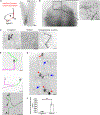
Figure 5.. Lingual neurons that terminate in different types of papillae exhibit distinct terminal shapes and branching patterns
(A) Left: schematic of unilateral viral injection into the mandibular branch of the left TG in Vglut2Cre mice. Right: neurons at the mandibular branch of the TG were labeled by tdTom. Scale bar: 300 μm. (B) tdTom-labeled trigeminal afferents at the surface of the tongue. Scale bar: 800 μm. Inset: An enlarged view of the tip of the tongue. Scale bar: 300 μm. (C) Single TG afferent terminals sparsely labeled by tdTom. Scale bars (left to right): 50, 100, and 50 μm. (D) Multiple TG afferents simultaneously labeled by tdTom. Top: two clusters of nerve fibers (green and magenta) co-innervating a single fungiform papilla. Scale bar: 100 μm. Center: two independent afferents (green and magenta) innervating two separate fungiform papillae. Scale bar: 50 μm. Bottom: two independent afferents (green and magenta) innervating several filiform papillae had overlap in their terminal fields. Arrowheads: endings in the same filiform papillae. Scale bar: 100 μm. (E) Clusters of filiform papillae innervated by separate TG afferent aggregates (blue arrows). Red arrowheads: fungiform papillae. Scale bar: 100 μm. (F) The terminal field size of TG afferents innervating fungiform (0.0036 ± 0.0015, n = 20 from 5 mice) or filiform papillae (0.0435 ± 0.0096, n = 14 from 6 mice). ***p = 9.59 × 10⁻¹⁰; two-sided Welch’s t test.
Figure 6.
Figure 6.. Lingual LTMRs are insensitive to rapid cooling or chemicals that can induce astringent or numbing sensations
(A) Activity of an example single lingual LTMR unitin the TG in response to indentation, rapid cooling, or tannic acid. (B) Lingual LTMRs did not respond to rapid cooling (n = 8 from 6 mice). (C) Lingual LTMRs did not respond to EGCG/tannicacid in a series of concentrations (n = 6 from 5 mice). (D) The average firing rates of three example LTMRs before and during Kava/sanshool treatment (from 2 mice). (E) Lingual LTMRs did not respond to sanshool or Kava (n = 6 from 4 mice).
Figure 7.
Figure 7.. A working model of the organization of tactile innervation in the tongue
Schematic hypothesis of how the lingual papillae at the surface of the tongue could be innervated by lingual LTMRs in the TG. A single fungiform papilla is innervated by LTMRs ranging from IA to RA (red, purple, and blue), while several adjacent filiform papillae are simultaneously innervated by single RA-LTMRs (blue).

Update of

References

    1. Pippi R, Spota A, and Santoro M (2018). Medicolegal Considerations Involving Iatrogenic Lingual Nerve Damage. J. Oral Maxillofac. Surg. 76, 1651.e1–1651.e13. 10.1016/j.joms.2018.03.020. - DOI - PubMed
    1. Tei K, Yamazaki Y, Kobayashi M, Izumiyama Y, Ono M, and Totsuka Y (2004). Effects of bilateral lingual and inferior alveolar nerve anesthesia effects on masticatory function and early swallowing. Oral Surg. Oral Med. Oral Pathol. Oral Radiol. Endod. 97, 553–558. 10.1016/S1079210403006875. - DOI - PubMed
    1. Chee C, Arshad S, Singh S, Mistry S, and Hamdy S (2005). The influence of chemical gustatory stimuli and oral anaesthesia on healthy human pharyngeal swallowing. Chem. Senses 30, 393–400. 10.1093/chemse/bji034. - DOI - PubMed
    1. Stern JM, and Johnson SK (1989). Perioral somatosensory determinants of nursing behavior in Norway rats (Rattus norvegicus). J. Comp. Psychol. 103, 269–280. 10.1037/0735-7036.103.3.269. - DOI - PubMed
    1. Niemi M, Laaksonen J-P, Vähätalo K, Tuomainen J, Aaltonen O, and Happonen R-P (2002). Effects of transitory lingual nerve impairment on speech: an acoustic study of vowel sounds. J. Oral Maxillofac. Surg. Off. J. Am. Assoc. Oral Maxillofac. Surg. 60, 647–652. 10.1053/joms.2002.33113. - DOI - PubMed

LinkOut - more resources