. 2024 Nov;106(5):870-886.
doi: 10.1016/j.kint.2024.08.022. Epub 2024 Aug 30.

Myeloperoxidase-ANCA IgG induces different forms of small vessel vasculitis based on type of synergistic immune stimuli

Affiliations

Myeloperoxidase-ANCA IgG induces different forms of small vessel vasculitis based on type of synergistic immune stimuli

Peiqi Hu et al. Kidney Int. 2024 Nov.

Abstract

Anti-neutrophil cytoplasmic autoantibody (ANCA) vasculitis has diverse patterns of injury including microscopic polyangiitis (MPA), granulomatosis with polyangiitis (GPA), and eosinophilic granulomatosis with polyangiitis (EGPA). Necrotizing and crescentic glomerulonephritis (NCGN) occurs in all syndromes and as renal limited vasculitis (RLV). Single-dose intravenous ANCA IgG-specific for mouse myeloperoxidase (MPO) causes RLV in mice. Although multiple mouse models have elucidated ANCA-IgG induced necrotizing and crescentic glomerulonephritis (NCGN), pathogenesis of ANCA-induced granulomatosis and vasculitis outside the kidney has not been clarified. To investigate this, we used intravenous MPO-ANCA IgG in the same strain of mice to induce different patterns of lung disease mirroring patients with granulomatosis with polyangiitis (GPA), microscopic polyangiitis (MPA), and eosinophilic granulomatosis with polyangiitis (EGPA). Repeated intravenous MPO-ANCA IgG induced GPA with NCGN, lung capillaritis, arteritis and granulomatosis. Lung leukocyte phenotypes were evaluated by immunohistochemical image analysis and by flow cytometry. ANCA lung capillaritis and microabscesses began within one day and evolved into granulomas in under seven days. Influenza plus single-dose MPO-ANCA IgG induced MPA with NCGN, lung capillaritis and arteritis, but no granulomatosis. Allergic airway disease caused by house dust mites or ovalbumin plus single-dose intravenous MPO-ANCA IgG induced EGPA with eosinophilic bronchiolitis, NCGN, capillaritis, arteritis, and granulomatosis. Thus, our study shows that the occurrence and pattern of lung lesions are determined by the same ANCA IgG accompanied by different synergistic immune factors.

Keywords: ANCA; crescentic glomerulonephritis; granulomatosis; polyangiitis; vasculitis.


Conflict of interest statement

Disclosure

The authors have no relationships/activities/interests that conflict with the content of our manuscript.

Figures
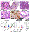
Figure 1.
Figure 1.. Kidney necrotizing and crescentic glomerulonephritis (NCGN), lung GPA-like granulomatosis, and arteritis are induced in female WT B6 mice 7 days after the first of two i.v. MPO-ANCA IgG doses on day 0 and day 1 with i.t. LPS.
(a) Glomerulus with cellular crescent (outlined) and segmental fibrinoid necrosis (arrow). (b) Low magnification of lung showing multiple granulomas (granulomatosis) (arrows) (H&E stain). (c) Granuloma with predominance of macrophages (H&E stain). (d) Higher magnification granuloma showing admixture of macrophages (including multinucleated giant cells, arrow) and few neutrophils (H&E). (e) Granuloma with numerous F4/80+ macrophages. (f) Early arteritis with transmural inflammation (arrow) (H&E). All scale bars 100μm. (g) Flow cytometric leukocyte immuno-phenotyping of lung digests in 6 B6 naive control mice, 6 B6 LPS alone mice, and 6 B6 mice 7 days after GPA induction with i.t. LPS and i.v. MPO-ANCA X2 (B6 mice). LPS alone did not induce a significant increase in leukocytes. LPS + MPO-ANCA X2 IgG induced increased numbers of neutrophils; and alveolar, interstitial, and inflammatory macrophages; T cells and B cells. Siglec-F+ neutrophils were detected only in lungs of mice given LPS+MPO-ANCA. Data represent mean ± SEM (n=5 mice per group). Statistical significance was determined using one-way ANOVA with Tukey’s test for multiple comparisons. * P <0.05, ** P <0.01, *** P <0.001, **** P <0.0001
Figure 2.
Figure 2.. Kidney NCGN and lung GPA-like granulomatosis are induced in 7 days in WT mice by i.v. MPO-ANCA IgG on day 0 and day 1 with or without i.t. LPS. Rag2 KO mice without T or B cells develop NCGN and lung granulomatosis, whereas C5 KO and FB KO mice develop lung granulomatosis but no NCGN.
One dose of MPO-ANCA induces NCGN but no lung disease. Data are from female mice except one group of mice receiving i.t. LP and MPO-ANCA x2. (a) % glomerular crescents, (b) lung lesion scores, (c) urine dipstick hematuria score, and (d) urine dipstick proteinuria score in female WT B6 mice receiving LPS alone (n=6), female WT mice receiving one dose of MPO-ANCA without LPS (n=12), female WT mice receiving X2 MPO-ANCA without LPS (n=9), male WT mice receiving X2 i.v. MPO-ANCA + i.t. LPS (n=4), female WT mice receiving X2 i.v. MPO-ANCA + i.t. LPS (n=14), female Rag2 KO mice receiving X2 i.v. MPO-ANCA + i.t. LPS (n=4), female C5 KO mice (n=4) and Factor B KO mice (n=3) receiving X2 i.v. MPO-ANCA + i.t. LPS, female WT mice receiving X2 i.v. anti-BSA + i.t. LPS (n=3), and female MPO KO mice receiving X2 i.v. MPO-ANCA + i.t. LPS (n=3). Data are expressed as the mean ± SEM. One-way ANOVA with Dunnett’s multiple comparisons test was used to test statistical significance between groups and shown as ns (not significant) P value >0.05, * P <0.05, ** P <0.01, *** P <0.001, **** P <0.0001.
Figure 3:
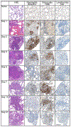
Figure 3:. Time course study reveals that repeat doses of MPO-ANCA induced granulomas evolve from alveolar capillaritis and microabscesses.
B6 mice (n=14) received i.t. LPS on day 0, 1st dose i.v. MPO-ANCA IgG ~30 min later, and 2nd dose i.v. on day 1. Pairs of mice were euthanized for histopathologic studies on days 1-21 beginning 6 hrs. after the 2nd MPO-ANCA dose. Naïve mice were used as controls. Lung neutrophil, macrophage, and T cell infiltration was evaluated by IHC using antibodies to mouse Nimp-R14 (neutrophil marker), F4/80 (macrophage marker), and CD3 (T cell marker). Naïve mice had open alveolar airspaces and delicate alveolar walls. Day 1 lungs had neutrophilic capillaritis, hemorrhage into airspaces, and scattered neutrophilic microabscesses. Day 2 and Day 3 consolidated lesions had an admixture of neutrophils and macrophages (also see Figure 4). Day 5, Day 7 and Day 14 lesions predominantly contained macrophages (i.e. are granulomas). Day 21 lesions contained only a few residual leukocytes. (Scale bar 200μm).
Figure 4:
Figure 4:. B6 mice that received i.t. LPS on day 0, 1st dose i.v. MPO-ANCA IgG ~30 min later, and 2nd dose i.v. on day 1 developed acute lung lesions within 6 hours that evolved into granulomas by 7 days; accompanied by necrotizing and crescentic glomerulonephritis.
B6 mice (n=14) received i.t. LPS on day 0, 1st dose i.v. MPO-ANCA IgG ~30 min later, and 2nd dose i.v. on day 1. Pairs of mice were euthanized for histopathologic studies on days 1-21 beginning 6 hrs. after the 2nd MPO-ANCA dose. (a) Capillaritis with focal hemorrhage into alveolar spaces 1 day after the 1st dose and approximately 6 hours after the 2nd dose of MPO-ANCA. (b) Day 1 alveolar wall capillaries containing Ly6g+ neutrophils. (c) Day 1 artery with focal marginating neutrophils (arrow). (d) Day 1 microabscesses. (e) Day 1 microabscesses with neutrophils staining for Ly6g by IHC. (f) Day 7 transitional lesions with residual central zone of neutrophils (long arrow) surrounded by macrophages (short arrow) (g). Glomerulus with segmental fibrinoid necrosis. (h) Glomerulus with cellular crescent (outlined) and segmental fibrinoid necrosis (arrows). (i) Cellular crescents rich in F4/80+ macrophages. All scale bars 100μm.
Figure 5:
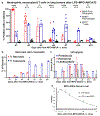
Figure 5:. Quantitative analysis of lung and glomerular lesions, and uranalysis in B6 mice that received i.t. LPS on day 0, 1st dose i.v. MPO-ANCA IgG ~30 min later, and 2nd dose i.v. on day 1 developed acute lung lesions with 6 hours that evolve into granulomas by 7 days; accompanied by necrotizing and crescentic glomerulonephritis.
Female B6 mice (n=14) received i.t. LPS on day 0, 1st dose i.v. MPO-ANCA IgG ~30 min later, and 2nd dose i.v. on day 1. Pairs of mice were euthanized for histopathologic studies on days 1-21 beginning 6 hrs. after the 2nd MPO-ANCA dose. (a) Neutrophils, macrophages, and T cells in consolidated lesions were measured by whole slide image analysis of IHC positive cells using Definiens Architect XD 64 software. Neutrophils predominated in lesions on day 1 and 2 after the first dose of MPO-ANCA, consistent with microabscesses. Macrophages predominated thereafter consistent with granulomas. By day 21, the size of lesions and the numbers of leukocytes in lesions diminished substantially. Data expressed as the mean ± SEM. (b) Glomerular necrosis peaked before crescents, and crescent formation plateaued at day 5. (c) Urinalysis by dipstick demonstrated that proteinuria was the earliest and most persistent marker of kidney injury, and hematuria and pyuria corresponded with active inflammation. All data expressed as the mean ± SEM. (d) Decline in MPO-ANCA serum levels corresponded with remission of additional crescent formation and remission of acute neutrophilic lung injury.
Figure 6.
Figure 6.. Induction of lung granulomatosis by i.t. LPS and i.v. two-dose MPO-ANCA IgG can be monitored by microcomputed tomography (micro-CT).
MCT was used to monitor GPA lung lesions in 2 female WT B10 mice two, four and six days after IT LPS and the first of two MPO-ANCA IgG doses. Mice were sacrificed on day 6 for pathologic examination. Observations were comparable in both mice. All images in this Figure are from the same mouse. (a) By day 2 after the 1st dose of MPO-ANCA IgG, focal ground-glass opacities consistent with alveolar hemorrhage appeared (dotted circle). (b) On day 4 the same position CT image demonstrated multiple small nodules and a large pulmonary mass (arrow) in the right lung. (c) On day 6 the dominant mass increased in size (arrow). (d) Gross inspection of lungs revealed multiple variably sized nodules with adjacent hemorrhage. (e) Histopathology of a granuloma (long arrow) with adjacent hemorrhage (short arrow). (H&E, scale bar 200μm). (f) High magnification of a granuloma with giant cell (arrow), numerous macrophages and a few neutrophils (H&E, scale bar 50μm).
Figure 7:
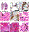
Figure 7:. One-dose i.v. MPO-ANCA IgG alone without synergistic influenza causes renal limited NCGN, influenza infection alone causes lung bronchiolitis without NCGN, and single dose MPO-ANCA with synergistic influenza causes MPA-like pulmonary-renal syndrome with lung hemorrhagic capillaritis, arteritis, and NCGN.
Female WT B6 mice 9–10-week-old (n=8) were inoculated intranasally with 1,000 PFU influenza A (H1N1) strain A/WSN/33 virus on day 0, injected i.v. with 50ug/g MPO-ANCA IgG on day 1 and euthanized on day 5. Comparison groups were B6 mice receiving MPO-ANCA IgG alone (n=9), or influenza virus alone (n=9). (a,d) Unremarkable lung histology in a mouse receiving i.v. MPO-ANCA alone. (b,e) Diffuse bronchiolitis with no hemorrhagic capillaritis in a mouse receiving influenza alone. (c,f) Bronchiolitis as well as extensive alveolar capillaritis with alveolar hemorrhage in a mouse receiving influenza and i.v. MPO-ANCA. (g) Severe alveolar capillaritis with numerous neutrophils (arrow) in a mouse with influenza and MPO-ANCA. (h) Arteritis (arrow) in a mouse with influenza and MPO-ANCA. (i) Necrotizing and crescentic GN in a mouse with influenza and MPO-ANCA (crescent outlined, arrow on necrosis). (j). NCGN had no statistical difference in severity in mice receiving MPO-ANCA alone, or concurrent with viral lung disease. Mice with virus alone developed no GN. Sale bars 100μm. Data expressed as the mean ± SEM. Statistical analysis by one-way ANOVA with Dunnett’s multiple comparisons test. The criterion for statistical significance was P value <0.05. All scale bars 100μm.
Figure 8:
Figure 8:. Sensitization and challenge (S&C) with OVA causes mouse asthma-like eosinophil-rich allergic airway disease that synergizes with one-dose i.v. MPO-ANCA to cause EGPA-like granulomatosis, arteritis, hemorrhagic capillaritis and NCGN.
(a) On the left, low magnification of lung from a mouse that received OVA S&C without i.v. MPO-ANCA and developed allergic airway disease with peribronchial inflammation but no alveolar hemorrhage or consolidation. On the right, low magnification of lung from a mouse that received OVA S&C and i.v. MPO-ANCA and developed enhanced peribronchial inflammation as well as focal hemorrhagic capillaritis (long arrows) and focal consolidation by granulomatous inflammation (short arrows). (b) Allergic bronchiolitis caused by OVA alone with numerous EPX+ eosinophils. (c) Allergic bronchiolitis caused by OVA plus MPO-ANCA with enhanced influx of EPX+ eosinophils and additional lung consolidation by granulomatous inflammation with fewer eosinophils. (d) Granulomatous inflammation with multinucleated giant cells (arrows) caused by OVA+ANCA. (e) Granulomatous inflammation with numerous F4/80+ macrophages caused by OVA+ANCA. (f) Early arteritis (endothelialitis) caused by OVA+ANCA. (g and h) Hemorrhagic capillaritis caused by OVA+ANCA. (i) NCGN with fibrinoid necrosis (arrow) and crescent (outlined) in a mouse receiving OVA+ANCA. (Scale bar 100μm) (Refer to Figures 7a and 7d for histologically unremarkable lung histology)
Figure 9:
Figure 9:. Sensitization and challenge with HDM causes mouse asthma-like eosinophil-rich allergic airway disease that synergizes with one dose i.v. MPO-ANCA to cause EGPA-like granulomatosis, arteritis, hemorrhagic capillaritis, and NCGN.
(a) On the left, low magnification of lung from a mouse that received HDM S&C without i.v. MPO-ANCA and developed allergic airway disease with peribronchial inflammation but no alveolar hemorrhage or consolidation. On the right, low magnification of lung from a mouse that received HDM S&C and i.v. MPO-ANCA and developed enhanced peribronchial inflammation as well as focal hemorrhagic capillaritis (long arrows) and focal consolidation by granulomatous inflammation (short arrows). (b) Mice that received HDM alone had asthma-like allergic airway disease with peribronchial inflammation but no alveolar hemorrhage or consolidation. Mice with HDM induced lung disease that received i.v. MPO-ANCA IgG developed (c) enhanced peribronchial inflammation as well as focal hemorrhagic capillaritis (long arrow) and arteritis (short arrow), (d) granulomatous inflammation with multinucleated giant cells (arrows) with (e) numerous F4/80+ macrophages, and (f) scattered EPX+ eosinophils. (g) Acute transmural arteritis caused by HDM+ANCA. (h) NCGN with crescent (between arrows) in a mouse receiving HDM+ANCA. (i) Percent glomeruli with necrosis and crescents in mice receiving MPO-ANCA alone, HDM or OVA alone, or HDM (closed circles) or OVA (open circles) with i.v. MPO-ANCA. (Scale bar 100μm) (Refer to Figures 7a and 7d for histologically unremarkable lung histology)
Figure 10.
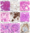
Figure 10.. In a mouse model of granulomatosis with polyangiitis, two doses of intravenous MPO-ANCA IgG given on day 0 and day 1 induced lung alveolar hemorrhagic capillaritis within 6 hours after the 2nd dose, multiple microabscesses within 2 days, and multiple granulomas within 7 days.
(a) Normal lung tissue has empty alveolar air spaces surrounded by thin alveolar walls comprising a single layer of pneumocytes overlying capillaries. (b) Withing 6 hours after the second of two MPO-ANCA doses on day 0 and day 1, alveolar capillaries have extensive neutrophil margination and diapedesis (long arrow) and airspaces contain red blood cells and neutrophils. (c) Within 2 days, multiple microabscesses form with focal replacement of alveolar walls by clustered neutrophils and leukocytoclastic debris. (d) Within 7 days, microabscesses are replaced by clusters of macrophages (granulomas) some of which have residual admixed neutrophils. (Scale bar 50 μm)

References

    1. Godman GC, and Churg J. Wegener’s granulomatosis: pathology and review of the literature. AMA Arch Pathol. 1954;58(6):533–53. - PubMed
    1. van der Woude FJ, van der Woude FJ, Rasmussen N, et al. Autoantibodies against neutrophils and monocytes: tool for diagnosis and marker of disease activity in Wegener’s granulomatosis. Lancet. 1985;1(8426):425–9. - PubMed
    1. Falk RJ, and Jennette JC. Anti-neutrophil cytoplasmic autoantibodies with specificity for myeloperoxidase in patients with systemic vasculitis and idiopathic necrotizing and crescentic glomerulonephritis. N Engl J Med. 1988;318(25):1651–7. - PubMed
    1. Guillevin L, Noel LH, Pourrat J, et al. Antineutrophil cytoplasm antibodies in systemic polyarteritis nodosa with and without hepatitis B virus infection and Churg-Strauss syndrome--62 patients. J Rheumatol. 1993;20(8):1345–9. - PubMed
    1. Lionaki S, Blyth ER, Hogan SL, et al. Classification of antineutrophil cytoplasmic autoantibody vasculitides: the role of antineutrophil cytoplasmic autoantibody specificity for myeloperoxidase or proteinase 3 in disease recognition and prognosis. Arthritis Rheum. 2012;64(10):3452–62. - PMC - PubMed

MeSH terms

LinkOut - more resources