. 2025 Jul:73:341-356.
doi: 10.1016/j.jare.2024.08.033. Epub 2024 Aug 30.

A new 1,2,3-triazole-indirubin hybrid suppresses tumor growth and pulmonary metastasis by mitigating the HGF/c-MET axis in hepatocellular carcinoma

Affiliations

A new 1,2,3-triazole-indirubin hybrid suppresses tumor growth and pulmonary metastasis by mitigating the HGF/c-MET axis in hepatocellular carcinoma

Shalini V Gowda et al. J Adv Res. 2025 Jul.

Abstract

Introduction: Hepatocellular carcinoma (HCC) is a fatal cancer that is often diagnosed at the advanced stages which limits the available therapeutic options. The interaction of HGF with c-MET (a receptor tyrosine kinase) results in the activation of c-MET which subsequently triggers the PI3K/Akt/mTOR axis. Overexpression of c-MET in HCC tissues has been demonstrated to contribute to tumor progression and metastasis.

Objectives: We aimed to synthesize triazole-indirubin conjugates, examine their growth suppressor efficacy in cell-based assays, and investigate the antitumor as well as antimetastatic activity of lead cytotoxic agent in the orthotopic mice model.

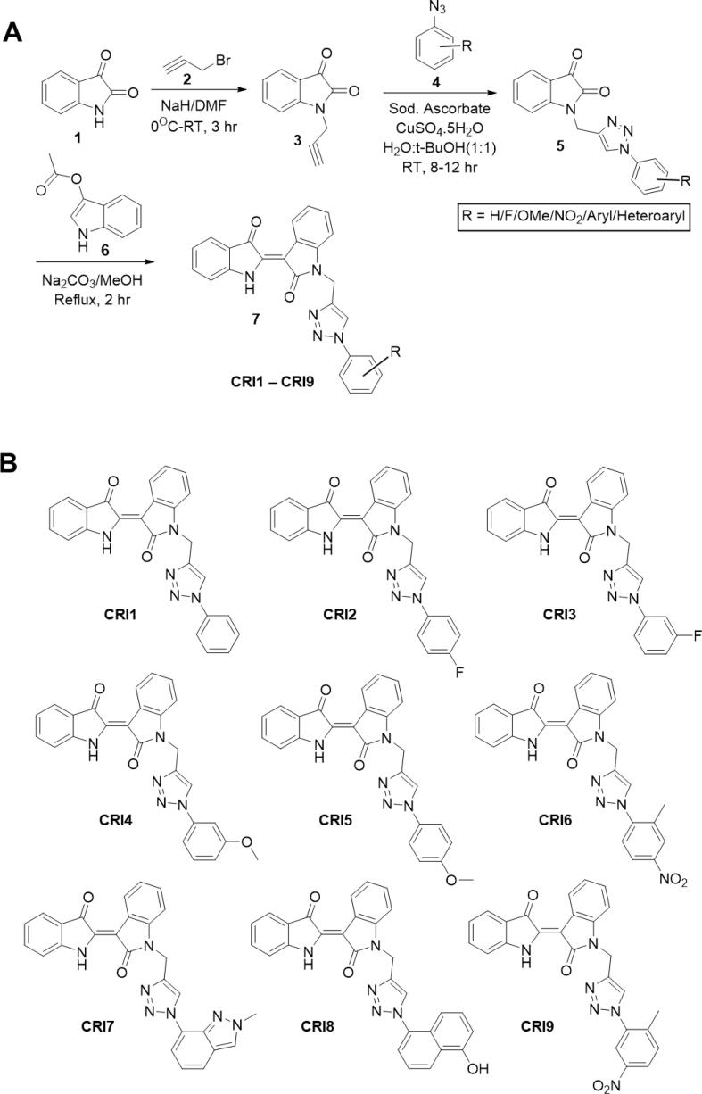
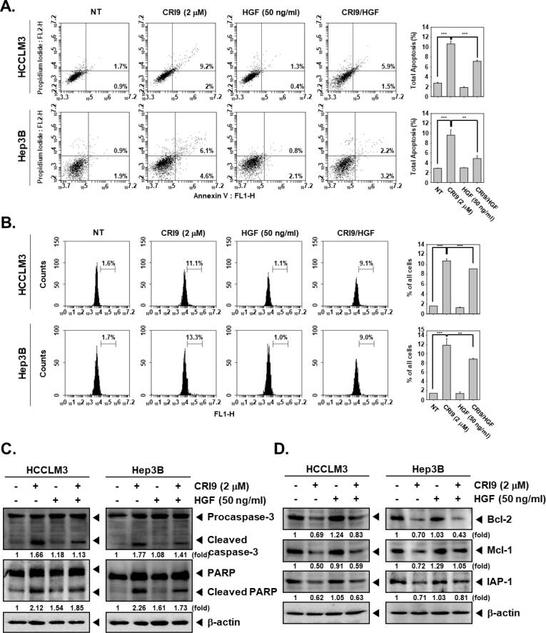
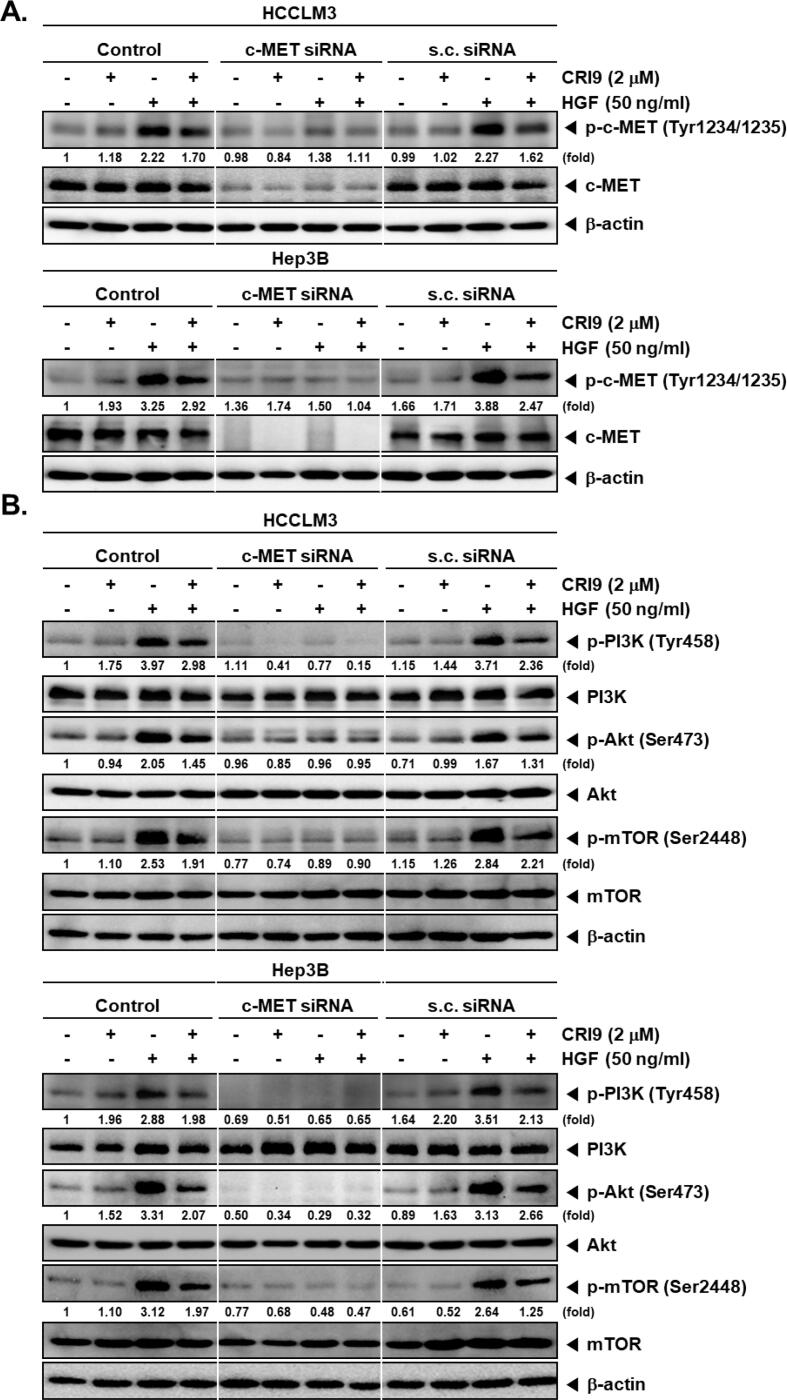
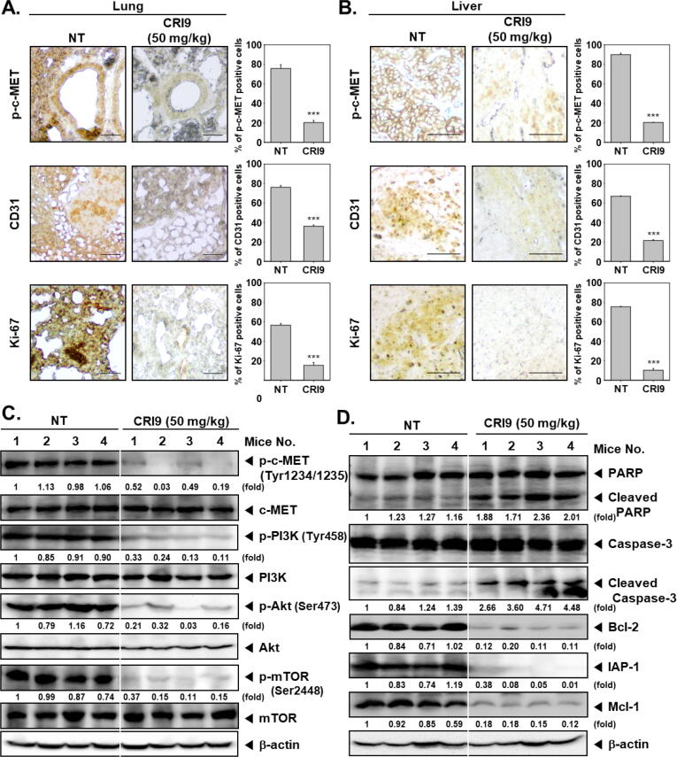
Methods: A series of triazole-indirubin hybrids were synthesized and cytotoxicity, apoptogenic, and antimigratory effect of the lead compound (CRI9) was evaluated using MTT assay, cell cycle analysis, annexin-V/PI assay, TUNEL assay, and wound healing assay. The effect of CRI9 on the operation of the HGF/c-MET/PI3K/Akt/mTOR axis was examined using western blotting and transfection experiments. Acute toxicity, antitumor, and antimetastatic activity of CRI9 were examined in NCr nude mice. The expression of c-MET/PI3K/Akt/mTOR, CD31, and Ki-67 was examined using immunohistochemistry and western blotting.

Results: Among the new compounds, CRI9 consistently displayed potent cytotoxicity against HGF-induced HCC cells. CRI9 induced apoptosis as evidenced by increased sub G1 cells, annexin-V+/PI+ cells, TUNEL+ cells, and cleavage of procaspase-3 and PARP. CRI9 inhibited HGF-induced phosphorylation of c-METY1234/1235 and subsequently suppressed the PI3K/Akt/mTOR axis. Also, depletion of c-MET or inhibition of c-MET by CRI9 resulted in suppression of the PI3K/Akt/mTOR axis. CRI9 showed no toxic effects in NCr nude mice and displayed a potent antitumor and antimetastatic effect in the orthotopic HCC mice model. CRI9 also reduced the levels of phospho-c-MET, CD31, and Ki-67 and suppressed the activation of the PI3K/Akt/mTOR axis in tumor tissues.

Conclusion: CRI9 has been identified as a new inhibitor of the c-MET/PI3K/Akt/mTOR axis in HCC preclinical models.

Keywords: Hepatocellular carcinoma; Indirubin; Orthotopic mice model; Triazole; c-MET.


Conflict of interest statement

Declaration of competing interest The authors declare that they have no known competing financial interests or personal relationships that could have appeared to influence the work reported in this paper.

Figures

None
Graphical abstract
Fig. 1
Fig. 1
(A) Synthetic route for the preparation of triazole-indirubin derivatives. (B) Chemical structure of newly synthesized triazole-indirubin derivatives (CRI1-CRI9).
Fig. 2
Fig. 2
(A) HCCLM3 and Hep3B cells were treated with indicated doses of either CRI9 alone or in combination with HGF for 48 h and the cell viability was measured using MTT assay as described in the methods. ** p < 0.05 and *** p < 0.001. (B and C) Western blotting was performed using the lysates of HCCLM3 and Hep3B as per the indicated experimental conditions to the study expression of protein-of-interest. (D) HCCLM3 and Hep3B cells were treated with the indicated dose of either CRI9 alone or in combination with HGF and flow cytometric analysis was performed as indicated in the methods section to analyze the distribution of cells in different phases of the cell cycle. *** p < 0.001 vs. non-treated (NT) cells.
Fig. 3
Fig. 3
HCCLM3 and Hep3B cells were exposed to CRI9 alone or with HGF for 48 h and these cells were used for the following assays (A) Annexin-V/PI staining to analyze apoptosis as indicated in the methods. The X-axis represents Annexin-V staining and the Y-axis represents PI staining. ** p < 0.05 and *** p < 0.001 vs. CRI9 only treated cells. (B) TUNEL assay to analyze apoptosis as indicated in the methods. ** p < 0.05 and *** p < 0.001 vs. CRI9 only treated cells. (C and D) Whole cell lysate was prepared using these cells followed by subjecting the lysate to SDS-PAGE and Western Blot as indicated in methods to study the expression of protein-of-interest.
Fig. 4
Fig. 4
(A) The wound healing assay was performed in the presence and absence of CRI9 and HGF as detailed in the methods. *** p < 0.001, ** p < 0.05, and ### p < 0.001 vs. each group. (B) The in vitro invasion assay was performed in the presence and absence of CRI9 and HGF using the Boyden chamber as detailed in the methods. *** p < 0.001 and ### p < 0.001 vs. HGF-treated cells. The scale bar represents 100 × magnification.
Fig. 5
Fig. 5
(A and B) HCCLM3 and Hep3B cells were subjected to transfection with scrambled siRNA/c-MET-siRNA (100 nM) for 24 h and then exposed to indicated doses of CRI9 and HGF for 48 h. Subsequently, lysates prepared from these cells were subjected to SDS-PAGE and Western Blot as indicated in methods to study the expression of protein-of-interest.
Fig. 6
Fig. 6
HCCLM3 and Hep3B cells were subjected to transfection with scrambled siRNA/c-MET-siRNA (100 nM) for 24 h and then exposed to indicated doses of CRI9 and HGF for 48 h and these cells were used for the following assays (A) Cell viability was measured using MTT assay as indicated in methods. * p < 0.01, ** p < 0.05, and *** p < 0.001 vs. NT cells. # p < 0.01, ## p < 0.05, and ### p < 0.001 vs. s.c. siRNA-transfected cells. (B) Cells were stained with Annexin/PI and analyzed using a flow cytometer for apoptosis as indicated in the methods. (C) Whole cell lysates were prepared using these cells followed by subjecting the lysate to SDS-PAGE and Western Blot as indicated in methods to study the expression of protein-of-interest.
Fig. 7
Fig. 7
Acute toxicity of CRI9 was examined by intraperitoneally administering the indicated doses of CRI9 or DMSO (1 %) and (A) the weight of the animals was recorded throughout the tenure and represented graphically, (B) the levels of serum markers of the liver as well as kidney functions were quantified. (C) The tumors were implanted and grown in the nude mice as indicated in the methods and the body weight of tumor-carrying mice was recorded at the indicated time intervals and expressed as relative body weight. (D) The tumor-bearing animals were administered with CRI9 (50 mg/kg) thrice/week for continuous 4 weeks and tumor burden was examined by capturing bioluminescence images as indicated in methods. Lungs were assessed for pulmonary HCC metastasis. Representative bioluminescence images of tumor-bearing mice are provided. (E) The development of tumor was quantified in control or CRI9-treated animals and expressed as relative tumor burden. * p < 0.01 and ** p < 0.05 vs. control group. (F) A scattered plot was generated using the tumor burden values taken from different animals of each group.
Fig. 8
Fig. 8
(A and B) The tumor tissues harvested from the lungs and liver tissues of the experimental animals were subjected to immunohistochemistry analysis for the quantification of phospho-c-MET, CD31, and Ki-67 as indicated in the methods. (C and D) The protein lysate was prepared using tumor tissues harvested from the liver of the experimental animals and was subjected to SDS-PAGE and Western Blot as indicated in methods to study the expression of protein-of-interest.

References

    1. Burton A., Wilburn J., Driver R.J., Wallace D., McPhail S., Cross T.J.S., et al. Routes to diagnosis for hepatocellular carcinoma patients: predictors and associations with treatment and mortality. Br J Cancer. 2024;130(10):1697–1708. - PMC - PubMed
    1. Tejeda-Maldonado J., García-Juárez I., Aguirre-Valadez J., González-Aguirre A., Vilatobá-Chapa M., Armengol-Alonso A., et al. Diagnosis and treatment of hepatocellular carcinoma: An update. World J Hepatol. 2015;7(3):362–376. - PMC - PubMed
    1. Philips C.A., Rajesh S., Nair D.C., Ahamed R., Abduljaleel J.K., Augustine P. Hepatocellular carcinoma in 2021: an exhaustive update. Cureus. 2021;13(11) - PMC - PubMed
    1. Mohan C.D., Bharathkumar H., Bulusu K.C., Pandey V., Rangappa S., Fuchs J.E., et al. Development of a novel azaspirane that targets the Janus kinase-signal transducer and activator of transcription (STAT) pathway in hepatocellular carcinoma in vitro and in vivo. J Biol Chem. 2014;289(49):34296–34307. - PMC - PubMed
    1. Harding J.J. Immune checkpoint blockade in advanced hepatocellular carcinoma: an update and critical review of ongoing clinical trials. Future Oncol. 2018;14(22):2293–2302. - PMC - PubMed

MeSH terms