. 2024 Sep 1;15(8):639.
doi: 10.1038/s41419-024-07005-w.

Human neural stem cells derived from fetal human brain communicate with each other and rescue ischemic neuronal cells through tunneling nanotubes

Affiliations

Human neural stem cells derived from fetal human brain communicate with each other and rescue ischemic neuronal cells through tunneling nanotubes

D L Capobianco et al. Cell Death Dis. .

Abstract

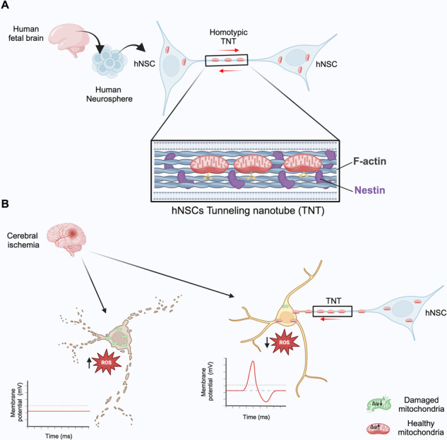
Pre-clinical trials have demonstrated the neuroprotective effects of transplanted human neural stem cells (hNSCs) during the post-ischemic phase. However, the exact neuroprotective mechanism remains unclear. Tunneling nanotubes (TNTs) are long plasma membrane bridges that physically connect distant cells, enabling the intercellular transfer of mitochondria and contributing to post-ischemic repair processes. Whether hNSCs communicate through TNTs and their role in post-ischemic neuroprotection remains unknown. In this study, non-immortalized hNSC lines derived from fetal human brain tissues were examined to explore these possibilities and assess the post-ischemic neuroprotection potential of these hNSCs. Using Tau-STED super-resolution confocal microscopy, live cell time-lapse fluorescence microscopy, electron microscopy, and direct or non-contact homotypic co-cultures, we demonstrated that hNSCs generate nestin-positive TNTs in both 3D neurospheres and 2D cultures, through which they transfer functional mitochondria. Co-culturing hNSCs with differentiated SH-SY5Y (dSH-SY5Y) revealed heterotypic TNTs allowing mitochondrial transfer from hNSCs to dSH-SY5Y. To investigate the role of heterotypic TNTs in post-ischemic neuroprotection, dSH-SY5Y were subjected to oxygen-glucose deprivation (OGD) followed by reoxygenation (OGD/R) with or without hNSCs in direct or non-contact co-cultures. Compared to normoxia, OGD/R dSH-SY5Y became apoptotic with impaired electrical activity. When OGD/R dSH-SY5Y were co-cultured in direct contact with hNSCs, heterotypic TNTs enabled the transfer of functional mitochondria from hNSCs to OGD/R dSH-SY5Y, rescuing them from apoptosis and restoring the bioelectrical profile toward normoxic dSH-SY5Y. This complete neuroprotection did not occur in the non-contact co-culture. In summary, our data reveal the presence of a functional TNTs network containing nestin within hNSCs, demonstrate the involvement of TNTs in post-ischemic neuroprotection mediated by hNSCs, and highlight the strong efficacy of our hNSC lines in post-ischemic neuroprotection. Human neural stem cells (hNSCs) communicate with each other and rescue ischemic neurons through nestin-positive tunneling nanotubes (TNTs). A Functional mitochondria are exchanged via TNTs between hNSCs. B hNSCs transfer functional mitochondria to ischemic neurons through TNTs, rescuing neurons from ischemia/reperfusion ROS-dependent apoptosis.


Conflict of interest statement

The authors declare no competing interests.

Figures

None
Human neural stem cells (hNSCs) communicate with each other and rescue ischemic neurons through nestin-positive tunneling nanotubes (TNTs). A Functional mitochondria are exchanged via TNTs between hNSCs. B hNSCs transfer functional mitochondria to ischemic neurons through TNTs, rescuing neurons from ischemia/reperfusion ROS-dependent apoptosis.
Fig. 1
Fig. 1. Nestin-positive TNT-like structures inside neurospheres and nestin supramolecular assembly state inside TNTs.
A and B Localization of Sox-2 (A), F-actin (red) with Nestin (green) (B) in 60 μm-thick neurosphere sections analyzed using laser-scanning confocal microscopy, followed by 3D reconstruction and XYZ slicing analysis. A single XY plane within the neurosphere is displayed (B, right). The zoomed-in box highlights nestin-positive TNTs-like structures (indicated by yellow arrows). Representative images were obtained from three different hNSCs donors. C Localization of nestin (green), Musashi-1 (magenta), and DAPI (cyano) in 60 μm-thick neurosphere sections analyzed via laser-scanning confocal microscopy, followed by 3D reconstruction. A 3D maximum projection is presented. The inset indicates the presence of nestin-positive TNTs-like structures connecting Musashi-1-positive cells. Representative images were obtained from three different hNSC donors. D 3D maximum projection of 97 z-planes displaying nestin localization in 60 μm-thick neurosphere sections analyzed using laser-scanning confocal microscopy, followed by 3D reconstruction. These nestin-positive connections form an extensive network that spans the entire volume of the neurosphere. Representative images were obtained from three different hNSC donors. E Nestin localization in neurospheres analyzed through Tau-STED super-resolution confocal microscopy. Discrete nestin supramolecular assemblies are visible. Analysis of nestin cluster dimensions reveals nestin assemblies within 0.01 and 0.05 μm2. Data were obtained using three different hNSC donors. F and G. SEM images at high resolution show the protrusions (arrows) on the neurospheres area (F). TEM images show the presence of elongated mitochondria (arrows) inside the protrusions (G).
Fig. 2
Fig. 2. TNTs-like structures during neurosphere migration.
A A migrating neurosphere was analyzed using time-lapse phase contrast microscopy. Highly dynamic TNTs-like structures were clearly visible starting two hours after seeding (yellow arrows). Representative images were obtained from three different hNSC donors. B and C Three hours after seeding, a migrating neurosphere was stained for mitochondria and F-actin and analyzed using live-cell time-lapse fluorescence microscopy. Highly dynamic TNTs-like structures, containing functional mitochondria (B) and F-actin (C), were present during neurosphere migration (yellow arrows). Supplementary Videos 1–4. Representative images were obtained from three different hNSC donors.
Fig. 3
Fig. 3. Tau-STED super-resolution microscopy reveals nestin assembly inside TNTs.
A Human neural stem cells (hNSCs) in 2D culture were stained for F-Actin (green) and Nestin (blue), with nuclei labeled using DAPI (pink). hNSCs formed connections through elongated F-actin-rich TNTs, which also displayed Nestin positivity (3D reconstruction created from deconvoluted XYZ confocal planes). Representative images were obtained from three different hNSC donors. B To obtain a more detailed view of the region marked as inset 1 in panel A, we employed Tau-STED super-resolution microscopy to examine a single XY plane. In inset 2, at a higher magnification, discernible and organized nestin structures are clearly visible within discrete clusters. Representative images were obtained from three different hNSC donors. C Utilizing 3D XYZ Tau-STED microscopy, we investigated TNTs connecting hNSC cells. Z-projections illustrate the spatial distribution of F-actin and nestin within TNTs. F-actin and nestin do not co-localize within TNTs. D Tau-STED analysis distinctly demonstrates the presence of supramolecular nestin clusters ~0.01–0.05 μm2 area inside TNTs. Representative images and data were obtained from three different hNSC donors.
Fig. 4
Fig. 4. Mitochondrial transport in F-actin and nestin-positive functional TNTs between hNSCs.
A Live-cell time-lapse widefield phase-contrast microscopy reveals that hNSCs rapidly generate TNTs when cultured in 2D. B hNSCs were stained with ∆Ψ-dependent MitoTracker Deep Red and analyzed using live-cell time-lapse fluorescence microscopy. The presence of functional mitochondria traveling through TNTs between hNSCs is indicated (Supplementary Videos 5–7). C and D hNSCs were stained with ∆Ψ-dependent MitoTracker Deep Red and analyzed through laser-scanning confocal microscopy, along with XY and Z-projection analysis. F-actin-positive (C) and Nestin-positive (D) TNTs, in which functional mitochondria were observed, are highlighted in zoomed insets. Representative images were obtained from three different hNSC donors.
Fig. 5
Fig. 5. TNT-mediated intercellular transfer of functional mitochondria between hNSCs.
A hNSCs were stained with ΔΨ-dependent MitoTracker Deep Red and co-cultured in direct contact with acceptor hNSCs stained with DiO. Live-cell time-lapse fluorescence microscopy revealed functional mitochondria traveling through TNTs from donor to acceptor hNSCs, as indicated by the yellow arrows (Supplementary videos 8–10). Representative images were obtained from three different hNSC donors. B and C The co-culture described in A was fixed, stained for F-actin (B and C) or Nestin (D), and analyzed using laser scanning confocal microscopy. This was followed by 3D reconstruction and XY and Z-projection analysis. The 3D reconstruction and slicing analysis of the DiO-acceptor cell clearly demonstrated the presence of mitochondria originating from the donor hNSC inside the acceptor DIO-positive hNSC. Representative images were obtained from three different hNSC donors. D hNSCs were stained with ΔΨ-dependent MitoTracker Deep Red and co-cultured without direct contact with acceptor hNSCs using the Transwell system. Donor cells were seeded on the Transwell, while acceptor cells were analyzed for Nestin and mitochondria localization using laser scanning confocal microscopy. This was followed by 3D reconstruction and XY and Z-projection analysis. Notably, there was a complete absence of mitochondria inside acceptor cells when the co-culture was not in direct contact. E The intercellular transfer of functional mitochondria was evaluated using non-contact transwell-based coculture (TW), conditioned medium (CM), or direct contact (DC). Donor hNSCs were stained with ΔΨ-dependent MitoTracker Deep Red (red), acceptor hNSCs were stained with DiO (green), and F-actin staining (cyan) was performed using a cell-permeable CellMask actin, avoiding cell permeabilization. The number of receiving cells containing mitochondria was measured in TW, CM, and DC. Only the direct contact coculture allows a robust transfer of mitochondria from donor to acceptor hNSCs (yellow arrows), while CM allows only rare and isolated mitochondrial signals inside receiving cells. Data are presented as mean percentage ± SEM. Data was obtained using three different hNSC donors. ****p < 0.0001. Representative images were obtained from three different hNSC donors.
Fig. 6
Fig. 6. Direct contact is essential for the complete rescue of post-ischemic dSH-SY5Y from ROS-triggered apoptosis.
A Normoxic, OGD, and OGD/R differentiated SH-SY5Y (dSH-SY5Y) were stained with Mitotracker Green AM, indicating mitochondria mass, and ΔΨ-dependent MitoTracker Red CMXRos, which selectively stained active and functional mitochondria. The Mitotracker Red/Green fluorescent ratio served as a semiquantitative analysis of mitochondria activity. OGD and OGD/R strongly affect mitochondria activity. OGD/R induces highly frequent changes in cell morphology in dSH-SY5Y, suggesting an apoptosis process (yellow arrows). ****p < 0.0001. B hNSCs were stained with fixable ΔΨ-dependent MitoTracker Deep Red (red) and cocultured in direct contact or in non-contact with human dSH-SY5Y stained with DiI and subject to oxygen-glucose deprivation (OGD/R). C Dil-labeled human dSH-SY5Y (Cyano) were cultured in normoxic conditions or exposed to OGD, washed, and reoxygenated (OGD/R) with or without a constant number of healthy hNSCs previously stained with the ∆Ψ-dependent MitoTracker Deep Red (hNSC-Mito) and co-cultured as reported in (B). After 24 h, co-cultures were analyzed for the levels of ROS production in dSH-SY5Y (C) and the percentage of apoptotic dSH-SY5Y by assessing caspase 3/7 activity (D). The quantitative analysis of ROS production and the measure of apoptotic dSH-SY5Y shows that hNSCs strongly reduced ROS production and apoptosis in OGD/R dSH-SY5Y only when cells are in direct contact with hNSCs. Data are presented as mean ± SEM or as percentage. Data were obtained using three different hNSC donors. *p = 0.0132; ****p < 0.0001.
Fig. 7
Fig. 7. TNT-mediated intercellular transfer of functional mitochondria from healthy hNSCs to OGD/R dSH-SY5Y.
A The intercellular transfer of functional mitochondria between healthy hNSCs and OGD/R dSH-SY5Y was evaluated using non-contact transwell-based coculture (TW), conditioned medium (CM), or direct contact (DC). Donor hNSCs were stained with ΔΨ-dependent MitoTracker Deep Red (red), acceptor OGD/R dSH-SY5Y were stained with DiO (green), and F-actin staining (cyan) was performed using a cell-permeable CellMask actin, avoiding cell permeabilization. The percentage of receiving OGD/R dSH-SY5Y containing mitochondria from hNSCs was measured in TW, CM, and DC. Only the direct contact coculture allows a robust transfer of mitochondria from donor hNSCs to acceptor OGD/R dSH-SY5Y, while CM allows only rare and isolated mitochondrial signals inside receiving cells. Data are presented as mean ± SEM or as percentage. Data were obtained using three different hNSC donors. In direct contact, we found heterotypic TNTs in which functional mitochondria run from hNSCs to OGD/R dSH-SY5Y. *p < 0.05; ****p < 0.0001. Representative images were obtained from three different hNSC donors. B Live-cell time-lapse fluorescence microscopy revealed functional mitochondria traveling through TNTs from donor-healthy hNSCs to acceptor OGD dSH-SY5Y stained with DiO, as indicated by the yellow arrows. (Supplementary Video 11–13). C Direct-contact coculture stained for caspase 3/7 activity and analyzed by confocal microscopy. The XY analysis demonstrated the presence of TNTs containing functional mitochondria connecting hNSCs with OGD-dSH-SY5Y and the Z-projections clearly show mitochondria originating from donor hNSCs (red, yellow arrows) inside OGD/R dSH-SY5Y which was negative for caspase 3/7 activity. D Non-contact coculture stained for caspase 3/7 activity and analyzed by XY confocal microscopy at high magnification. E The direct contact co-culture was analyzed for stemness and neuronal markers. In this direct-contact coculture hNSCs and OGD/R dSH-SY5Y preserved the expression of stemness (Sox-2) and neuronal (β3-tubulin) markers, respectively. Representative images and data were obtained from three different hNSC donors.
Fig. 8
Fig. 8. The direct contact with hNSCs is essential for the complete rescue of evoked action potentials, resting potential, and evoked inward current in post-ischemic dSH-SY5Y.
A Normoxic differentiated SH-SY5Y (dSH-SY5Y) was analyzed by patch clamp whole-cell experiments. Normoxic dSH-SY5Y generates evoked action potentials (Evoked APs) and shows both are evoked inward current and a sustained evoked outward current (evoked currents). Representative traces are reported. Complete data and statistics are reported in panel E and Table 1. B OGD/R dSH-SY5Y are not able to generate evoked action potentials and show a complete loss of the inward current or a significantly decreased inward current and a significant decrease in the outward current. Representative traces are reported. Complete data and statistics are reported in panel E and Table 1. C OGD/R dSH-SY5Y cocultured in direct contact with hNSCs were almost completely rescued. In this condition, OGD-R dSH-SY5Y exhibited the ability to generate evoked action potentials in 72.7% of analyzed cells and a conspicuous inward current in all cells analyzed, comparable with the control normoxic condition. Representative traces are reported. Complete data and statistics are reported in panel E and Table 1. D OGD/R dSH-SY5Y cocultured non-contact with hNSCs were not able to generate evoked action potentials in the 63.7% of analyzed cells and the inward current evoked remained not fully rescued. Representative traces are reported. Complete data and statistics are reported in panel E and Table 1. E Graphs and statistics of dSH-SY5Y resting potentials, peak inward and outward current in normoxia, OGD/R, OGDR+hNSCs in direct contact, and OGD/R+hNSCs in non-contact coculture are reported. n = 8 for normoxic dSH-SY5Y and n = 11 for other conditions. Data are presented as single data and mean ± SEM. Coculture data were obtained using three different hNSC donors. *p < 0.05; **p < 0.005; ***p < 0.0005.

References

    1. Vieira MS, Santos AK, Vasconcellos R, Goulart VAM, Parreira RC, Kihara AH, et al. Neural stem cell differentiation into mature neurons: mechanisms of regulation and biotechnological applications. Biotechnol Adv. 2018;36:1946–70. 10.1016/j.biotechadv.2018.08.002 - DOI - PubMed
    1. Hippenmeyer S. Principles of neural stem cell lineage progression: insights from developing cerebral cortex. Curr Opin Neurobiol. 2023;79:102695. 10.1016/j.conb.2023.102695 - DOI - PubMed
    1. Reynolds BA, Weiss S. Generation of neurons and astrocytes from isolated cells of the adult mammalian central nervous system. Science. 1992;255:1707–10. 10.1126/science.1553558 - DOI - PubMed
    1. Mazzini L, Gelati M, Profico DC, Sorarù G, Ferrari D, Copetti M, et al. Results from Phase I Clinical Trial with intraspinal injection of neural stem cells in amyotrophic lateral sclerosis: a long-term outcome. Stem Cells Transl Med. 2019;8:887–97. 10.1002/sctm.18-0154 - DOI - PMC - PubMed
    1. Mendez I, Sanchez-Pernaute R, Cooper O, Viñuela A, Ferrari D, Björklund L. et al. Cell type analysis of functional fetal dopamine cell suspension transplants in the striatum and substantia nigra of patients with Parkinson’s disease. Brain. 2005;128(Part 7):1498–510. 10.1093/brain/awh510 - DOI - PMC - PubMed

Substances

LinkOut - more resources